Impact of renal replacement therapy on the
respiratory function of patients under mechanical
ventilation
INTRODUCTION
Renal failure is an independent predictor of mortality in intensive care unit (ICU) patients, despite the technological advances in the management of critically ill patients and the new techniques of renal replacement therapy (RRT).(1) he mortality of renal failure remains high, especially when associated
with the dysfunction of other organs, such as acute lung injury (ALI). Patients with multiple organ dysfunction in need of dialysis and ventilatory support are common in the intensive care environment.
For a long time, certain alterations in the chest radiographs of patients with kidney injury have been attributed to the increased permeability of pulmonary Fernanda Maia Lopes1, José Roberval Ferreira1,
Dimitri Gusmao-Flores1,2
1. Intensive Care Unit, Hospital Geral Roberto Santos - Salvador (BA), Brazil.
2. Intensive Care Unit, Hospital Universitário
Professor Edgar Santos - Salvador (BA), Brazil. behavior and ventilatory mechanics Objective: To assess the oxygenation
after hemodialysis in patients under ventilatory support.
Methods: he present study was
performed in the general intensive care unit of a tertiary public hospital. Patients over 18 years of age under mechanical ventilation and in need of dialysis support were included. Each patient was submitted to 2 evaluations (pre- and post-dialysis) regarding the cardiovascular and ventilatory parameters, the ventilatory mechanics and a laboratory evaluation.
Results: Eighty patients with
acute or chronic renal failure were included. he analysis of the ventilatory mechanics revealed a reduction in the plateau pressure and an increased static compliance after dialysis that was independent of a reduction in blood volume. he patients with acute renal failure also exhibited a reduction in peak pressure (p=0.024) and an increase in the
dynamic compliance (p=0.026), whereas the patients with chronic renal failure exhibited an increase in the resistive pressure (p=0.046) and in the resistance of the respiratory system (p=0.044). he group of patients with no loss of blood volume after dialysis exhibited an increase in the resistive pressure (p=0.010) and in the resistance of the respiratory system (p=0.020), whereas the group with a loss of blood volume >2,000mL exhibited a reduction in the peak pressure (p=0.027). No changes in the partial pressure of oxygen in arterial blood (PaO2) or in the PaO2/the fraction
of inspired oxygen (PaO2/FiO2) ratio
were observed.
Conclusion: Hemodialysis was able to alter the mechanics of the respiratory system and speciically reduced the plateau pressure and increased the static compliance independent of a reduction in blood volume.
This study was conducted at the General Intensive Care Unit of the Hospital Geral Roberto Santos - Salvador (BA), Brazil.
Conflicts of interest: None. Submitted on March 28, 2013 Accepted on August 26, 2013
Corresponding author:
Fernanda Maia Lopes
Rua Rosalvo Firmino Lopes, 22 - Parque das Mangueiras - Centro
Zip code: 44530-000 - Sapeaçu (BA), Brazil E-mail: [email protected]
Impacto da terapia renal substitutiva na função respiratória de
pacientes sob ventilação mecânica
ABSTRACT
Keywords: Renal replacement therapy; Artiicial, respiration; Renal insuiciency
capillaries, known as the “uremic lung”.(2) However, recent
studies on experimental models of acute kidney injury that preserv body volume have demonstrated that the increased interstitial pulmonary edema correlates with the dysregulation of proteins involved in water and electrolyte transport,(3) and these changes take place within a few
hours.(4) In the past 50 years, the mortality of patients with
acute kidney injury has remained high despite the advances in intensive therapy, where the abnormalities observed in the lungs, which can also develop in the heart, brain, bone marrow and gastrointestinal tract,(5,6) might not be
completely reversible after the institution of dialysis therapy. Only a few studies have evaluated respiratory function after performing RRT.(7) Furthermore, an obvious limitation
of these studies was the use of less biocompatible dialysis membranes (such as cuprofan), thus causing pulmonary inlammation and worsening respiratory function.
Recently, patients under mechanical ventilation (MV) and on conventional intermittent hemodialysis (IHD) or sustained low-eiciency dialysis (SLED) were evaluated. No changes in the oxygenation or ventilatory mechanics were observed upon dialysis support.(8) However, that study(8)
evaluated only 31 patients and did not indicate how long after the dialysis therapy that the analyses were performed.
hus, given the lack of information on this subject, the objective of the present study was to evaluate the oxygenation behavior and the ventilatory mechanics after hemodialysis in patients under ventilatory support.
METHODS
he present study was performed in the general ICU of the Hospital Geral Roberto Santos (HGRS), in Salvador
(Bahia State, Brazil), a tertiary hospital of high complexity from the personal service network of the Brazilian Uniied Health System (Sistema Único de Saúde - SUS). he general ICU has 22 beds and assists adult clinical or surgical patients, and there are 7 beds that are preferentially occupied by renal failure patients with a referral for dialysis.
Patients over 18 years of age on MV who required dialysis support (IHD or SLED) were included. Only those patients for whom the informed consent form was signed by the responsible person were included, according to the ethical aspects of the 196/96 resolution of the National Health Council. his project was approved by the Research Ethics Committee of the HGRS under the protocol Nº 07/11.
he data acquisition was performed by one of the authors, who used a structured and pre-approved evaluation form especially developed for this survey that
was composed of questions regarding the demographic and clinical data, the cardiovascular and ventilatory parameters, the ventilatory mechanics, the laboratory evaluation and the dialysis procedure.
Initially, all patients were positioned in the dorsal decubitus position, with the head section inclined at an angle above 30° or according to the medical prescription. To avoid interference with the measured variables, no lung expansion therapy was performed at least 30 minutes prior to the data acquisition and up to the last measurement recorded (1 hour after dialysis). he patients were evaluated by a physiotherapist with respect to the need for bronchial clearance measures, which was conirmed by the presence of snoring in the lung auscultation and/or the presence of a jagged pattern in the low-volume curve; if conirmed, a tracheal aspiration was performed according to the recommendations of the American Association for Respiratory Care.(9) Immediately prior to the beginning
of dialysis, the irst collection of data was performed and a blood sample was drawn for the arterial blood gas analysis. Because of the recirculation rate, all variables were re-evaluated, and a new blood sample was drawn 1 hour after the end of the dialysis procedure. Each patient was submitted to 2 evaluations (pre- and post-dialysis).
he cardiovascular parameters, assessed with a DX 2022 multiparameter monitor (Dixtal Biomédica, Manaus, Brazil), were the heart rate (HR), in beats/min, and the mean blood pressure (MBP), the systolic blood pressure (SBP) and the diastolic blood pressure (DBP), in mmHg.
he assessed ventilatory parameters were as follows: the inspiratory pressure (Pins) and positive and end-respiratory pressure (PEEP), in cm H2O; the tidal volume (Vt), in mL; the respiratory rate (RR), in breaths/min (bpm); the minute volume (Vm), in L/min; and the fraction of inspired oxygen (FiO2), as a percent. he values from the display of the mechanical ventilator were recorded.
All patients who participated in the study were ventilated with the Vela ventilator machine (Viasys Healthcare, Critical Care Division, California, USA). For the measurement of the ventilatory mechanics, the patients had their ventilatory modality changed to volume-controlled ventilation (VCV) with a Vt of 8mL/kg, a constant low of 60L/min (square wave), a base PEEP and a RR of 15 bpm, along with the performance of a manual inspiratory pause of 3s. he hyperventilation technique was applied to exclude spontaneous breathing eforts (RR>30bpm for 2 minutes).
he evaluated parameters of the ventilatory mechanics were as follows: the peak pressure (Ppeak), considering the value from the display of the mechanical ventilator; the intrinsic PEEP (PEEPi), obtained by performing a manual occlusion of the expiratory valve of the ventilator for 3s at the end of expiration; the plateau pressure (Pplateau), obtained from the display of the ventilator by means of a manual occlusion of the airways for 3s at the end of inspiration; the resistive pressure (Pres), obtained by calculating the diference between the Ppeak and the Pplateau; the static compliance (Cstat), calculated by dividing the Vt by the Pplateau minus the PEEP and PEEPi; the dynamic compliance (Cdyn), calculated by dividing the Vt by the Ppeak minus the PEEP and PEEPi; and the resistance of the respiratory system (Rrs), calculated by dividing the Pres by the low. he Ppeak, PEEPi, Pplateau and Pres were measured in cm H2O. he Cstat and Cdyn were calculated in mL/cm H2O, and the Rrs was calculated in cm H2O/L/s.
All ventilatory mechanics measurements were performed by a single physiotherapist.
Renal replacement therapy
he patients were divided into 2 groups according to the type of renal failure: acute or chronic. Acute renal failure (ARF) was deined as an acute change in serum creatinine levels (a total increase >0.3mg/dL or a relative increase of 50% with respect to base levels) or urinary output (a reduction to <0.5mL/kg/min for more than 6h), according to the criteria of the Acute Kidney Injury Network (AKIN). Chronic renal failure (CRF) was deined as proposed by the Kidney Disease Outcomes Quality Initiative (KDOQI), which establishes that any adult individual exhibiting glomerular iltration (GF) <60mL/min/1.73m2 or, in the cases of GF ≥60mL/
min/1.73m2, a marker of structural kidney injury (for
example albuminuria) for ≥3 months sufers from CRF. Dialysis was prescribed by a nephrologist according to the patient’s need and hemodynamic condition. here
was no interference from the authors regarding the referral or the dialysis method selected by the nephrologist. he patients who were submitted to RRT were divided according to the methods: SLED and IHD. A blood low (Qa) varying from 150 to 300mL/min and a dialysate low (Qd) of 300 to 500mL/min was used. Ultrailtration was calculated as the diference between the volume of the ultrailtered luid and the infused dialysis luid. he patients with no contraindications for anticoagulation used heparin. he Polylux 8 LR polysulfone capillary dialyzer (Gambro Dialysatoren GmbH, Hechingen, Germany) was used for both methods.
Statistical analysis
he data were analyzed with the Statistical Package for Social Sciences (SPSS) Software, version 11.0 for Windows. he quantitative data were expressed as the mean±standard deviation (SD), and the rate data were expressed as the number of individuals (N) and percent cases (%). he normality of the distribution of the variables was assessed by means of the Kolmogorov-Smirnov test. In the comparisons between groups, an ANOVA was performed to assess the diference between the means among more than 2 variables, and the Pearson’s chi-squared or Fisher’s exact test was performed to assess the association between the qualitative variables. he paired Student’s t test was used for the paired samples (pre- and post-dialysis periods), according to the type of renal failure and the blood volume reduction. Diferences were considered as statistically signiicant at p<0.05.
RESULTS
Eighty patients were included. Eight patients were excluded for the following reasons: 2 sufered from previous chronic pulmonary diseases (chronic obstructive pulmonary disease and asthma), 1 sufered from congestive heart failure, 3 patients exhibited incidents during dialysis (cardiorespiratory arrest and hemodynamic instability with the need to interrupt dialysis), 1 used PEEP >10cm H2O and 1 was tracheostomized.
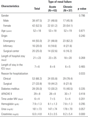
Table 1 - General patient characteristics
Characteristics
Type of renal failure Total Acute
(N=43)
Chronic
(N=37) p value
Gender 0.796
Male 38 (47.5) 21 (48.8) 17 (45.9)
Female 42 (52.5) 22 (51.2) 20 (54.1)
Age (years) 52±18 53±18 52±19 0.871
Origin 0.240
Emergency 44 (55.0) 21 (48.8) 23 (62.2)
Infirmary 16 (20.0) 8 (18.6) 8 (21.6)
Surgical center 20 (25.0) 14 (32.6) 6 (16.2) Length of hospital stay
(days) 21±23 23±25 18±20 0.269
Length of stay in the
ICU (days) 7±6 8±6 6±5 0.465
Reason for hospitalization 0.033
Clinical 53 (66.2) 24 (55.8) 29 (78.4)
Surgical 27 (33.8) 19 (44.2) 8 (21.6)
Diabetes mellitus 28 (35.0) 13 (30.2) 15 (40.5) 0.335
APACHE II 29±8 28±8 30±7 0.418
Time under MV (days) 6±4 7±5 5±4 0.201
Hemoglobin (g/dL) 7.9±1.3 8.1±1.3 7.8±1.3 0.295
Urea (mg/dL) 161±73 147±74 178±70 0.057
Creatinine (mg/dL) 6.0±4.8 4.3±3.5 8.2±5.4 0.000
ICU - intensive care unit; APACHE II - Acute Physiology and Chronic Health Evaluation II; MV - mechanical ventilation. The results are expressed as number (%) or the mean±standard deviation. ANOVA or chi-squared test.
Table 2 - Characteristics of the dialysis sessions
Dialysis
Type of renal failure Total Acute
(N=43)
Chronic
(N=37) p value
Type of dialysis 0.642
SLED 39 (48.8) 22 (51.2) 17 (45.9)
Conventional 41 (51.2) 21 (48.8) 20 (54.1)
Referral for dialysis 0.760
Uremia 49 (61.2) 27 (62.8) 22 (59.5)
Hypervolemia 31 (38.8) 16 (37.2) 15 (40.5)
Qa (mL/min) 195±32 195±36 195±28 0.998
Qd (mL/min) 362±93 355±90 370±96 0.493
Ultrafiltration (mL) 1,738±912 1,732±918 1,745±916 0.948
Anticoagulation 0.326
Yes 10 (12.5) 7 (16.3) 3 (8.1)
No 70 (87.5) 36 (83.7) 34 (91.9)
SLED - sustained low efficiency dialysis; Qa - blood flow; Qd - dialysate flow. The results are expressed as number (%) or the mean±standard deviation. ANOVA or chi-squared test (type of dialysis and referral for dialysis) and Fisher’s exact test (anticoagulation).
Regarding the hemodynamic parameters, the post-dialysis SBP was signiicantly higher in the CRF group (141±33mmHg versus 130±26mmHg; p=0.051) compared to the pre-dialysis period. With respect to the number and dose of vasopressors, both groups were similar and remained unchanged in the post-dialysis period.
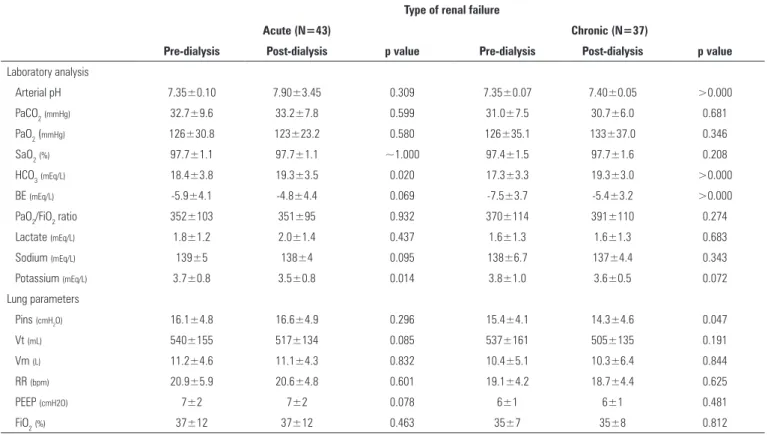
he changes observed after dialysis regarding the lung parameters and the laboratory analysis of arterial blood are shown in table 3. All patients used the pressure controlled ventilation (PCV) mode. here were no changes in the PaO2 or in the PaO2/FiO2 ratio in either group.
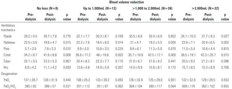
he changes in the ventilatory mechanics induced by dialysis are listed in table 4, whereas these changes with respect to the loss of volume after dialysis are itemized in table 5.
DISCUSSION
In the present study, an increase in the Cstat and a reduction in the Pplateau were observed after dialysis when the mechanical properties of the respiratory system were analyzed.
Another relevant inding was that the signiicant improvement in the ventilatory mechanics provided by dialysis in both patient groups did not lead to an improvement in oxygenation (the PaO2 and the PaO2/ FiO2 ratio). his result was observed even in the cases where a reduction in blood volume occurred upon ultrailtration. hese results are in agreement with the data observed in other studies(8,10,11) and can be explained by
the increased blood pH and the resulting left-shift of the oxyhemoglobin (HbO2) dissociation curve.
However, previous studies with a similar methodology have identiied a diferent behavior of ventilatory mechanics after dialysis. Steinhorst et al.(8) have studied 31 renal failure patients (acute and
chronic) on a dialysis program (IHD or SLED) and have observed no signiicant changes in the Rrs, the Cdyn or the Cstat, ascribing the obtained results to the low blood volume reduction. Huang et al.(10) have evaluated
14 patients and have observed an improvement in the auto-PEEP, the Rrs and the Cdyn, ascribing these results to the ultrailtration that was achieved. In an evaluation with 14 patients, Chen et al.(12) have observed an
Table 3 - Laboratory analysis and lung parameters, pre- and post-dialysis
Type of renal failure
Acute (N=43) Chronic (N=37)
Pre-dialysis Post-dialysis p value Pre-dialysis Post-dialysis p value
Laboratory analysis
Arterial pH 7.35±0.10 7.90±3.45 0.309 7.35±0.07 7.40±0.05 >0.000
PaCO2(mmHg) 32.7±9.6 33.2±7.8 0.599 31.0±7.5 30.7±6.0 0.681
PaO2 (mmHg) 126±30.8 123±23.2 0.580 126±35.1 133±37.0 0.346
SaO2(%) 97.7±1.1 97.7±1.1 ~1.000 97.4±1.5 97.7±1.6 0.208
HCO3(mEq/L) 18.4±3.8 19.3±3.5 0.020 17.3±3.3 19.3±3.0 >0.000
BE (mEq/L) -5.9±4.1 -4.8±4.4 0.069 -7.5±3.7 -5.4±3.2 >0.000
PaO2/FiO2 ratio 352±103 351±95 0.932 370±114 391±110 0.274
Lactate (mEq/L) 1.8±1.2 2.0±1.4 0.437 1.6±1.3 1.6±1.3 0.683
Sodium (mEq/L) 139±5 138±4 0.095 138±6.7 137±4.4 0.343
Potassium (mEq/L) 3.7±0.8 3.5±0.8 0.014 3.8±1.0 3.6±0.5 0.072
Lung parameters
Pins (cmH2O) 16.1±4.8 16.6±4.9 0.296 15.4±4.1 14.3±4.6 0.047
Vt (mL) 540±155 517±134 0.085 537±161 505±135 0.191
Vm (L) 11.2±4.6 11.1±4.3 0.832 10.4±5.1 10.3±6.4 0.844
RR (bpm) 20.9±5.9 20.6±4.8 0.601 19.1±4.2 18.7±4.4 0.625
PEEP (cmH2O) 7±2 7±2 0.078 6±1 6±1 0.481
FiO2(%) 37±12 37±12 0.463 35±7 35±8 0.812
Table 4 - Ventilatory mechanics, pre- and post-dialysis
Ventilatory mechanics
Type of renal failure
Acute (N=43)) Chronic (N=37)
Pre-dialysis Post-dialysis p value Pre-dialysis Post-dialysis p value
Ppico 33.0±8.8 31.3±7.8 0.024 30.0±6.7 29.7±6.4 0.708
Pplatô 22.5±6.1 20.1±5.9 0.000 21.2±5.7 19.3±5.6 0.000
Pres 10.5±6.3 11.0±5.1 0.374 8.9±4.1 10.4±3.7 0.046
Cest 36.8±14.9 43.4±18.4 >0.000 36.2±13.5 42.2±17.1 >0.000
Cdin 20.7±8.1 22.2±9.0 0.026 21.2±6.6 21.2±6.0 ~1.000
Rsr 14.9±10.1 15.2±8.0 0.777 12.6±6.5 14.7±6.0 0.044
p<0.005). he authors suggest that the negative balance produced by hemodialysis could lead to a decrease in peribronchial edema.
In the present study, a reduction in the Pplateau and an increase in the Cstat after hemodialysis were observed. his behavior can be explained by the redistribution of pulmonary ventilation that occurs after volume removal by ultrailtration and, perhaps, by the improvement of uremia through hemodialysis, thereby allowing the ventilation of alveoli that were previously illed with luid. hese indings might also suggest that the dialysis procedure favors the luid dynamics occurring within the interstitial and intravascular spaces, resulting in a reduction of the edema. Notably, an improvement of the Cstat was observed in the present
study, independent of the blood volume loss provided by ultrailtration. However, the group of patients with a volume loss >2,000mL also exhibited a reduced Ppeak, which suggests that the volume reduction provided by ultrailtration generates a signiicant reduction of the airway resistance component. he group with no blood volume loss exhibited an increase in the Pres and the Rrs, most likely due to the maintenance of a positive hydric balance.
Table 5 - Ventilatory mechanics and oxygenation, pre- and post-dialysis, according to blood volume reduction
Blood volume reduction
No loss (N=8) Up to 1,000mL (N=12) >1,000 to 2,000mL (N=38) >2,000mL (N=22)
Pre-dialysis
Post-dialysis
p value
Pre-dialysis
Post-dialysis
p value
Pre-dialysis
Post-dialysis
p value
Pre-dialysis
Post-dialysis
p value
Ventilatory mechanics
Ppeak 29.2±4.5 28.7±7.8 0.770 32.1±7.7 30.3±6.1 0.109 30.5±6.8 30.4±6.9 0.852 34.1±10.5 31.7±8.3 0.027
Pplateau 23.5±5.6 19.6±4.7 0.015 22.2±7.6 19.5±6.0 0.014 21±4.7 19.2±5.5 0.000 22.6±7.1 20.9±6.5 0.002
Pres 5.7±2.6 7.8±2.3 0.010 9.9±3.0 10.8±3.5 0.224 9.8±6.1 11.3±5.0 0.070 11.0±5.4 10.8±4.4 0.815
Cstat 34.2±8.7 41.6±8.8 0.008 36.8±17.2 46±19.8 0.002 35.7±10.8 42.5±17.1 0.000 38.5±19.1 42.3±20.7 0.015
Cdyn 23.1±3.5 23.3±5.3 0.857 20.4±8.2 22.3±7.7 0.110 21.0±6.7 21.6±8.2 0.441 20.5±9.2 21.2±8.1 0.396
Rrs 8.0±4.2 11.1±4.2 0.020 13.8±4.8 14.9±5.0 0.307 14.0±9.9 15.6±8.1 0.172 15.7±8.5 15.3±6.9 0.756
Oxygenation
PaO2 137±28.7 128±31.9 0.440 108±25.3 133±39.2 0.093 126±33.9 125±29.0 0.931 132±32.9 129±29.5 0.553
PaO2/FiO2 385±82 366±57 0.521 301±113 351±87 0.062 369±104 380±117 0.564 369±116 362±102 0.655
higher serum hemoglobin (Hb) levels and lower urea and creatinine levels. he CRF group exhibited an increased Rrs after dialysis, for which no explanation was found. Bianchi et al.(13) have reported that
repeated lung injury due to an overload of luid can damage the alveolar capillary membrane and induce a reduced difusion capacity. hese repeated episodes of subclinical edema that occur during each hemodialysis session might induce interstitial ibrosis in chronic renal patients who are under hemodialysis treatment for a longer period of time.(13) In addition to interstitial
ibrosis, other abnormalities, such as hyperemia and bronchitis, are commonly found in the autopsies of patients on chronic hemodialysis,(14) explaining the
increased Rrs.
Renal failure can compromise respiratory function in many ways. Perhaps acute edema is the most common and severe pulmonary complication in renal failure patients. Furthermore, the presence of luid in the pleural and abdominal compartments can restrict thoracic expansion, leading to changes in the ventilatory mechanics and gas exchange. he accumulation of luid around the small airways can also be found, resulting in premature closing and air entrapment, thus leading to increased respiratory work and PEEPi. hese pulmonary changes lead to reduced compliance and to increased airway resistance.(10) Finally, hypervolemia
can determine the diminish of alveolar ventilation, with CO2 retention and consequent acute respiratory acidosis, thus hampering the weaning from MV.(15)
he sequential evaluation of the ventilatory response during dialysis treatment suggests indicators for the progression of the disease and allows for an adequate adjustment of the ventilatory parameters within physiological limitations. Such evaluation aims to improve patient assistance, facilitate synchronization of the patient with the breather, assist in the MV removal program and provide a better quality of life to the patient.
he present study has some limitations that must be addressed. his study was a unicentric study with a small number of patients and no sample-size calculation. he clinical interventions during dialysis were not controlled. Furthermore, all pre- and post-dialysis evaluations were performed by an evaluator who knew to which group the patients belonged, thus raising the possibility of a bias in the analysis. he sample of CRF patients exhibited higher serum urea and creatinine levels than the ARF patients. his diference was because the CRF patient sample was from a hospital for dialysis referral through the Bahia State regulatory system; therefore, many patients were possibly admitted with no previous treatment of the disease and no possibility of previous dialysis and hence exhibited a dialysis urgency. his situation may have led to a bias in the present study.
CONCLUSION
Objetivo: Avaliar o comportamento da oxigenação e da mecânica ventilatória em pacientes com suporte ventilatório após a realização de hemodiálise.
Métodos: Estudo realizado na unidade de terapia intensiva geral de um hospital público terciário. Foram incluídos pacientes maiores de 18 anos, sob ventilação mecânica, com necessidade de suporte dialítico. Cada paciente foi submetido a duas avaliações (pré e pós-diálise) referentes a parâmetros cardiovasculares e ventilatórios, mecânica ventilatória e avaliação laboratorial.
Resultados: Foram incluídos 80 pacientes com insuiciência renal aguda e crônica. A análise da mecânica ventilatória demonstrou que houve redução da pressão de platô e aumento da complacência estática, após diálise, independentemente da redu-ção da volemia. Pacientes com insuiciência renal aguda também
apresentaram redução da pressão de pico (p=0,024) e aumento da complacência dinâmica (p=0,026), enquanto pacientes com insuiciência renal crônica apresentaram aumento da pressão resistiva (p=0,046) e da resistência do sistema respiratório (p=0,044). No grupo de pacientes sem perda volêmica, após diálise, observou-se aumento da pressão resistiva (p=0,010) e da resistência do sistema respiratório (p=0,020), enquanto no grupo com perda >2.000mL observou-se redução da pressão de
pico (p=0,027). Não houve alteração na PaO2 e nem na relação
PaO2/FiO2.
Conclusão: A hemodiálise foi capaz de alterar a mecânica do sistema respiratório, especiicamente reduzindo a pressão de platô e aumentando a complacência estática, independente da redução da volemia.
RESUMO
Descritores: Terapia de substituição renal; Respiração artiicial; Insuiciência renal
REFERENCES
1. Ko GJ, Rabb H, Hassoun HT. Kidney-lung crosstalk in the critically ill patient. Blood Purif. 2009;28(2):75-83. Review.
2. Bass HE, Singer E. Pulmonary changes in uremia. J Am Med Assoc. 1950;144(10):819-23.
3. Rabb H, Wang Z, Nemoto T, Hotchkiss J, Yokota N, Soleimani M. Acute renal failure leads to dysregulation of lung salt and water channels. Kidney Int. 2003;63(2):600-6.
4. Klein CL, Hoke TS, Fang WF, Altmann CJ, Douglas IS, Faubel S. Interleukin-6 mediates lung injury following ischemic acute kidney injury or bilateral nephrectomy. Kidney Int. 2008;74(7):901-9.
5. Kelly KJ. Distant effects of experimental renal ischemia/reperfusion injury. J Am Soc Nephrol. 2003;14(6):1549-58.
6. Liu M, Liang Y, Chigurupati S, Lathia JD, Pletnikov M, Sun Z, et al. Acute kidney injury leads to inflammation and functional changes in the brain. J Am Soc Nephrol. 2008;19(7):1360-70.
7. Munger MA, Ateshkadi A, Cheung AK, Flaharty KK, Stoddard GJ, Marshall EH. Cardiopulmonary events during hemodialysis: effects of dialysis membranes and dialysate buffers. Am J Kidney Dis. 2000;36(1):130-9. 8. Steinhorst CR, Vieira JM Jr, Abdulkader RC. Acute effects of intermittent
hemodialysis and sustained low-efficiency hemodialysis (SLED) on the pulmonary function of patients under mechanical ventilation. Ren Fail. 2007;29(3):341-5.
ACKNOWLEDGMENTS
he authors thank the patients who participated in this study, the team of the general ICU of the HGRS, especially the physiotherapists, for their collaboration,
afection and support, the nephrologists for their guidance, the hemodialysis and laboratory technicians for their collaboration in the data acquisition and all those from the Santa Casa de Misericórdia de São Félix for their encouragement to seek scientiic knowledge.
9. AARC clinical practice guideline. Endotracheal suctioning of mechanically ventilated adults and children with artificial airways. American Association for Respiratory Care. Respir Care. 1993;38(5):500-4.
10. Huang CC, Lin MC, Yang CT, Lan RS, Tsai YH, Tsao TC. Oxygen, arterial blood gases and ventilation are unchanged during dialysis in patients receiving pressure support ventilation. Respir Med. 1998;92(3):534-40. 11. Hoste EA, Vanholder RC, Lameire NH, Roosens CD, Decruyenaere JM,
Blot SI, et al. No early respiratory benefits with CVVHDF in patients with acute renal failure and acute lung injury. Nephrol Dial Transplant. 2002;17(12):2153-8.
12. Chen CW, Lee CH, Chang HY, Hsiue TR, Sung JM, Huang JJ. Respiratory mechanics before and after hemodialysis in mechanically ventilated patients. J Formos Med Assoc. 1998;97(4):271-7.
13. Bianchi PD, Barreto SS, Thomé FS, Klein AB. Repercussão da hemodiálise na função pulmonar de pacientes com doença renal crônica terminal. J Bras Nefrol. 2009;31(1):25-31.
14. Chan CH, Lai CK, Li PK, Leung CB, Ho AS, Lai KN. Effect of renal transplantation on pulmonary function in patients with end-stage renal failure. Am J Nephrol. 1996;16(2):144-8.