ABSTRACT
BACKGROUND AND OBJECTIVES: Trigeminal neuralgia is one of the most common neuropathic pains that compromise head and neck. It manifests as shock or burning pain normally evoked by non-noxious facial stimulations. Its etiopathology is not totally understood, but it is known that diferent mecha-nisms contribute to the establishment and maintenance of pain. his study aimed to address current contexts of epidemiology, diagnosis, management and pathophysiological mechanisms un-derlying trigeminal neuralgia in peripheral and central nervous systems.
CONTENTS: Inlammation and release of inlammatory me-diators, neuropeptides and neurotrophic factors, as well as de-generative changes of nervous ibers caused by direct nervous injury are relevant peripheral mechanisms which lead to al-tered sensitivity of nociceptive neurons, development of spon-taneous and exacerbated activity, allodynia and hyperalgesia. Among central mechanisms, exacerbated activation of central nociceptive neurons, neuroplasticity, changes in electrophysi-ological properties and neuronal hyperexcitability, in addition to changes in modulatory pain controls, lead to pain establish-ment and maintenance.
CONCLUSION: Several mechanisms are involved in neuro-pathic pains, both in peripheral and central levels, although spe-ciic trigeminal neuralgia events are not totally described. Studies concerning its speciic neurobiology are needed to understand functional and behavioral changes, which can contribute to tri-geminal neuralgia clinical management and treatment.
Keywords: Central sensitization, Etiopathology, Neuropathic pain, Peripheral sensitization, Trigeminal nerve, Trigeminal neuralgia.
Trigeminal neuralgia: peripheral and central mechanisms
Neuralgia trigeminal: mecanismos periféricos e centrais
Grazielle Mara Ferreira Costa1, Camila Megale de Almeida Leite2
1. Universidade Federal de Minas Gerais, Faculdade de Medicina, Programa de Pós-Gradu-ação em Patologia, Belo Horizonte, MG, Brasil.
2. Universidade Federal de Minas Gerais, Departamento de Morfologia, Instituto de Ciên-cias Biológicas, Belo Horizonte, MG, Brasil.
Submitted in November 12, 2014. Accepted for publication in October 21, 2015.
Conlict of interests: none – Sponsoring sources: FAPEMIG, PRPq/UFMG and CNPq.
Correspondence to: Camila Megale de Almeida Leite
Laboratório Profa. Conceição Machado, Bloco O3-245
Departamento de Morfologia - Instituto de Ciências Biológicas - UFMG Av. Antônio Carlos, 6627 – Pampulha
31270-901 Belo Horizonte, MG, Brasil. E-mail: [email protected]
© Sociedade Brasileira para o Estudo da Dor
RESUMO
JUSTIFICATIVA E OBJETIVOS: A neuralgia do trigêmeo é uma das dores neuropáticas mais comumente encontradas na região de cabeça e pescoço e manifesta-se como crises de choque ou queimação geralmente desencadeadas por estímulos não do-lorosos na região da face. A sua etiopatogenia não é totalmente conhecida, mas sabe-se que diversos mecanismos contribuem para seu estabelecimento. O objetivo deste estudo foi abordar os contextos atuais de epidemiologia, diagnóstico, tratamento e mecanismos isiopatológicos subjacentes à neuralgia do trigêmeo nos sistemas nervoso periférico e central.
CONTEÚDO: A inlamação e a liberação de mediadores inla-matórios, neuropeptídeos e fatores neurotróicos, assim como alterações degenerativas das ibras nervosas decorrentes da lesão nervosa direta são mecanismos periféricos relevantes que, em conjunto ou isoladamente, levam à sensibilidade alterada dos neurônios nociceptivos, com desenvolvimento de atividade es-pontânea e exacerbada e, consequentemente, dor eses-pontânea e hiperalgesia. Dentre os mecanismos centrais, a ativação exacer-bada de neurônios nociceptivos centrais, a neuroplasticidade, as alterações nas propriedades eletroisiológicas e a hiperexcitabi-lidade neuronal, além das modiicações nos controles modu-latórios da dor, são eventos que levam à instalação e à manuten-ção da dor.
CONCLUSÃO: Diversos mecanismos estão envolvidos nas dores neuropáticas, tanto a nível periférico quanto central, ape-sar dos eventos especíicos da neuralgia do trigêmeo não estarem totalmente elucidados. Estudos que abordem a sua neurobiolo-gia especíica são necessários para a compreensão das alterações funcionais e comportamentais presentes, com claras repercussões no tratamento e manuseio clínico da neuralgia do trigêmeo. Descritores: Dor neuropática, Etiopatologia, Nervo trigêmeo, Neuralgia do trigêmeo, Sensibilização central, Sensibilização periférica
INTRODUCTION
According to the International Association for the Study of Pain (IASP), pain is deined as an “unpleasant sensory and emotional experience associated to real or potential injuries or described in terms of such injuries”1. Neuropathic pain is caused by central or peripheral somatosensory nervous system injury or dysfunction and afects approximately 8% of the population1,2. Its etiopathology is complex and involves sev-eral biological mechanisms still not totally explained3. Trigeminal nerve is the ifth cranial nerve pair, being
sible for general head and face sensitivity. Trigeminal neural-gia (TN) is one of the most common neuropathic pains found in head and neck and is manifested as shock or burning crises in undeined intervals, in general triggered by non-painful stimulation in the face (allodynia)4.
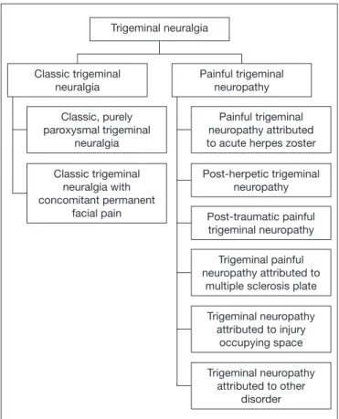
In general population, TN has incidence of 12.6 to 27/100,000 inhabitants5, being uncommon in people below 40 years of age and more common after 60 years of age. he incidence on people above 80 years of age is 25.9/100,000 inhabitants6. Recent data show that 55 to 70% of TN patients are females, being that 45% of patients refer maxillary pain7. TN diagnosis is essentially clinic, based on patients’ history and diagnostic criteria of IASP and of the International Head-ache Society (IHS)1,8. Pain attacks are paroxysmal lasting from fractions of seconds to two minutes in one or more trigeminal nerve innervation territories, being that the frequency may vary between hundreds of attacks per day to years of remis-sion between one crisis and the other. Pain shall mandatorily have the following criteria: 1) severe, acute and supericial pain; 2) pain described as similar to electric shock, cutting or burning; 3) pain with spontaneous start in trigger zone or triggered by innocuous stimulations in the trigger zone5,9. According to the International Classiication of Headache Disorders (ICHD-III, 2013)8, there are two major types of TN: classic and painful, which are subdivided as shown in igure 1.
Classic TN includes all cases without known etiology, as well as those amenable to having vascular trigeminal nerve com-pression. Painful TN, in turn, is diagnosed in cases second-ary to herpetic infection, trauma, tumors, multiple sclerosis or structural skull base deformities. Clinical presentation might be similar, by painful TN results from a structural injury difer-ent from vascular compression9-12. Studies point to a third type of TN, family TN, which is an extremely uncommon condi-tion, with few literature reports and represents less than 1% of all TN cases13.
Etiopathogenic mechanisms of TN are still not totally known, but the vast majority of classic TNs documented worldwide are related to trigeminal nerve compression at its exit from the pons by an aberrant arterial or venous loop. In cases of painful TN, as-sociated comorbidities seem to have etiopathogenic relationship with the establishment and maintenance of the disease14. herapeutic approaches for TN are divided in two modali-ties: pharmacological and surgical, according to the case15. For pharmacological treatment, anticonvulsants, such as carbam-azepine, oxcarbcarbam-azepine, baclofen, lamotrigine and pimozide are the most frequently prescribed drugs12. hese drugs block the propagation of the nervous impulse through changes in conductance of the nervous iber by blocking ion channels, al-lowing the control of neuronal excitability and of the synaptic activity16. Carbamazepine is successful in up to 70% of cases and has been the most frequently prescribed drug17.
Surgical treatment is indicated when patients do not respond to clinical treatments or when there is detection of vascular com-pression by imaging exams17,18. Among surgical techniques, there are vascular microdecompression, consisting in surgical devia-tion and isoladevia-tion of a vessel close to the nerve. Alcholizadevia-tion of peripheral branches is also a feasible alternative in refractory cases and consists in alcohol injection in trigeminal nerve pe-ripheral branches. Recently, some other procedures have been widely used, such as percutaneous electrocoagulation and ther-mocoagulation by radiofrequency. Both cause injury to the tri-geminal ganglion, preventing the passage of nociceptive stimuli and generating pain relief, associated to sensory deicits18.
ETIOPATHOGENY AND NEUROPATHOLOGY
Several factors contribute for the establishment of neuropathic pain without a speciic etiology. TN pathophysiology involves diferent neurophysiologic mechanisms, both in peripheral and central nervous systems, such as activation of receptors, transmis-sion and projection of nociceptive information and convergence of nociceptive aferents to central neurons, in addition to inter-actions between neurotransmitters and neuromodulators19,20 Recent studies have established some theories for TN etiol-ogy. One pathophysiological hypothesis is trigeminal conver-gence/projection. his theory proposes that nociceptive en-tries recurrent from head and neck converge to the trigeminal spinal nucleus (caudal sub-nucleus), leading to the release of neurotransmitters and vasoactive substances. hese mediators decrease second order neurons activation threshold, which also receive impulses from non-nociceptive ibers, generating in-Figure 1. Classiication of trigeminal neuralgia, according the
Interna-tional Classiication of Headache Disorders (ICHD-III)
Adapted from International Classiication of Headache Disorders, 20138.
Painful trigeminal neuropathy attributed to acute herpes zoster
Post-herpetic trigeminal neuropathy
Post-traumatic painful trigeminal neuropathy
Trigeminal painful neuropathy attributed to
multiple sclerosis plate
Trigeminal neuropathy attributed to injury
occupying space
Trigeminal neuropathy attributed to other
disorder Painful trigeminal
neuropathy Classic trigeminal
neuralgia
Trigeminal neuralgia
Classic, purely paroxysmal trigeminal
neuralgia
Classic trigeminal neuralgia with concomitant permanent
creased low of information transmitted to upper centers which interpret pain5,21-23. he bioresonance theory states that vibra-tion frequency of structures involving the trigeminal nerve may injury nervous ibers, leading to abnormal transmission of im-pulses and generating facial pain24. Another hypothesis is igni-tion. his suggests that injuries in trigeminal aferents of the root or of the trigeminal ganglion generate hyperexcitability of the axon and/or cell body, originating pain paroxysms as a function of exacerbated neuronal activity25.
In addition to such hypotheses, TN may also be induced by progressive dystrophy of trigeminal nerve peripheral branch-es which, in turn, may be evoked by comprbranch-ession syndrome and/or immunoallergic reactions26. Nervous and tissue injury caused by these processes leads to release of mediators which sensitize peripheral nervous terminations (peripheral sensitiza-tion), leading to neurochemical and phenotypic changes and increased excitability of trigeminal ganglion aferent neurons and trigeminal nuclei (central sensitization). In addition, there are changes in descending modulating pathways which change pain thresholds and perception27. Some studies also suggest that increased levels of IgE and histamine, common in some allergic processes, may be involved with the origin of TN. In these cases, the build-up of IgE in regions close to the trigemi-nal nerve may promote degranulation of mast cells with release of substances such as histamine and serotonin, which seem to play a relevant role in TN etiology28-30.
All proposed theories for TN etiology are based on central and peripheral cellular, molecular and electrophysiological mecha-nisms, described below.
hese mechanisms were, in their majority, shown through ex-perimental models of trigeminal nerve neuropathic pain, due to the diiculty in studying such mechanisms in humans.
Peripheral mechanisms
After the injury of a trigeminal nerve branch, as well as of other nerves, inlammatory process leads to the release of pro-inlam-matory cytokines, growth factors, hydrolytic enzymes and nitric oxide (NO), with consequent decrease in nociceptors activa-tion threshold and increase in nervous iber excitability31,32. In addition to inlammation and inlammatory mediators release, neuropeptides and neutrophic factors, degenerative nervous i-bers changes caused by direct damage, such as axonal injury and demielination, are also relevant peripheral mechanisms. Cytokines tumor necrosis factor alpha (TNF-α), interleukins 1 beta (IL-1β) and 6 (IL-6) have been implied both in central and peripheral sensitization33. TNF-α is released by virtually all immune system cells and by glial cells and causes metabolic and hemodynamic changes, in addition to distally activating other cytokines. his cytokine is able to promote neuronal hyperex-citability, to increase excitatory transmission and to promote inlammation in several nervous system levels, becoming an important mediator for chronic neuropathic pain, in addition to an excellent therapeutic target33.
IL-1β produces systemic inlammation, induces substance P (SP) and NO production, having important function in pain development and maintenance. here are strong evidences that
IL-1β reinforces synaptic transmission and neuronal activity in several nervous system sites33. IL-6, in turn, promotes neutro-phils maturation and activation, maturation of macrophages and diferentiation of cytotoxic T and natural killer lympho-cytes34. IL-6 is predominantly pro-inlammatory in neuropath-ic pain, promoting inlammation exacerbation through the ac-tivation of glial cells in the central nervous system33.
However, at injury site, it may promote antinociceptive action, as well as peripheral axon regeneration35. Due to this diferent efect, IL-6 is an attractive therapeutic target to treat neuro-pathic pain, as compared to other cytokines33. Notwithstand-ing described mediators beNotwithstand-ing involved with peripheral sensi-tization in neuropathic pain, few studies have established their inal role speciically for trigeminal neuralgia. While some au-thors suggest little relevance of IL-6 for trigeminal neuropathic pain36, others report important correlation of this mediator and of IL-1β for the generation of trigeminal neuropathic pain37,38. In addition to inlammatory mediators, neuropeptides have also been associated to neuropathic pain. Nervous injury of some trigeminal nerve branches has caused changes in the ex-pression of neuropeptides such as SP, peptide related to cal-citonin gene (CGRP), intestinal vasoactive polypeptide (VIP) and neuropeptide Y (NPY) in trigeminal nerve and trigeminal ganglion. So, it has been suggested that the build-up of neu-ropeptides at injury site may be related to the development of ectopic neural activity and to the development and modulation of neuropathic pain, although well-deined correlations with the development of pain along time have not yet been totally evidenced. In addition, injured nervous terminations release vasoactive neuropeptides, perpetuating inlammation (neuro-genic inlammation) and peripheral sensitization39.
Diferent painful conditions, including neuropathic pain, are associated to deregulation of neurotrophic factors expression37. Anderson & Rao37 have observed increased nervous growth fac-tor (NGF) expression in trigeminal nerve and nuclei and have suggested a role for this mediator in the mechanical allodynia observed after nervous injury in a trigeminal neuropathic pain model in rats40. Additionally, decreased production of neuro-trophic factors, among them glia-derived neuroneuro-trophic factor (GDNF), has been associated to the development of peripheral neuropathies. his factor is also involved in neuronal survival and plasticity mechanisms in response to injuries, in nervous injury repair, in addition to having a regulating role in the ac-tivity of nociceptive pathways41-43. More recent studies suggest that GDNF acts in a paracrine manner in trigeminal hyperal-gesia and may be a potential therapeutic target44.
de-mielination of trigeminal sensory ibers4.
Focal demielination leads to the apposition of demielinated axons with absence of glial cells processes between them, with possibility of generating ectopic or spontaneous nervous im-pulses and ephactic transmission between ibers, strengthening the role of cross excitation as relevant pathogenic mechanism4. Changes in anisotropy and difusivity in trigeminal nerve and gray matter were observed in TN patients, suggesting that microstructure abnormalities, not detectable by conventional imaging exams, and demielination without axonal injury are important factors in the pathogenesis of TN46-48.
All peripheral mechanisms, together or isolated, lead to changed sensitivity of nociceptive neurons, with the develop-ment of spontaneous and exacerbated activity and, as a conse-quence, spontaneous pain and hyperalgesia.
Central mechanisms
Central sensitization is the pathological and increased activa-tion of central primary nociceptive neurons, anatomic reor-ganization (neuroplasticity), changes in electrophysiological properties with development of hyperexcitability and modii-cations in pain modulation controls.
In the central nervous system, inlammatory mediators also participate in peripheral sensitization, interfere with hippo-campus cognitive, memory and mood functions and are able to excessively sensitize nociceptive neurons through excessive release of neurotrasmitters49, thus showing the role of inlam-mation in pain generation also at central level.
Molecular and cellular changes lead to the change of broad spectrum neurons phenotype into nociceptive neurons. Ad-ditionally, there are neuroplasticity phenomena with the es-tablishment of new synapses between nociceptive and non-nociceptive neurons. hese changes lead to allodynia, because changes in non-nociceptive stimuli pathways activate the noci-ceptive pathway, triggering pain43. In addition to mechanical allodynia, central changes lead to modulation controls changes and result in pain ampliication, increasing the ield where it is perceived and prolonging response to painful impulses50. here is facilitation, potentiation and ampliication of central responses with decreased inhibitory pathways and permanent activation of excitatory pathways, even in the absence of noci-ceptive stimuli51.
Neuronal hyperexcitability is a response to the remodeling of some transmembrane ion channels involved in the beginning of action potentials generation, such as sodium channels. In the neuronal membrane of the injured cell, increased sodium channels density, decreased potassium channels expression and increased expression of the auxiliary subunit of voltage-depen-dent calcium channels lead to depolarization of the membrane with consequent calcium inlow and action potential genera-tion3,18. Changes in channels density increase the period of neuronal depolarization and repetitive discharge52 and start central sensitization by several mechanisms, among them N-methyl-D-Aspartate receptors (NMDA), c-fos expression and transcription of genes coding dynorphin and encephalin, with consequent functional cell changes3,41.
Described mechanisms for central sensitization in neuropathic pain in general have not yet been fully tested for trigeminal neu-ropathic pain, which is highly relevant since pain in brain seg-ments has diferent mechanisms from pain outside the brain36. Studies have observed activation of astrocytes with increased ex-pression of IL-1β in trigeminal nuclei, associated to the develop-ment of allodynia and hyperalgesia and pain chronicity53,54. Kinase phosphorylation regulated by extracellular signal (ERK) seems to be involved with central sensitization leading to ther-mal orofacial hypersensitivity, showing the role of cell signal-ing cascades involvsignal-ing kinase proteins activated by mitogen (MAPK) in trigeminal neurons sensitization55. A recent study has shown that astrocytes are involved in NMDA glutamate receptors changes, important for TN central sensitization, through the release of D-serine neuromodulator aminoacid56. Speciically with regard to TN, painful stimulations were as-sociated to increased activity of the spinal tract nucleus of trigeminal, thalamus, primary and secondary somatosensory cortex, among other central areas. he activation of trigeminal innervation areas by non-nociceptive stimuli has induced exac-erbated activity in many of these central regions, showing the maintenance of a trigeminal nociceptive system sensitization status57. Decreased gray matter volume observed in primary and secondary somatosensory cortex, thalamus and other cen-tral structures of TN patients was correlated to longer disease duration, suggesting the adaptation of neuroplastic phenom-ena in response to chronic TN pain58.
CONCLUSION
Diferent mechanisms are involved with neuropathic pain installation and maintenance, both at peripheral and central levels. Speciic mechanisms underlying TN are not totally ex-plained, in spite of simultaneously and interdependently act-ing in diferent types of neuropathic pain. So, future studies addressing speciic TN neurobiology in its morphological, electrophysiological and molecular approaches are critically relevant for the understanding of functional and behavioral changes, with clear repercussions on TN treatment and clinical management.
REFERENCES
1. IASP. Part III: Pain Terms, a current list with deinitions and noter or usage. 2011; ISSN 978-0-931092-05-3.
2. Bouhassira D, Attal N, Alchaar H, Boureau F, Brochet B, Bruxelle J, et al. Compari-son of pain syndromes associated with nervous or somatic lesions and development of a new neuropathic pain diagnostic questionnaire (DN4). Pain. 2005;114(1)29-36. 3. Kraychete DC, Gozzani JL, Kraychete AC. [Neuropathic pain--neurochemical
as-pects]. Rev Bras Anestesiol. 2008;58(5)492-505. English, Portuguese.
4. Love S, Coakham HB. Trigeminal neuralgia: pathology and pathogenesis. Brain. 2001;124(Pt 12):2347-60.
5. Montano N, Conforti G, Di Bonaventura R, Meglio M, Fernandez E, Papacci F.
Advances in diagnosis and treatment of trigeminal neuralgia. her Clin Risk Manag. 2015;24(11):289-99.
6. Katusic S, Beard CM, Bergstralh E, Kurland LT. Incidence and clinical features of trige-minal neuralgia, Rochester, Minnesota, 1945-1984. Ann Neurol, 1990;27(1):89-95. 7. Ibrahim S. Trigeminal neuralgia: diagnostic criteria, clinical aspects and treatment
ou-tcomes. A retrospective study. Gerodontology. 2014;31(2):89-94.
8. he International Classiication of Headache Disorders, 3rd ed. Cephalalgia.
9. Zakrzewska JM, Linskey ME. Trigeminal neuralgia. BMJ Clin Evid. 2014;(10):pii1207. 10. he Internacional Classiication of Headache Disorders, 3nd ed. Cephalalgia.
2004;4(Suppl 1): 126-7.
11. Obermann M. Treatment options in trigeminal neuralgia. her Adv Neurol Disord. 2010;3(2):107-15.
12. Gronseth G, Cruccu G, Alksne J, Argof C, Brainin M, Burchiel K, et al. Practice pa-rameter: the diagnostic evaluation and treatment of trigeminal neuralgia (an evidence--based review): report of the Quality Standards Subcommittee of the American Acade-my of Neurology and the European Federation of Neurological Societies. Neurology, 2008;71(15):1183-90.
13. Ebner FH, Tatagiba M, Roser F. Familiar trigeminal nevralgia--microsurgical expe-rience and psychological observations, Acta Neurochir. 2010;152(2):381-2. 14. Kraft RM. Trigeminal neuralgia. Am Fam Physician. 2008;77(9):1291-6. 15. Mattos JM, Bueno FV, Mattos LR. Nevralgia do trigêmeo: um novo protocolo de
tratamento clinico. Rev Dor. 2005;6(4)652-5.
16. Dworkin RH, Backonja M, Rowbotham MC, Allen RR, Argof CR, Bennett GJ, et al. Advances in neuropathic pain: diagnosis, mechanisms, and treatment recommen-dations. Arch Neurol. 2003;60(11):1524-34.
17. Cheshire WP. Trigeminal neuralgia: for one nerve a multitude of treatments. Expert Rev Neurother. 2007;7(11):1565-79.
18. Frizzo HM, Hasse PN, Veronese RM. Neuralgia do trigêmeo: revisão bibliográica analítica. Rev Cir Traumatol Buco-Maxilo-Fac. 2004;4(4):212-7.
19. Svensson E. Pain mechanisms in myogenous temporomandibular disorders. Pain Fo-rum. 1997(6):158-65.
20. Siddall PJ, Cousins MJ. Pain mechanisms and management: an update. Clin Exp Pharmacol Physiol. 1995;22(10):679-88.
21. Sessle BJ, Hu JW. Mechanisms of pain arising from articular tissues. Can J Physiol Pharmacol. 1991;69(5):617-26.
22. Sessle BJ, Ho JW, Yu XM. New trends in referred pain and hyperalgesia, pain rese-arch and clinical management. In: Vecchiet D, Albe-Fessard D, Limblom U, editors. Brainstem Mechanisms of Referred Pain and Hyperalgesia in the Orofacial and Tem-poromandibular Region. 7th ed. Amsterdam: Elsevier; 1993. 59-71p.
23. Bonica JL. he Management of Pain. 2nd ed. Malvern, PA: Lea and Febiger; 1990. 180p.
24. Jia DZ, Li G. Bioresonance hypothesis: a new mechanism on the pathogenesis of trigeminal neuralgia. Med Hypotheses. 2010;74(3):505-7.
25. Devor M, Amir R, Rappaport ZH. Pathophysiology of trigeminal neuralgia: the igni-tion hypothesis. Clin J Pain. 2002;18(1):4-13.
26. Kumar S, Rastogi S, Kumar S, Mahendra P, Bansal M, Chandra L. Pain in trigeminal neuralgia: neurophysiology and measurement: a comprehensive review. J Med Life. 2013;6(4):383-8.
27. Jarvis MF, Boyce-Rustay JM. Neuropathic pain: models and mechanisms. Curr Pharm Des. 2009;15(15):1711-6.
28. Sabalis GI, Karlov VA, Morkunas RM, Stropus RA. Peripheral mechanisms of the pathogenesis of trigeminal neuralgia. Zh Nevropatol Psikhiatr Im S S Korsakova. 1982;82(4):25-9.
29. Hanes WJ. Tic douloureux: a new allergyc approach to the etiology and treatment. A reporto f seventy-six cases. Ann Allergy. 1962;20:635-48.
30. Smirnov VA. Odontologic and other etiologic factors in trigeminal neuralgia. Zh Ne-vropatol Psikhiatr Im S S Korsakova. 1976;76(11):1639-42.
31. Julius D, Basbaum AI. Molecular mechanisms of nociception. Nature. 2001;413(6852):203-10.
32. Daemen MA, Kurvers HA, Kitslaar PJ, Slaaf DW, Bullens PH, Van den Wildenberg FA. Neurogenic inlammation in an animal model of neuropathic pain. Neurol Res. 1998;20(1):41-5.
33. Austin PJ, Moalem-Taylor G. he neuro-immune balance in neuropathic pain: in-volvement of inlammatory immune cells, immune-like glial cells and cytokines. J Neuroimmunol. 2010;229(1-2):26-50.
34. de Oliveira CM, Sakata RK, Issy AM, Gerola LR, Salomão R. [Citokines and pain]. Rev Bras Anestesiol. 2011;61(2):260-5. English, Portuguese, Spanish.
35. Singh JA, Beg S, Lopez-Olivo MA. Tocilizumab for rheumatoid arthritis. Cochrane
Database Syst Rev. 2010;7(7):CD008331.
36. Latrémolière A, Mauborgne A, Masson J, Bourgoin S, Kayser V, Hamon M, et tal. Diferential implication of proinlammatory cytokine interleukin-6 in the de-velopment of cephalic versus extracephalic neuropathic pain in rats. J Neurosci. 2008;28(34):8489-501.
37. Anderson LC, Rao RD. Interleukin-6 and nerve growth factor levels in periphe-ral nerve and brainstem after trigeminal nerve injury in the rat. Arch Operiphe-ral Bird. 2001;46(7):633-40.
38. Takeda M, Takahashi M, Matsumoto S. Contribution of activated interleukin recep-tors in trigeminal ganglion neurons to hyperalgesia via satellite glial interleukin-1beta paracrine mechanism. Brain Behav Immun. 2008;22(7):1016-23.
39. De Corato A, Lisi L, Capuano A, Tringali G, Tramutola A, Navarra P, et al. Trigemi-nal satellite cells express functioTrigemi-nal calcitonin gene-related peptide receptors, who-se activation enhances interleukin-1β pro-inlammatory efects. J Neuroimmunol. 2011;237(1-2):39-46.
40. Ossipov MH. Growth factors and neuropathic pain. Curr Pain Headache Rep. 2011;15(3):185-92.
41. Takeda M, Takahashi M, Hara N, Matsumoto S. Glial cell line-derived neurotrophic factor modulates the excitability of nociceptive trigeminal ganglion neurons via a pa-racrine mechanism following inlammation. Brain Behav Immun. 2013;28:100-7. 42. Airaksinen MS, Saarma M. he GDNF family: signalling, biological functions and
therapeutic value. Nat Rev Neurosci. 2002;3(5):383-94.
43. Woolf CJ. Somatic pain--pathogenesis and prevention. Br J Anaesth. 1995;75(2):169-76. 44. Suzuki R, Dickenson AH. Neuropathic pain: nerves bursting with excitement.
Neu-roreport. 2000;11(12):R17-21.
45. Moalem G, Tracey DJ. Immune and inlammatory mechanisms in neuropathic pain. Brain Res Rev. 2006;51(2):240-64.
46. Aguiar de Sousa D, Geraldes R, Gil-Gouveia R, de Sá JC. New daily persistent heada-che and radiologically isolated syndrome. J Neurol. 2013;260(8):2179-81. 47. Liu Y, Li J, Butzkueven H, Duan Y, Zhang M, Shu N, et al. Microstructural
abnor-malities in the trigeminal nerves of patients with trigeminal neuralgia revealed by multiple difusion metrics. Eur J Radiol. 2013;82(5):783-6.
48. Hodaie M, Chen DQ, Quan J, Laperriere N. Tractography delineates microstructural changes in the trigeminal nerve after focal radiosurgery for trigeminal neuralgia. PLoS One. 2012;7(3):e32745.
49. Kim KH, Kim JI, Han JA, Choe MA, Ahn JH. Upregulation of neuronal nitric oxide synthase in the periphery promotes pain hypersensitivity after peripheral nerve injury. Neuroscience. 2011;8(190):367-78.
50. Dickenson AH. Recent advances in the physiology and pharmacology of pain: plasti-city and its implications for clinical analgesia. J Psychopharm, 1991 (5)342-351. 51. Aydinli I, Keskinbora K. New immune system approach to pain pathology-interaction
with the sensory system. Agri. 2004, 16(2):7-16.
52. Nagano M, Sakai A,Takahashi N, Umino M, Yoshioka K, Suzuki H. Decreased ex-pression of glial cell line-derived neurotrophic factor signaling in rat models of neuro-pathic pain. Br J Pharmacol. 2003;140(7)1252-60.
53. Lee S, Zhao YQ, Ribeiro-da-Silva A, Zhang J. Distinctive response of CNS glial cells in oro-facial pain associated with injury, infection and inflammation. Mol Pain. 2010;6:79.
54. Suzuki I, Tsuboi Y, Shinoda M, Shibuta K, Honda K, Katagiri A, et al. Involvement of ERK phosphorylation of trigeminal spinal subnucleus caudalis neurons in thermal hypersensitivity in rats with infraorbital nerve injury. PLoS One. 2013;8(2):e57278. 55. Xu Q, Yaksh TL. A brief comparison of the pathophysiology of inlammatory versus
neuropathic pain. Curr Opin Anaesthesiol. 2011;24(4):400-7.
56. Dieb W, Haidi A. Astrocytes are involved in trigeminal dynamic mechanical allo-dynia: potentialrole of D-serine. J Dent Res. 2013.92(9):808-13.
57. Moisset X, Villain N, Ducreux D, Serrie A, Cunin G, Valade D, et al. Functional brain imaging of trigeminal neuralgia. Eur J Pain. 2011;15(2):124-31.