Ro le o f se nso ry ne rvo us syste m
vaso active pe ptide s in hype rte nsio n
Clinical Center, Michigan State University, East Lansing, MI, USA R.E. Watson, S.C. Supowit,
H. Zhao, K.A. Katki and D.J. DiPette
Abstract
The goal of the present research was to elucidate the roles and mechanisms by which the sensory nervous system, through the actions of potent vasodilator neuropeptides, regulates cardiovascular function in both the normal state and in the pathophysiology of hypertension. The animal models of acquired hypertension studied were deoxycor-ticosterone-salt (DOC-salt), subtotal nephrectomy-salt (SN-salt), and Nw
-nitro-L-arginine methyl ester (L-NAME)-induced hypertension during pregnancy in rats. The genetic model was the spontaneously hypertensive rat (SHR). Calcitonin gene-related peptide (CGRP) and substance P (SP) are potent vasodilating neuropeptides. In the ac-quired models of hypertension, CGRP and SP play compensatory roles to buffer the blood pressure (BP) increase. Their synthesis and release are increased in the DOC-salt model but not in the SN-salt model. This suggests that the mechanism by which both models lower BP in SN-salt rats is by increased vascular sensitivity. CGRP functions in a similar manner in the L-NAME model. In the SHR, synthesis of CGRP and SP is decreased. This could contribute to the BP elevation in this model. The CGRP gene knockout mouse has increased baseline mean arterial pressure. The long-term synthesis and release of CGRP is increased by nerve growth factor, bradykinin, and prostaglandins and is decreased by a
2-adrenoreceptor agonists and glucocorticoids. In several animal models, sensory nervous system vasoactive peptides play a role in chronic BP elevation. In the acquired models, they play a compensatory role. In the genetic model, their decreased levels may contribute to the elevated BP. The roles of CGRP and SP in human hypertension are yet to be clarified.
Co rre spo nde nce R.E. Watson B 338 Clinical Center Michigan State University East Lansing, MI 48824-1315
USA
Fax: + 1-517-432-1326 E-mail: ralph.watson@ ht.msu.edu
Presented at the IV International Symposium on Vasoactive Peptides, Belo Horizonte, MG, Brazil,
O ctober 19-21, 2001.
Research supported by the National Institutes of Health (NIH # HL44277).
Received November 28, 2001 Accepted January 22, 2002
Ke y wo rds
·Hypertension
·Calcitonin gene-related
peptide
·Substance P ·Neurons (afferent)
Intro ductio n
Traditionally, the role of the sensory ner-vous system in blood pressure regulation has been through afferent baroreceptor-mediated mechanisms. There is now increasing evi-dence that, in addition to afferent mechan-isms, the sensory nervous system plays a role in multiple areas (e.g., nociception and
CGRP is a 37-amino acid neuropeptide derived from the tissue-specific splicing of the primary RNA transcript of the calcito-nin/CGRP gene (1,2), which will be referred to as the a-CGRP gene. Calcitonin is
pro-duced mainly in the parafollicular cells of the thyroid, but CGRP synthesis occurs al-most exclusively in regions of the central and peripheral nervous systems (3). There is a second CGRP gene (ß-CGRP) that does not produce calcitonin, and also produces CGRP primarily in central neuronal tissues (3,4). The two CGRP genes, a- and ß- in the
rat and I and II in humans, differ in their protein sequences by one and three amino acids, respectively, and the biological activi-ties of the two peptides are quite similar in most vascular beds (5).
SP is an 11-amino acid peptide sensory neurotransmitter that mediates pain, touch, and temperature. Like CGRP, it is involved in many physiologic activities including smooth muscle contraction and vasodilation (5-8). SP is a member of the tachykinin family. The three major mammalian tachyki-nins are SP, neurokinin A (NKA), and neu-rokinin B (NKB) (9). SP and NKA are en-coded by the preprotachykinin A gene, and NKB is encoded by the preprotachykinin B gene (5,9,10). SP is often released from the same sensory nerve terminals as CGRP. SP has been shown to regulate blood flows of various organs.
D istributio n and lo calizatio n o f immuno re active CGRP and substance P
Immunoreactive (that which can react to the antibody in the assay) CGRP and SP and their receptors are widely distributed in the nervous and cardiovascular systems (5,8,11). In the peripheral nervous system, prominent sites of CGRP and SP synthesis are in the spinal nerve dorsal root ganglia (DRG). These structures contain the cell bodies of sensory nerves that terminate centrally in laminae I/
II of the dorsal horn of the spinal cord and peripherally in blood vessels (11-13). Blood vessels in all vascular beds are surrounded by a dense perivascular CGRP and SP neural network. In these vessels CGRP- and SP-con-taining nerves are found at the junction of the adventitia and the media, passing into the muscle layer (3,5). These peptides are often co-localized in the same peripheral nerve ter-minals. Circulating CGRP and SP are believed to be a spillover phenomenon from these perivascular nerve terminals caused by the release of these peptides to promote vasodila-tion or other funcvasodila-tions (3). Receptors for CGRP have been identified both in the me-dia and intima of resistance vessels. SP re-ceptors and neurokinin-1 (NK-1) rere-ceptors, are found in endothelial cells (9).
Cardio vascular actio ns o f CGRP and substance P
of activating ATP-activated potassium chan-nels of vascular smooth muscle (3). There is additional evidence that the vasodilator re-sponse evoked by CGRP is mediated, in part, by nitric oxide (NO) release and that various vascular beds differ in their dependence on the endothelium for the dilator response to CGRP (3). The vasodilator response medi-ated by NO is endothelium dependent, while the other mechanisms directly affect the vas-cular smooth muscle. Therefore, CGRP can dilate blood vessels through endothelium-dependent and -inendothelium-dependent mechanisms.
The vasodilator action of SP is always endothelium dependent (8,13) and is medi-ated by NK-1 receptors locmedi-ated in endotheli-al cells (9). There are three tachykinin recep-tor subtypes known as 1, 2, and NK-3. SP is the preferred ligand (binding mole-cule) for NK-1 receptors, NKA for NK-2, and NKB for NK-3, but all three tachykinins have some affinity for all three NK receptors if a high enough dose is given. The three receptors all belong to the superfamily of G-protein-coupled receptors. Phosphoinositol is a cell membrane phospholipid. Each of these three receptors causes phosphoinositol breakdown, which initiates the vascular ac-tions of the tachykinins. NK-1 receptors mediate tachykinin-induced vasodilation by an endothelium-dependent mechanism in-volving the release of both NO and a hyper-polarizing/vasodilator factor different from NO (9).
Re le ase o f ne uro pe ptide s fro m se nso ry ne rve te rminals
Sensory nerve fibers are classified as cap-saicin (the active ingredient in pepper) sensi-tive and capsaicin insensisensi-tive. The capsai-cin-sensitive nerves have the receptor for capsaicin. When capsaicin-sensitive sensory nerve fibers are exposed to capsaicin, they release CGRP and SP. CGRP- and SP-rich nerve fibers are components of the sensory nervous system, comprising principally
cap-saicin-sensitive C- and Ad-fiber nerves that
respond to chemical, thermal, and mechani-cal stimuli (6,7,17,18). These stimuli cause release of CGRP and SP. If the stimulation is powerful enough, it also causes propagation of sensory information back to the central nervous system. Although these nerves have traditionally been thought to sense stimuli in the periphery and transmit the information centrally, there was early evidence that they also have an efferent function. It is clear that DRG neuron-derived peptides are released at peripheral sensory nerve terminals in the absence of afferent nerve stimulation (17). The continuous release of peptides from DRG neurons may reflect a paracrine function, with the released peptides binding to nearby receptors. This implies that these neurons participate in the continuous regulation of blood flow and other tissue activities. In-deed, it has been postulated that some DRG neurons are specialized in controlling pe-ripheral effector mechanisms, but have no role in sensation (17). Sensory nerve termi-nals can release CGRP and SP in response to local factors including nerve growth factor (NGF) (19), vascular wall tension (6,7), bradykinin/prostaglandins (20,21), endothe-lin, and the sympathetic nervous system (21). We have demonstrated that these same fac-tors which alter acute release of CGRP can also modulate the long-term production and release of this peptide. Using primary cul-tures of adult rat DRG neurons we have reported that NGF or bradykinin/prostaglan-dins (20,22) can stimulate CGRP synthesis and release, whereas glucocorticoids (22) or
a2-adrenoreceptor agonists (23) inhibit the
stimulatory effects of NGF on CGRP. Thus, alterations in these factors, some of which are known to occur in hypertension, may mediate any changes seen in CGRP or SP synthesis and release.
Ro le o f CGRP in hype rte nsio n
sig-nificantly decrease high blood pressure in humans (3,11), it is not clear what role CGRP plays in human hypertension. The reported levels of circulating immunoreactive CGRP in hypertensive humans have been conflict-ing (3,11). This has been attributed to sev-eral factors including the assay itself, hetero-geneity of the disease, severity and duration of the hypertension, the degree of end organ damage, and the variety of treatment regi-mens used in these patients (3). In contrast, a direct role for CGRP in experimental hyper-tension has now been established. Earlier reports demonstrated that CGRP can attenu-ate chronic hypoxic pulmonary hyperten-sion (24). We studied CGRP in three animal models of hypertension: 1) deoxycorticos-terone-salt (DOC-salt) model, a model in which the rat undergoes a uninephrectomy followed by excess mineralocorticoid and salt administration (25); 2) subtotal nephrec-tomy-salt (SN-salt) model, in which the rat undergoes a uninephrectomy plus surgical removal of 66% of the remaining kidney followed by excess salt administration (25), and 3) L-NAME-induced hypertension dur-ing pregnancy, in which the rat is given the NO inhibitor Nw-nitro-L-arginine methyl
es-ter (25). We have demonstrated for the first time that CGRP acts as a compensatory de-pressor mechanism to partially attenuate the blood pressure increase in these three mod-els of experimental hypertension (26-30). Because these studies were done acutely, the important question regarding the long-term participation of CGRP in hypertension has not been answered.
Ro le o f substance P in hype rte nsio n
SP is often co-localized and co-released with CGRP in sensory nerve terminals. The spontaneously hypertensive rat-stroke-prone strain (SHRSP) is a genetic model of exper-imental hypertension (25). Reduced levels of SP have been described in SHRSP and in human essential hypertension (31).
There-fore, SP could contribute to the elevated blood pressure through the decreased activ-ity of a counterregulatory mechanism. Kohl-mann et al. (31) have employed a non-pep-tide SP receptor (NK-1) antagonist to assess the hemodynamic role of this peptide in five models of experimental hypertension. In ad-dition to the DOC-salt and SN-salt models, they studied the SHR and two renovascular models, the two-kidney, one-clip and one-kidney, one-clip. In the first, both native kidneys are intact and a partially constricting clip is placed on one renal artery. In the second, the rat undergoes uninephrectomy, and a partially constricting clip is placed on the remaining renal artery (25). In conscious unrestrained rats the SP antagonist induced significant increases in mean arterial pres-sure (MAP) in three salt-dependent models, DOC-salt, SN-salt, and one-kidney, one-clip hypertensive rats. In contrast, the antagonist produced nonsignificant effects on the MAP in two-kidney, one-clip and SHR (salt-inde-pendent models). Therefore, SP may act as a partial compensatory mechanism to counter-act the blood pressure increase in salt-pendent hypertension. The experiments de-scribed above were performed acutely, so the long-term participation of SP in hyper-tension is unknown.
He mo dynamic e ffe cts o f CGRP in e xpe rim e ntal hype rte nsio n
D e o xyco rtico ste ro ne -salt hype rte nsio n
correspond-ingly immunoreactive CGRP levels were el-evated in laminae I/II of the spinal cord compared to the control groups (26). Fur-thermore, this increase in neuronal CGRP expression in the DOC-salt rats was specific for the DRG, since we did not observe any alterations in the brain or brain stem. In order to determine if these changes in CGRP were playing an important hemodynamic role, groups of rats had intravenous (for drug administration) and arterial (for continuous MAP monitoring) catheters surgically placed and were studied in the conscious, unre-strained state. Injection of saline did not alter MAP in any of the five groups and adminis-tration of the CGRP receptor antagonist CGRP8-37 did not significantly increase MAP in any of the four normotensive groups. How-ever, administration of the antagonist to the DOC-salt rats rapidly induced, in a dose-dependent manner, a further increase of the elevated MAP (Figure 1). In light of the rapid onset of the hypertensive effects of CGRP8-37 and because the antagonist prob-ably does not penetrate the central nervous system, it is likely that the pressor activity of CGRP8-37 results from a direct interaction of the antagonist with vascular CGRP recep-tors (32-35). These data support the hypo-thesis that in DOC-salt hypertension CGRP is acting as a compensatory depressor to buffer the increased blood pressure (27).
Subto tal ne phre cto my-salt hype rte nsio n
We examined the effect of endogenous CGRP on blood pressure in SN-salt-induced hypertension, another model of low-renin, salt-dependent hypertension (28). SN-salt and normotensive controls were instrumented and given saline or CGRP8-37 as described above (Figure 2). The effects of two different doses of CGRP8-37 in the control group were simi-lar to those observed with saline, which did not significantly alter the MAP. In contrast, administration of antagonist to the SN-salt rats produced a dose-dependent increase of
the elevated MAP, similar to what was ob-served in the DOC-salt rats. These results suggest that, in this setting, CGRP is also playing a compensatory depressor role. Sur-prisingly, when the CGRP mRNA and pep-tide levels were quantified in the DRG from hypertensive and control rats there were no detectable changes. These results suggested a second mechanism by which CGRP exerts its counterregulatory action. As opposed to increased CGRP synthesis and release, this effect may be mediated through an increase in the vascular responsiveness to CGRP. Indeed, additional experiments indicate that vascular reactivity to CGRP is enhanced in SN-salt rats (36).
L-NAME-induce d hype rte nsio n
The purpose of the following experiments was to determine whether CGRP is involved in the vascular adaptations that occur in normal pregnancy and its role in the hyper-tension of L-NAME-treated female rats. In-hibition of NO synthesis with L-NAME in pregnant rats has been shown to cause hy-pertension, proteinuria, fetal growth retarda-tion, and increased fetal mortality (37). The co-administration of CGRP with L-NAME prevented the gestational, but not the post-partum hypertension (37). It also prevented the proteinuria and significantly decreased pup mortality (37). Further studies revealed that this differential effect of CGRP on blood
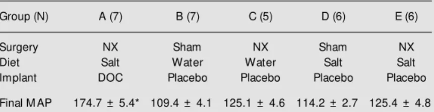
Table 1. Summary of the DOC-salt (group A) and normotensive control groups (B-E) studied.
Group (N) A (7) B (7) C (5) D (6) E (6)
Surgery NX Sham NX Sham NX
Diet Salt Water Water Salt Salt Implant DOC Placebo Placebo Placebo Placebo
Final M AP 174.7 ± 5.4* 109.4 ± 4.1 125.1 ± 4.6 114.2 ± 2.7 125.4 ± 4.8
pressure during gestation and postpartum is mediated by progesterone (38). Gestational progesterone is high, while postpartum pro-gesterone is low. Similar to the findings in gestational rats, CGRP reversed the hyper-tension in L-NAME-treated oophorecto-mized rats receiving progesterone injections. Therefore, these studies suggest that CGRP is anti-hypertensive in L-NAME-treated preg-nant and non-pregpreg-nant rats and that the va-sodilator effects of CGRP are modulated by progesterone. To determine whether endog-enous CGRP participates in blood pressure regulation in the L-NAME-treated pregnant rats, the CGRP antagonist (CGRP8-37) was given to L-NAME-treated control pregnant rats, starting on day 17 of gestation (Figure 3) (37). The effect of CGRP8-37 is brief (a few minutes), so each day has its own baseline blood pressure. The baseline blood pressure was higher in the L-NAME than in the con-trol rats on days 19, 20, and 21 of pregnancy and on postpartum day 1. CGRP8-37 did not change blood pressure in the control groups. However, antagonist administration to the L-NAME rats further increased blood pres-sure on days 19, 20, and 21 of pregnancy but had no effect on postpartum day 1. Further-more, CGRP mRNA and peptide levels in DRG were not different between the L-NAME and control rats at any of the time points studied. These data indicate that CGRP also plays a counterregulatory role in a salt-independent model. Although the mechan-ism by which this occurs has not been eluci-dated, it appears that the sensitivity of the vasculature to CGRP is enhanced in this model, with no increase in CGRP synthesis. This is mediated, at least in part, by proges-terone.
Spo ntane o usly hype rte nsive rats
We previously demonstrated that the age-related decrease in neuronal CGRP expres-sion in the SHR could contribute to the elevated blood pressure. NGF has been shown Figure 1. The calcitonin
gene-re-lated peptide receptor antago-nist CGRP8-37 increases mean
arterial pressure (M AP) in deoxy-corticosterone (DOC)-salt hyper-tensive rats but not normoten-sive controls. Rats w ere instru-mented for continuous M AP re-cording and antagonist adminis-tration as described in the text. With the rats fully aw ake and unrestrained, bolus doses of the indicated amounts of CGRP8-37
w ere given. * P>0.01, higher vs
low er dose of CGRP8-37.
* * P<0.001, DOC-salt (group A)
vs each of the four controls at both CGRP8-37 doses (ANOVA
follow ed by the Tukey-Kramer multiple comparisons test).
C G R P8 -3 7 -i n d u c e d M A P i n c re a s e ( m m H g ) 12 10 8 6 4 2 0
CGRP8-37 (3.2 x 10⁴ pmol/l)
CGRP8-37 (6.4 x 10⁴ pmol/l)
SN Control
* * *
Figure 2. The calcitonin gene-related peptide receptor antago-nist CGRP8-37 increases mean
arterial pressure (M AP) in the subtotal nephrectomy (SN)-salt hypertensive rats but not nor-motensive controls. Rats w ere instrum ented for continuous M AP recording and antagonist administration. With the rats fully aw ake and unrestrained, bolus doses of the indicated am ount s of CGRP8-37 w ere
given intravenously. * P< 0.05, SN-salt hypertensive vs control rats at the low er CGRP8-37 dose.
* * P<0.01, SN-hypertensive vs
cont rol rat s at t he higher CGRP8-37 dose (ANOVA
fol-low ed by the Tukey-Kramer mul-tiple comparisons test).
C G R P8 -3 7 -i n d u c e d M A P i n c re a s e ( m m H g ) 16 * * *
Control L-NAM E D19 D20 D21 P1 14 12 10 8 6 4 2 0 Figure 3. The calcitonin
gene-related peptide receptor antago-nist CGRP8-37 increases mean
arterial pressure (M AP) in the Nw-nitro-L-arginine methyl ester
(L-NAM E)-treated pregnant rats. Animals w ere instrumented for continuous M AP recording and CGRP8-37 adm inist rat ion on
days 19, 20, and 21 (D19-21) of pregnancy and postpartum day 1 (P1). With the animals in a fully aw ake and unrestrained state, bolus doses of 100 µg CGRP8-37 w ere given. * P<0.05
compared to control animals (ANOVA follow ed by the Tukey-Kramer multiple comparisons test). C G R P8 -3 7 -i n d u c e d M A P in c re a s e ( m m H g ) 18 16 14 12 10 8 6 4 2 0 Groups
A B C D E
* * * * *
CGRP8-37 (3.2 x 10⁴ pmol/l)
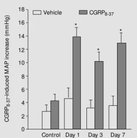
to be a stimulant of CGRP synthesis in DRG. We therefore proposed that NGF adminis-tration to SHR would decrease blood pres-sure through the stimulation of CGRP syn-thesis in DRG. NGF (10 ng kg-1 day-1) was
administered by intraperitoneal injection to 12-week-old SHR (39). NGF was given once a day for 1, 3, and 7 days. A separate group of control SHR received vehicle only. At the end of each treatment period the animals were instrumented for continuous MAP re-cording and infusion of saline or CGRP8-37. After a single NGF treatment (day 1, N = 8), the MAP was reduced by 21 ± 2 mmHg compared to the control SHR. The MAP was still reduced by 22 ± 3 mmHg on day 3 (N = 9); however, by day 7 (N = 11) the MAP was back up to control levels (169 ± 5 mmHg). To determine whether any of the MAP re-duction observed on days 1 and 3 was due to CGRP, during the course of the MAP deter-minations each animal was treated intrave-nously (iv) with either saline or the CGRP receptor antagonist (200 µg). Saline treat-ment produced a negligible increase in MAP at all the time points studied (Figure 4). In the control SHR, CGRP8-37 treatment resulted in a 3.4 ± 1.2 mmHg increase in MAP which was similar to what was seen with saline. However, on days 1 and 3 of NGF treatment CGRP8-37 produced a 12.7 ± 2.2 and 11.6 ± 2.1 mmHg increase in MAP, respectively. Unexpectedly, on day 7 when the MAP was back up to control levels, antagonist treat-ment still resulted in an 11.7 ± 2.4 mmHg increase in MAP. When we examined CGRP mRNA levels in DRG from the control and NGF-treated rats, there was a significant two-fold increase on days 1, 3, and 7 in the NGF-treated group only (Figure 5). CGRP peptide levels in DRG displayed a similar increase (39). Therefore, NGF treatment on days 1 and 3 produces a significant 21 mmHg decrease in MAP. About half of this MAP reduction is due to CGRP as determined by blockade of the CGRP receptor. This action of CGRP most likely results from the
en-CGRP mRNA C G R P m R N A /1 8 S r R N A 5 4 3 2 1 0
Control Day 1 Day 3 Day 7
18S rRNA
Control Day 1 Day 3 Day 7
*
* * A
B
Figure 5. Northern blot analysis of RNA samples from the control and nerve grow th factor-treated spontaneously hypertensive rats.
A, Total cellular RNA samples w ere fractionated on denaturing formaldehyde-agarose gel and transferred to a nylon membrane. The membrane w as hybridized w ith the 32P-labeled calcitonin
gene-related peptide (CGRP) ge-nomic DNA insert. The CGRP probe w as removed from the membrane, w hich w as subse-quently hybridized w ith the 32
P-labeled 18S rRNA probe. After hybridization w ith each probe, the membrane w as w ashed and placed in a phosphor screen cas-sette. The exposed phosphor screen w as then placed in a phos-phor imager to quantify the hy-bridization signals as show n in panel B. * P<0.05 compared to control animals (ANOVA follow ed by the Tukey-Kramer multiple comparisons test).
hanced release and production of this pep-tide. These results (days 1 and 3) strongly suggest that the decreased production of CGRP that is observed in SHR could contri-bute to the elevated blood pressure. After a week of NGF treatment CGRP synthesis is elevated. Antagonist administration indi-cates that CGRP is continuing to play a compensatory role, but the MAP has re-turned to control levels. The continued ac-tivity of CGRP8-37 argues against
down-regu-C G R P8 -3 7 -i n d u c e d M A P i n c re a s e ( m m H g ) 18 16 14 12 10 8 6 4 2 0
Control Day 1 Day 3 Day 7
*
*
*
Vehicle CGRP8-37
Figure 4. Calcitonin gene-related peptide (CGRP) receptor antago-nist adm iantago-nist rat ion increases mean arterial pressure (M AP) in the nerve grow th factor (NGF)-treated but not the control spon-t aneously hyperspon-t ensive raspon-t s (SHR). Control (N = 8) and NGF-treated (days 1, N = 9; 3, N = 10, and 7, N = 11) SHR w ere instru-m ented for continuous M AP recording and intravenous drug adm inistration. W ith the rats fully aw ake and unrestrained, bolus doses of saline (200 µl) or
CGRP8-37 (800 µg/kg in 200 µl
lation of the CGRP receptor as the cause for the increase in MAP on day 7. One possibil-ity is that the enhanced production of CGRP is acting to decrease the MAP on days 1, 3, and 7. However, by day 7, NGF may have stimulated a pressor system to counteract the depressor effects of CGRP and bring the MAP back up to control levels. Two candi-dates are the sympathetic nervous system and neuropeptide Y, both of which are up-regulated by NGF (39).
Characte rizatio n o f the a-CGRP
kno cko ut mo use
Dr. Robert Gagel (Baylor College of Medicine) provided us with homozygous a
-CGRP knockout (KO) mice from breeding pairs generated by gene targeting of the cal-citonin/a-CGRP gene (40). The ß-CGRP
gene is intact in KO mice; however, a-CGRP
is by far the predominant CGRP form pro-duced in DRG neurons (3). For our initial studies, systolic blood pressures were deter-mined using a mouse tail-cuff blood pres-sure apparatus. Using this protocol it was determined that the systolic blood pressure was significantly (P<0.01) higher in KO mice (N = 9; 160 ± 6.1 mmHg) compared to controls (N = 10; 125 ± 4.8 mmHg) (41). Having observed that the systolic blood
pres-sure was elevated in KO mice, it was neces-sary to then determine whether this was re-flected in the MAP. KO (N = 9) and wild-type control (N = 9) mice (25-30 g males) were anesthestized with ketamine/xylazine and the right carotid artery was cannulated with P-10 tubing that was connected to a Gould pressure transducer and recorder. Con-tinuous MAP measurements were made with the mice fully awake and unrestrained. As shown in Figure 6, the MAP was significant-ly elevated in KO mice (139 ± 4.9 mmHg) compared to controls (118 ± 4.9 mmHg) (41). After the MAP determinations the mice were sacrificed and the DRG were removed and frozen for later quantification of a
-CGRP, ß--CGRP, and SP mRNA and peptide levels.
To address the question of whether the KO mice would have an enhanced sensitivi-ty to CGRP, pilot studies were performed in which the mice were instrumented as de-scribed above, except that the jugular vein was also cannulated for administration of saline or CGRP. Administration of saline (0.1 ml) produced a negligible increase in MAP in both the KO (N = 4) and wild-type (N = 4) mice. Administration of an iv bolus dose of rat a-CGRP (1.5 ng/g body weight)
resulted in a rapid 16.3 ± 4.3 mmHg de-crease in MAP in the wild-type mice and a 29.5 ± 4.4 mmHg reduction in the KO mice. These results indicate that the KO mice have an increased sensitivity to CGRP. DRG tis-sue from the two groups of mice was then processed to evaluate a-CGRP, ß-CGRP,
and SP mRNA levels. There was no a-CGRP
mRNA in the KO mice following Northern blot analysis, whereas the 18S rRNA can be seen in the DRG RNA samples from both groups of mice. This demonstrates that the Northern blot analysis was performed cor-rectly. Experiments were also performed to determine if ß-CGRP and SP mRNA levels were altered between the KO and wild-type mice in the absence of a challenge to blood pressure homeostasis. As expected, the
M
A
P
(
m
m
H
g
)
160
150
140
130
120
110
100
90
80
M ale Female Wild type a-CGRP-KO Figure 6. M ean arterial pressure
(M AP) det erm inat ions m ade w ith an indw elling carotid arteri-al catheter in carteri-alcitonin gene-re-lated peptide knockout (CGRP-KO) (N = 7/group) and w ild-type (N = 7/group) mice of either sex, in a fully aw ake and unrestrained st at e. * P< 0.05 com pared t o w ild-type animals (ANOVA fol-low ed by the Tukey-Kramer mul-tiple comparisons test).
*
mRNA species for ß-CGRP and SP were present in both groups and normalization of the hybridization signals (ß-CGRP and SP) to 18S rRNA levels indicated that there was no significant change in the synthesis of these mRNAs. This indicates that only the a
-CGRP gene is missing in these KO mice. Immunostaining for immunoreactive CGRP was performed in the spinal cords of both KO and control mice. There was intense staining of CGRP in laminae I/II of the dor-sal horn of the spinal cord in the wild-type mice. Even though the antibody used in these studies recognizes both a- and ß-CGRP, no
staining is observed in the cords of the KO mice, because of the lack of a-CGRP
ex-pression and the very low levels of ß-CGRP that are produced in DRG (41).
Ro le o f substance P in subto tal ne phre cto my-salt and de o xyco rtico s-te ro ne -salt induce d hype rs-te nsio n in the rat
As described previously, Kohlmann et al. (31) reported that SP plays a compensa-tory role to buffer the blood pressure in-crease in SN-salt, DOC-salt, and one-kid-ney, one-clip rats. They did not determine whether SP expression in DRG was signifi-cantly increased in any of these models. We have since confirmed an antihypertensive role for SP in the SN- and DOC-salt rats (42). We utilized the SP receptor antagonist Spantide-II (Span-II). In DOC-salt rats iv Span-II caused a rapid increase in MAP compared to controls (9.3 ± 1.5 vs 0.9 ± 2.2 mmHg) (42). In SN-salt rats, iv Span-II also significantly elevated MAP (13.9 ± 0.8 vs 1.7 ± 1.7 mmHg) (43). Our studies also suggested that the depressor effect of SP is mediated, at least in part, through the inter-action of SP with vascular NK-1 receptors. Quantification of DRG SP mRNA and pep-tide levels revealed no significant increase in SP mRNA or protein in the SN-salt rats (N = 8) compared to two groups (N = 8/group) of
controls (sham operated with either tap wa-ter or saline drinking wawa-ter) (43). This is similar to what was found for CGRP expres-sion in this setting. In contrast, SP mRNA in DRG was significantly increased in the DOC-salt rats (N = 9) compared with the control group (sham operated with tap water to drink, N = 7) (6.5 ± 0.4 vs 4.4 ± 0.7 arbitrary units) (42). SP peptide content in DRG was also significantly increased in DOC-salt rats com-pared to controls (0.26 ± 0.03 vs 0.18 ± 0.01 pg SP/µg protein) (42). Again, this is similar to what was found for CGRP, although the increase in SP expression was not nearly as robust as that for CGRP (4.5-fold). These results suggested that an increase in vascular reactivity to SP was responsible, at least in part, for the antihypertensive activity of this peptide in both models. To evaluate this possibility, additional groups of SN-salt (N = 6) and control (N = 6) rats were instru-mented for MAP recording and drug admin-istration. With the animals fully awake and unrestrained, SP (12 nM/kg) administration produced a 17 ± 3% fall in baseline MAP in the control group and a significant 26 ± 4% decrease in the SN-salt rats. Also, adminis-tration of the same dose of SP to DOC-salt (N = 7) and control (N = 7) rats caused a decrease in MAP of 14 ± 4 and 29 ± 5%, respectively. This indicates that an increase in vascular responsiveness to SP is one mech-anism by which SP exerts its depressor ef-fects in both SN- and DOC-salt hyperten-sion. These results also provide additional evidence for a counterregulatory role for SP.
D e ve lo pme nt o f the adult rat do rsal ro o t ganglio n ne uro n primary culture mo de l syste m
mechanisms that modulate long-term expres-sion of CGRP and SP in vivo, it was neces-sary to develop an in vitro model system using primary cultures of DRG neurons. Lind-say (44) previously reported that in cultured adult DRG neurons, NGF stimulates the ex-pression of CGRP and SP. Importantly, the induction of these peptides was not medi-ated indirectly through the non-neuronal cells nor was it the result of a survival-promoting effect of NGF. We have also established the conditions for the dissociation and mainte-nance of adult DRG neurons in both serum and serum-free conditions. All of the in vitro studies described herein were performed un-der serum-free conditions. Using immuno-cytochemical staining we initially showed that CGRP synthesis is restricted to approxi-mately 40% of the neuronal cell population in the DRG (22). This is comparable to the values reported by other investigators. To determine if our preparations of neurons were also responsive to NGF, we treated the cultures with NGF (50 ng/ml) (Supowit SC and Zhao H, unpublished results). We found that CGRP mRNA content was increased 3.4-fold at 24 h and 5.7-fold after 48 h. NGF treatment also increased immunoreactive CGRP release 3.1- and 4.6-fold at these same time points. Further, in vitro characteriza-tion of the accharacteriza-tions of NGF revealed a strict dose-dependent (5-200 ng/ml) increase in CGRP mRNA and peptide release.
To determine the effect of activation of two primary intracellular signal transduction pathways on CGRP production and release, neuronal cultures were treated with dibutyryl cAMP or phorbol 12-myristate 13-acetate (PMA), a protein kinase A activator and pro-tein kinase C activator, respectively (22). Ex-posure of the cultures to either dibutyryl cAMP or PMA for 24 h increased CGRP mRNA accumulation 2.2- and 3.0-fold while immu-noreactive CGRP release was stimulated 2.0-and 4.8-fold. This was the first demonstration that these agents could directly regulate CGRP mRNA production and immunoreactive CGRP
release in DRG neurons. These data indicate that local or circulating factors that act to regulate neuronal cell function through pro-tein kinase A and C signal transduction path-ways also modulate CGRP expression. These second messenger pathways have been impli-cated in NGF, bradykinin/prostaglandin, and
a
2-adrenoreceptor agonist actions (45). Since
all of these factors have been shown to be altered in either SHR and/or DOC-salt hyper-tension, and have also been implicated in the regulation of CGRP expression, it was pos-sible to determine in vitro if these agents could directly modulate CGRP synthesis and/or re-lease.
Mechanisms by which nerve growth factor, the sympathetic nervous system, and glucocorticoids interact to regulate neuronal CGRP expression
Several lines of evidence indicate that a
2
-adrenoreceptor agonists and glucocorticoids inhibit the acute release of CGRP from sen-sory nerve terminals (23). To test this possibil-ity in vitro, we employed primary cultures of adult rat DRG neurons. Neuronal cultures were initially exposed (24 h) to either the a2
-adrenoceptor agonist UK 14,304 (1 µM) or vehicle; however, no changes in CGRP mRNA content or immunoreactive CGRP release were observed (23). Using the rationale that in vivo DRG neurons receive a continuous supply of target tissue-derived NGF, which stimulates CGRP synthesis, the neurons were treated (24 h) with vehicle, NGF (25 ng/ml) alone, or NGF plus UK. NGF treatment increased CGRP mRNA accumulation 5.5-fold and immunore-active CGRP release 3.0-fold over control levels. The stimulatory effects of NGF were markedly inhibited but not abolished by UK (NGF + UK vs control, CGRP mRNA 2.9-fold; immunoreactive CGRP, 1.7-fold). These values were also significant when compared to NGF treatment alone. Experiments using the a
2-antagonist yohimbine confirmed that
the effects of UK were mediated by the a
-adrenoceptor. Likewise, the inhibitory effects of dexamethasone on CGRP expression were mediated by the attenuation of the stimulatory effects of NGF. Although the mechanism that mediates this phenomenon is not known, these data suggest that inhibition of stimulated rather than basal expression of CGRP (and possibly SP) is a common theme underlying the nega-tive regulation of neuropeptide synthesis and release in sensory neurons. In addition, the results of the a2-adrenoceptor agonist studies
support the argument that in the SHR the hyperinnervation of the vasculature by the sympathetic nervous system is responsible, at least in part, for the reduction of CGRP pro-duction that is observed in this setting.
Re gulatio n o f ne uro nal CGRP e xpre s-sio n by the bradykinin/pro staglandin syste m
Several lines of evidence suggest that the
enhanced activity of the kallikrein/kinin sys-tem observed during the onset phase of DOC-salt hypertension is partly responsible for the up-regulation of CGRP expression that is observed in this model. In vitro testing of this hypothesis using the DRG primary cul-ture system showed for the first time that bradykinin and selective prostanoids were potent stimulators of long-term CGRP syn-thesis and release. Moreover, these studies demonstrated that the effects of bradykinin were mediated entirely through the cyclo-oxygenase-mediated increase in prostaglan-din production. These data have been pub-lished in abstract form (20).
Co nclusio n
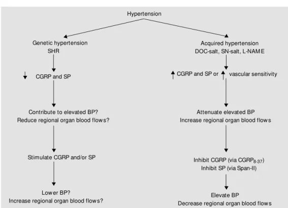
In several animal models, sensory ner-vous system vasoactive peptides seem to play a role in chronic blood pressure eleva-tion. In the acquired models, they play a Hypertension
Genetic hypertension Acquired hypertension DOC-salt, SN-salt, L-NAM E
CGRP and SP or vascular sensitivity
Attenuate elevated BP Increase regional organ blood flow s
Inhibit CGRP (via CGRP8-37)
Inhibit SP (via Span-II)
Elevate BP
Decrease regional organ blood flow s Low er BP?
Increase regional organ blood flow s? Stimulate CGRP and/or SP Contribute to elevated BP? Reduce regional organ blood flow s?
CGRP and SP
Figure 7. Proposed role(s) of calcitonin gene-related peptide (CGRP) and substance P (SP) in experimental hypertension. SHR: spontaneously hypertensive rats; BP: blood pressure; DOC-salt, SN-salt and L-NAM E-induced hypertension: deoxycorticosterone, subtotal nephrectomy and Nw-nitro-L-arginine methyl ester, respectively;
Re fe re nce s
1. Amara SG, Jonas V, Rosenfeld M G & Evans RM (1982). Alternative RNA pro-cessing in calcitonin gene expression gen-erates mRNA encoding different polypep-tide products. Nature, 298: 240-244. 2. Rosenfeld M G, M ermod JJ, Amara SG,
Sw anson LW, Rivier PE, Vale WW & Evans RM (1983). Production of a novel neuropeptide encoded by the calcitonin gene via tissue-specific RNA processing.
Nature, 304: 129-133.
3. Wimalaw ansa SJ (1996). Calcitonin gene-related peptide and its receptors: molecu-lar genetics, physiology, pathophysiology, and therapeutic potentials. Endocrine Re-view s, 17: 533-585.
4. Breimer LH, M acIntyre I & Zaidi M (1988). Peptides from the calcitonin genes: mo-lecular genetics, structure, and function.
Biochemical Journal, 255: 377-390. 5. Dockray GJ (1994). Physiology of enteric
neuropeptides. In: Johnson LR (Editor),
Physiology of the Gastrointestinal Tract. Raven Press, New York, NY, USA. 6. Lembeck F & Holzer P (1979). Substance
P as a neurogenic mediator of antidromic vasodilation and neurogenic plasma ex-travasation. Naunyn-Schmiedeberg’s Ar-chives of Pharmacology, 310: 175-183. 7. Holzer P (1988). Local effector functions
of capsaicin-sensitive sensory nerve end-ings: involvement of tachykinins, calcito-nin gene-related peptide, and other neu-ropeptides. Neuroscience, 24: 739-768. 8. Yaksh TL, Bailey JE & Roddy DR (1988).
Peripheral release of substance P from primary afferents. In: Dubner R, Gebhart GF & Bond M R (Editors), Proceedings from the Vth World Congress on Pain. Elsevier, Amsterdam, Netherlands. 9. M aggi CA (1995). The mammalian
tachy-kinin receptors. General Pharmacology, 26: 911-944.
10. Brow n M J & M orice AH (1987). Clinical pharmacology of vasodilator peptides.
Journal of Cardiovascular Pharmacology, 10: 582-590.
11. DiPette DJ & Wimalaw ansa SJ (1994). Cardiovascular actions of calcitonin gene-related peptide. In: Crass J & Avioli LV
(Editors), Calcium Regulating Hormones and Cardiovascular Function. CRC Press, Ann Arbor, M I, USA.
12. Gibson SJ, Polak JM , Bloom SR, Sabate IM , M ulderry PM , Ghatei M A, M cGregor GP, M orrison JF, Kelly JS & Evans RM (1984). Calcitonin gene-related peptide immunoreactivity in the spinal cord of man and of eight other species. Journal of Neu-roscience, 4: 3101-3111.
13. M arti E, Gibson SJ, Polak JM , Facer P, Springall DP, Aitchison M & Koltzenburg M (1987). Ontogeny of peptide- and amine-containing neurones in motor, sen-sory and autonomic regions of rat and human spinal cord, dorsal root ganglia and rat skin. Journal of Comparative Neurol-ogy, 226: 332-359.
14. Asimakis GK, DiPette DJ, Conti VR, Hol-land OB & Zw ishenberger JB (1987). He-modynamic action of calcitonin gene-re-lated peptide in the isogene-re-lated rat heart. Life Sciences, 41: 597-603.
15. DiPette DJ, Schw arzenberger K, Kerr N & Holland OB (1989). Dose dependent sys-temic and regional hemodynamic effects of calcitonin gene-related peptide. Ameri-can Journal of the M edical Sciences, 297: 65-70.
16. Brain SD, Williams TJ, Tippins JR, M orris HR & M acIntyre I (1985). Calcitonin gene-related peptide is a potent vasodilator.
Nature, 313: 54-56.
17. Holzer P & M aggi CA (1998). Dissociation of dorsal root ganglion neurons into affer-ent and efferaffer-ent-like neurons. Neurosci-ence, 86: 389-398.
18. Rang HP, Bevan SJ & Dray A (1994). Noci-ceptive peripheral neurones: cellular prop-erties. In: Wall PD & M elzak R (Editors),
Textbook of Pain. Churchill-Livingstone, Edinburgh, UK.
19. Lindsay RM , Lockett C & Sternberg J (1989). Neuropeptide expression in cul-tures of adult sensory neurons: modula-tion of substance P and calcitonin gene-related peptide levels by nerve grow th factor. Neuroscience, 33: 53-65. 20. Supow it SC, Hallman DM , Zhao H &
DiPette DJ (1995). Bradykinin regulates
neuronal calcitonin gene-related peptide expression and release. Hypertension, 26: 564 (Abstract).
21. Kaw asaki H, Nuki C & Saito A (1990). Ad-renergic modulation of calcitonin gene-related peptide (CGRP) containing nerve-mediated vasodilation in the rat mesen-teric resistance vessels. Brain Research, 506: 287-292.
22. Supow it SC, Christensen M D, Westlund KN, Hallman DM & DiPette DJ (1995). Dexamethasone and activators of the pro-tein kinase A and C signal transduction pathw ays regulate neuronal calcitonin gene-related peptide expression and re-lease. Brain Research, 686: 77-86. 23. Supow it SC, Hallman DM , Zhao H &
DiPette DJ (1998). Alpha2-adrenoreceptor activation inhibits neuronal calcitonin gene-related peptide expression. Brain Research, 782: 184-193.
24. Looi S, Ekman R, Lippton JC & Keith I (1992). CGRP and somatostatin modulate chronic pulmonary hypertension. Ameri-can Journal of Physiology, 263: H681-H690.
25. DiPette DJ (1999). Experimental models of hypertension. In: Izzo JL & Black HR (Editors), Hypertension Primer: The Es-sentials of High Blood Pressure. Lippin-cott Williams & Wilkins, Baltimore, M D, USA.
26. Supow it SC, Guraraj A, Ram ana CV, Westlund KN & DiPette DJ (1995). En-hanced neuronal expression of calcitonin gene-related peptide in mineralocorticoid-salt hypert ension. Hypert ension, 25: 1333-1338.
27. Supow it SC, Zhao H, Hallman DM & DiPette DJ (1997). Calcitonin gene-related peptide is a depressor of deoxycorticos-terone-salt hypertension in the rat. Hyper-tension, 29: 945-950.
28. Supow it SC, Zhao H, Hallman DM & DiPette DJ (1998). Calcitonin gene-related peptide is a depressor in subtotal ne-phrectomy hypertension. Hypertension, 31: 391-396.
29. Gangula PR, Supow it SC, Wimalaw ansa SJ, Zhao H, Hallman DM , DiPette DJ & compensatory role. In the genetic model,
their decreased levels may contribute to the elevated blood pressure, as illustrated in Fig-ure 7. The roles of CGRP and SP in human hypertension are yet to be clarified.
Ackno wle dgm e nts
Yallampalli C (1997). Calcitonin gene-re-lated peptide is a depressor in NG
-nitro-L-arginine methyl ester (L-NAM E)-induced preeclampsia. Hypertension, 29: 248-253. 30. Supow it SC, Zhao H, Wang DH & DiPette DJ (1995). Regulation of calcitonin gene-related peptide expression: role of in-creased blood pressure. Hypertension, 26: 1177-1180.
31. Kohlmann O, Cesaretti M L, Ginoza M , Tavares A, Zanella M T, Ribeiro AB, Ramos OL, Leeman SE, Gavras I & Gavras H (1997). Role of substance P in blood pres-sure regulation in salt-dependent experi-mental hypertension. Hypertension, 29: 506-509.
32. Quirion R, VanRossum D, Dumont Y, St Pierre S & Fournier A (1992). Character-ization of CGRP1 and CGRP2 receptor subtypes. Annals of the New York Acade-my of Sciences, 657: 88-105.
33. Gardiner SM , Compton AM , Kemp PA, Bennett T, Bose C, Foulkes R & Hughes B (1990). Antagonistic effect of human al-pha-CGRP (8-37) on the in vivo regional hemodynamic actions of human alpha-CGRP. Biochemical and Biophysical Re-search Communications, 171: 938-943. 34. Gardiner SM , Compton AM , Kemp PA,
Bennett T, Bose C, Foulkes R & Hughes B (1991). Human alpha-calcitonin gene-re-lated peptide (CGRP)-(8-37), but not
-(28-37), inhibits carotid vasodilator effects of human alpha-CGRP in vivo. European Journal of Pharmacology, 199: 375-378. 35. Hughes SR & Brain SD (1991). A
calcito-nin gene-related peptide (CGRP) antago-nist (CGRP8-37) inhibits microvascular re-sponses induced by CGRP and capsaicin in skin. British Journal of Pharmacology, 104: 738-742.
36. Supow it SC, Watts SW, Zhao H, Wang D & DiPette DJ (2000). Vascular reactivity to calcitonin gene-related peptide is en-hanced in subtotal nephrectomy-salt hy-pertension. Hypertension, 36: 701 (Ab-stract).
37. Yallampalli C, Dong YL & Wimalaw ansa SJ (1996). Calcitonin gene-related peptide reverses the hypertension and significant-ly decreases the fetal mortality in pre-eclampsia rats induced by N(G)-nitro-L-ar-ginine methyl ester. Human Reproduc-tion, 11: 895-899.
38. Gangula PR, Wimalaw ansa SJ & Yallam-palli C (1997). Progesterone up-regulates vasodilator effects of calcitonin gene-re-lated peptide in N(G)-nitro-L-arginine me-thyl ester-induced hypertension. Ameri-can Journal of Obstetrics and Gynecol-ogy, 176: 894-900.
39. Supow it SC, Zhao H & DiPette DJ (2001). Nerve grow th factor enhances calcitonin gene-related peptide expression in the
spontaneously hypertensive rat. Hyper-tension, 37: 728-732.
40. Hoff AO, Thomas PM , Cote GJ, Qui H, Bain H & Gagel RF (1998). Generation of a calcitonin knockout mouse model. Bone, 23: S64 (Abstract).
41. Gangula PR, Zhao H, Supow it SC, Wima-law ansa SJ, DiPette DJ, Westlund KN, Gagel RF & Yallampalli C (2000). Increased blood pressure in alpha-calcitonin gene-related peptide/calcitonin gene knockout mice. Hypertension, 35: 470-475. 42. Katki KA, Supow it SC & DiPette DJ (2000).
Enhanced expression of substance P in deoxycorticosterone acetate-induced hy-pertension. Circulation, 102 (Suppl II): II-348-II-349 (Abstract).
43. Katki KA, Supow it SC & DiPette DJ (2002). Substance P in subtotal nephrectomy-salt hypertension. Hypertension, 39: 389-393. 44. Lindsay RM (1988). Nerve grow th factors (NGF, BDNF) enhance axonal regenera-tion but are not required for survival of adult sensory neurons. Journal of Neuro-science, 8: 2394-2405.
45. DiPette DJ & Supow it SC (2000). Calcito-nin gene-related peptide and hyperten-sion. In: Oparil S & Weber M A (Editors),