Meiotic Chromosome Synapsis-Promoting
Proteins Antagonize the Anti-Crossover
Activity of Sgs1
Lea Jessop1, Beth Rockmill2, G. Shirleen Roeder2,3,4, Michael Lichten1*
1Center for Cancer Research, National Cancer Institute, Bethesda, Maryland, United States of America,2Department of Molecular, Cellular, and Developmental Biology, Yale University, New Haven, Connecticut, United States of America,3Department of Genetics, Yale University, New Haven, Connecticut, United States of America,4Howard Hughes Medical Institute, Yale University, New Haven, Connecticut, United States of America
Sgs1, the budding yeast homolog of the mammalian BLM helicase, has been implicated in preventing excess recombination during both vegetative growth and meiosis. Most meiotic crossover (CO) recombination requires full function of a set of yeast proteins (Zip1, Zip2, Zip3, Zip4/Spo22, Mer3, Msh4, and Msh5, termed the SIC or ZMM proteins) that are also required for homologous chromosome synapsis. We report here genetic and molecular assays showing thatsgs1single mutants display relatively modest increases in CO recombination (less than 1.6-fold relative to wild-type). In contrast, a much greater CO increase is seen when an sgs1 mutation is introduced into the CO- and synapsis-deficientzip1, zip2, zip3, mer3,ormsh4mutants (2- to 8-fold increase). Furthermore, close juxtaposition of the axes of homologous chromosomes is restored. CO restoration in the mutants is not accompanied by significant changes in noncrossover (NCO) recombinant frequencies. These findings show that Sgs1 has potent meiotic anti-CO activity, which is normally antagonized by SIC/ZMM proteins. Our data reinforce previous proposals for an early separation of meiotic processes that form CO and NCO recombinants.
Citation: Jessop L, Rockmill B, Roeder GS, Lichten M (2006) Meiotic chromosome synapsis-promoting proteins antagonize the anti-crossover activity of Sgs1. PLoS Genet 2(9): e155. DOI: 10.1371/journal.pgen.0020155
Introduction
DNA double-strand breaks (DSBs) pose a significant risk to cells. Failure to repair DSBs can result in death, while imprecise repair can form translocations, deletions, and other chromosome rearrangements. DSBs are repaired by two distinct mechanisms: end-joining, in which the ends of breaks are ligated, often imprecisely, and homologous recombination, in which breaks are repaired using homolo-gous sequences as a template to form recombinants that are either crossover (CO) or noncrossover (NCO) with regard to flanking parental sequences. Although repair by homologous recombination is generally considered nonmutagenic (but see [1]), the CO outcome has the potential for deleterious genome rearrangement, loss of heterozygosity, or both. Perhaps as a consequence, the rare interhomolog recombi-nation events that do occur during the mitotic cell cycle are infrequently accompanied by crossing over [2].
In contrast, COs are frequent in meiosis, with at least one per homolog pair [3]. COs are an integral part of the interhomolog connections that are necessary for homolog alignment and spindle assembly at metaphase I [4,5]. As a consequence, mutants with either general meiotic recombi-nation defects or specific defects in meiotic COs undergo frequent homolog mis-segregation and gamete death. Even a single pair of chromosomes that fails to cross over is at increased risk of nondisjunction at meiosis I [6–8]. In most organisms where these events have been examined, the total number of interhomolog recombination events is consider-ably greater than the number of COs [9], and both COs and NCOs are needed to facilitate meiotic homolog pairing [10,11].
The molecular mechanism of meiotic recombination and
the factors that determine whether events will produce NCO or CO products have been studied most extensively in the budding yeastSaccharomyces cerevisiae. Studies in this organism show that meiotic recombination is initiated by DSBs, formed by the meiosis-specific endonuclease Spo11 [12]. Breaks are subsequently resected to generate single-stranded DNA tails with free 39ends [13]. Most COs are produced via formation of a semi-stable single end invasion intermediate in which one DSB end interacts with the homolog [14], followed by capture of the second DSB end to form a double Holliday junction (dHJ) intermediate [14–17]. By contrast, most NCOs form via processes that do not appear to involve stable dHJ intermediates [16,17] and a synthesis-dependent strand-annealing mechanism has been suggested [2,16,18].
Evidence for mechanistic separation of CO and NCO recombination comes from molecular studies of two classes of mutants that block CO formation without reducing NCOs. Cells lacking the Ndt80 transcription factor or the Cdc5
Editor:James E. Haber, Brandeis University, United States of America
ReceivedApril 5, 2006;AcceptedAugust 2, 2006;PublishedSeptember 22, 2006
A previous version of this article appeared as an Early Online Release on August 2, 2006 (DOI: 10.1371/journal.pgen.0020155.eor).
DOI:10.1371/journal.pgen.0020155
This is an open-access article distributed under the terms of the Creative Commons Public Domain declaration which stipulates that, once placed in the public domain, this work may be freely reproduced, distributed, transmitted, modified, built upon, or otherwise used by anyone for any lawful purpose.
Abbreviations:AA, axial association; CO, crossover; dHJ, double Holliday junction; DSB, double-strand break; NCO, noncrossover; SC, synaptonemal complex; SIC, synapsis initiation complex; ZMM, Zip Msh Mer proteins
kinase produce NCO recombinant DNA molecules at normal levels, but lack COs and accumulate dHJ intermediates [16,19]. Mutants in any of several budding yeast genes (ZIP1, ZIP2, ZIP3, ZIP4/SPO22, MSH4, MSH5,or MER3,referred to here collectively asZMM genes) also show CO loss without apparent NCO defects (reviewed in [9]). In strains of the SK1 genetic background that are sporulated at 338C,zmmmutants display severe defects in single-end invasion and dHJ-intermediate formation as well as CO defects, and this CO loss is not accompanied by an increase in NCO DNA molecules [17]. This finding is consistent with an earlier suggestion that the CO/NCO decision is made at an early step, possibly at or soon after DSB formation [20]. If the decision were made later, blocking CO formation might allow CO-designated DSBs to be repaired as NCOs, which would result in increased NCO production. These two classes of CO-defective mutants also differ in their effect on homolog synapsis, in thatndt80andcdc5mutants show normal synapsis [19,21], while zmm mutants display synapsis defects ([17,22], and references within).
Of the ZMM proteins, Msh4, Msh5, and Mer3 have known biochemical activities that could stabilize early recombina-tion structures and promote the formarecombina-tion of dHJ inter-mediates [23,24]. The other ZMM proteins appear to participate less directly. Zip1 is a major component of the synaptonemal complex (SC) that forms between homolog axes during prophase of meiosis I [7]. It has been suggested that Zip2 and Zip3 are part of a meiosis-specific ubiquitin- or SUMO-conjugating complex [25–27]. In zip1D mutants,
homolog axes are no longer tightly paired, but instead associate at a few sites per chromosome that are marked by foci of Zip2 and Zip3 [7,22,28,29]. Accumulating data suggest that, in wild-type budding yeast, these Zip2/Zip3 foci, which also contain Msh4 and Msh5 [30,31], mark sites both of CO recombination and of Zip1 polymerization initiation
[22,28,29,32,33]. These foci, whose protein contents are termed the synapsis initiation complex (SIC), may correspond to the late recombination nodules that mark CO sites in higher eukaryotes [22,34].
Sgs1, a budding yeast RecQ-type helicase, has been implicated in regulating the CO/NCO decision and in maintaining genome stability. The absence of Sgs1 causes increased mitotic recombination [35], especially when mis-matches are present in the recombining partners [36,37]. Mutants lacking Sgs1 also show increased chromosomal rearrangement [38] and reduced sporulation efficiency and spore viability [35]. Similarly, vertebrate cells lacking BLM, an Sgs1 homolog, show elevated rates of sister-chromatid exchange and chromosome rearrangements [39–41]. These mutant phenotypes are consistent with the Sgs1/BLM helicase having a direct anti-CO activity, although many of these observations can also be explained by suggesting that Sgs1/ BLM prevents the formation of lesions that provoke recombination or rearrangement.
Consistent with a role for these helicases in directing events away from COs and towards NCOs, human BLM and TOP3a
together can dissolve synthetic dHJ substrates in vitro to produce NCOs [42]. While limited solubility has prevented a similar study of Sgs1 [43], two observations support the suggestion that it has anti-CO activity. First, two separate studies, one of spontaneous mitotic recombination and the other of the mitotic repair of a DSB formed by the HO endonuclease, both found about a 2-fold increase in CO recombinants insgs1mutants relative to wild-type, although the vast majority of repair products in both cases were NCOs [44,45]. Second, Rockmill et al. found that, in cells of the BR strain background, the frequency of meiotic COs among tetrads and the number of Zip3–green fluorescent protein (GFP) foci per pachytene nucleus were both about 1.4-fold greater insgs1mutants than in wild-type [32]. Rockmill et al. also showed thatsgs1mutation accelerates homolog synapsis in otherwise wild-type cells, and restores close, end-to-end association between homolog axes in zip1 mutants. They referred to the axial association (AA) seen in zip1 sgs1 as pseudosynapsis, to distinguish it from true synapsis, where end-to-end SC is present.
In order to learn more about the role of Sgs1 in meiotic recombination, we examined the effect of sgs1 mutants on meiotic recombination, using both tetrad analysis and an assay that directly scores recombination at the DNA level (Figure 1). Our findings indicate that, in wild-type cells, Sgs1 activity has a limited role in CO formation and does not play a unique role in NCO formation. However, inzmmmutants, where CO formation is markedly reduced and synapsis is impaired,sgs1mutants restore COs, in some cases to nearly wild-type levels, and also restore tight homolog AA. These data demonstrate that Sgs1 has anti-CO activity, and suggest that an important role for the SIC/ZMM proteins is to protect nascent CO-designated recombination intermediates from dissolution by Sgs1.
Results
Previous studies examinedsgs1Dandsgs1DC795mutants in the BR strain background and found a modest (0%–60%) increase in allelic crossing over, but did not directly evaluate the effect on NCO recombinants ([32]; B. Rockmill, K.
Synopsis
Most eukaryotic cells are diploid (two copies of each chromosome per cell), but gametes (in animals, sperm and eggs) are haploid (one chromosome copy). Gametes are produced from diploid cells during meiosis. The two copies of each chromosome are brought together in end-to-end alignment (synapsis), and then are connected by crossover recombination, which involves the joining of DNA from one chromosome copy to DNA of the other. Crossovers are critical for chromosome separation in the diploid-to-haploid transition, and also promote genetic diversity by shuffling parental genotypes.
In contrast, during mitotic cell growth, crossovers create genome rearrangements and loss of heterozygosity, which are associated with cancer and other diseases. A DNA-unwinding enzyme, called BLM in mammals and Sgs1 in budding yeast, prevents mitotic crossover recombination by taking apart intermediates that would otherwise give rise to crossovers.
Voelkel-Meiman, and G. S. Roeder, unpublished data). In the SK1 background, homozygous sgs1D diploids display high
chromosome instability, and mating-type heterozygosity cannot be maintained at levels that ensure sporulation in liquid culture. To extend evaluation of the meiotic role of Sgs1 to SK1 strains, where recombination can be readily scored at the DNA level, we used two sgs1 mutant alleles,
sgs1DC795andsgs1-mn. Neither allele displays the same extent of chromosome instability assgs1D, and both support efficient premeiotic growth and sporulation. The sgs1DC795 allele expresses only the first 652 amino acids of the protein and is lacking both the helicase domain [46] and a region called the HRDC domain, which in BLM interacts with Holliday junctions [42]. A DNA fragment containing SGS1 or
sgs1DC795 coding sequences along with 600 nucleotides of
upstream sequences was integrated atTRP1in strains where the endogenous SGS1 gene was deleted (see Materials and Methods). We will refer to strains withsgs1DC795atTRP1as
sgs1DC795,and to isogenic control strains withSGS1atTRP1
asTRP1:SGS1. ‘‘Wild-type’’will be reserved for strains with
SGS1 at its normal locus. In the sgs1-mn allele, SGS1 is transcribed from aCLB2promoter, which is expressed during the mitotic cell cycle but not during meiosis.TRP1:SGS1and
wild-type strains show similar spore viability (98%; see also Figure 2A) and resistance to methyl methane sulfonate (unpublished data). Bothsgs1DC795andsgs1-mnshow reduced
spore viability (Figure 2A), with spore inviability patterns typical of random spore death. A substantial fraction of this spore death is likely to be due to premature separation of sister chromatids, associated with recombination near cen-tromeres (B. Rockmill, K. Voelkel-Meiman, and G. S. Roeder, unpublished data).
Sgs1 Has a Limited Effect on CO Recombination in Wild-Type Cells
We examined meiotic recombination in SK1 strains carrying a 3.5-kilobase URA3-ARG4 recombination interval inserted athis4on one copy of Chromosome III and atleu2on the homolog (Figure 1). COs in three intervals can be scored in these strains: ectopic COs between the his4::URA3-ARG4
and leu2::URA3-ARG4 inserts, allelic COs in theHIS4-LEU2
interval, and allelic COs in the LEU2-MAT interval.
Figure 1.Recombination Interval Used for Molecular Analyses (A) The 3.5-kilobase ectopic recombination interval contains coding sequences forURA3(gray) andARG4(black). DSBs form in the promoter regions ofURA3(DSB1) andARG4(DSB2). The lollipop inarg4represents a palindromic sequence inserted atþ9 of the open reading frame; this mutation is used to score gene conversion [16].
(B) The ectopic recombination interval is inserted atHIS4(blue) on one copy of Chromosome III and atLEU2 (red) on the homolog.HIS4and LEU2are 16.7 kilobases apart. Inrad50Sstrains, where DSBs persist, 5% of chromosomes have a DSB inhis4::URA3-ARG4and 0.7% have a DSB in leu2::URA3-ARG4 [16]. The centromere (black circle) and MAT locus (green) are also indicated. Allelic COs can be scored in theHIS4-LEU2and LEU2-MATintervals.
(C) Ectopic COs can occur between his4::URA3-ARG4 and leu2::URA3-ARG4.
DOI: 10.1371/journal.pgen.0020155.g001
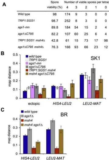
Figure 2.Loss of Full Sgs1 Activity Restores Spore Viability and Crossing Over tozmmMutants
(A) Overall spore viability and patterns of spore lethality in tetrads from SK1 strains.
(B) Map distance (cM; error bars denote standard error of map distance) in three intervals on Chromosome III in SK1 (see Figure 1B and 1C for details). Values for wild-type are from [16].
(C) Map distance in two allelic intervals on Chromosome III in BR strains. Values for wild-type andsgs1Dare from [32].
DOI: 10.1371/journal.pgen.0020155.g002
TRP1:SGS1strains displayed a 1.4-fold greater frequency of ectopic COs when compared with wild-type (p , 0.001, G -test), but CO frequencies in the two allelic intervals were similar to those seen in wild-type (Figure 1B). Both sgs1
mutants produced ectopic COs at frequencies similar to those seen inTRP1:SGS1and about 1.7-fold greater than in wild-type. Allelic CO frequencies in sgs1 mutants were not consistently greater or less than those seen in wild-type or
TRP1:SGS1,and in all cases were within 30% of wild-type or
TRP1:SGS1values (Figure 2B). In the BR background,sgs1D
-null mutants and sgs1DC795 display consistent increases of
30%–40% in allelic crossing over in the HIS4-LEU2 and
LEU2-MATintervals [32].
The Zip3 protein forms foci on pachytene chromosomes, and it has been suggested that, in wild-type cells, these foci mark sites of COs [22]. In the BR strain background,sgs1D -null mutants andsgs1DC795 mutants display a 1.5- and
1.3-fold increase, respectively, in the number of Zip3 foci, detected using a Zip3-GFP fusion protein ([32]; B. Rockmill, K. Voelkel-Meiman, and G. S. Roeder, unpublished data). Loss of functional Sgs1 protein has a similar impact on Zip3-focus formation in the SK1 background. Comparable numbers of Zip3-GFP foci were detected in wild-type and TRP1:SGS1
nuclei (wild-type 63 6 8 foci/nucleus, 34 nuclei scored;
TRP1:SGS16667 foci/nucleus, 40 nuclei scored), while about 25% more foci were detected insgs1DC795 nuclei (82 6 9
foci/nucleus, 39 nuclei scored;p,0.001,t-test).
Most tetrad analyses consider only data from four-spore viable tetrads. Because COs promote spore viability, this can overestimate CO frequencies in mutant backgrounds in which spore viability is reduced. To examine every meiotic product regardless of viability, and to determine whether or not Sgs1 function affects NCO formation, we scored recombinants in the ectopic URA3-ARG4 interval at the molecular level, using DNA from SK1 cultures undergoing synchronous meiosis (Figure 3).
Timing of DSB appearance and disappearance, maximal levels of DSBs, and timing of meiotic divisions did not differ substantially among wild-type,TRP:SGS1, sgs1DC795,and sgs1-mn (Figure 3D and unpublished data). Molecular analysis revealed no statistically significant difference among wild-type,TRP1:SGS1, sgs1DC795,orsgs1-mnstrains with regards to
the timing of formation or final levels of NCOs or COs in the ectopic recombination interval (Figures 3D and S2), although experiment-to-experiment variation would have obscured CO increases of 30% or less. These results indicate that, in otherwise wild-type SK1 cells, the majority of CO and NCO recombinant molecules form independently of full Sgs1 function.
We performed a similar analysis in strains containing a different recombination interval,URA3-tel-ARG4, integrated atHIS4andLEU2[18]. This interval differs from the URA3-ARG4interval described above in that DSBs occur at a single site in the interval and form more frequently (20% versus 5% of chromosomes). Wild-type, TRP1:SGS1, and sgs1DC795
strains produced similar levels of NCO and CO recombinant molecules in this interval (Figure S3).
Sgs1 Inhibits CO Formation inzmm Mutants
The finding that Sgs1 has a limited impact on meiotic recombination in wild-type SK1 cells stands in contrast to numerous reports suggesting a prominent anti-CO function
during the mitotic cell cycle. Because SIC/ZMM proteins promote meiotic CO formation, we reasoned that they might be masking the anti-CO activity of Sgs1. Therefore, we examined the effect ofsgs1mutation on meiotic recombina-tion in severalzmmmutants in SK1 and BR strains. The results of these studies are summarized below.
msh4D.SK1msh4mutants show relatively high sporulation
frequencies and spore viability [31], allowing tetrad-based measurements of genetic distances. Consistent with previous studies [31],TRP1:SGS1 msh4Dstrains showed about 2.5-fold
fewer COs compared with TRP1:SGS1 MSH4, in all three intervals illustrated in Figure 1, a marked reduction in spore viability, and a disproportionate increase in the number of tetrads with two or no viable spores (Figure 2A and 2B). All three phenotypes were suppressed by sgs1DC795 (Figure 2),
with similar spore viability patterns and CO frequencies seen in sgs1DC795 MSH4 and sgs1DC795 msh4D. A similar msh4D
CO defect, and suppression bysgs1D,was seen in BR strains
(Figure 2C).
Molecular assays confirm this CO defect, and its suppres-sion bysgs1mutation. COs inTRP1:SGS1 msh4Dwere reduced 3.7-fold relative to TRP1:SGS1 (Figure 3D). A similar reduction was seen inmsh4D(Figure S4). COs were increased nearly toMSH4levels in sgs1DC795 msh4Dorsgs1-mn msh4D
(2.7-fold greater than TRP1:SGS1 msh4D or msh4D alone; Figures 3 and S4). We found no substantial differences in the time of formation or in final levels of NCO recombinants between msh4Dand control strains. The msh4D mutant also
had no defects in DSB formation, DSB repair, or meiotic progression.
mer3D. Unlike msh4D, mer3D cells show DSB repair and
meiotic progression defects (Figure 3; [47]). DSBs formed normally in both TRP1:SGS1 mer3D and sgs1DC795 mer3D
strains, but some breaks persisted beyond the normal time of repair, with DSBs detectable in TRP1:SGS1 mer3Dcells after
12 h of sporulation. This DSB repair defect was partially suppressed by sgs1DC795, with all DSBs gone after 10 h of
sporulation. Meiotic progression was also defective in
TRP1:SGS1 mer3, with binucleate cells appearing 3 h later than normal (Figure 3D), and only about 40% of cells completing meiosis I by 12 h. A greater fraction ofsgs1DC795 mer3Dcells completed at least one division at 12 h, although
progression was still delayed.
The CO defect we observed inTRP1:SGS1 mer3Dstrains was
more severe than the 2- to 3-fold reduction previously reported [47]. COs could not be detected inTRP1:SGS1 mer3D
mutants 8 h after initiation of sporulation, a time when COs had reached a maximum inTRP1:SGS1 MER3. Even at 12 h, CO levels were 17-fold less than in control strains. This CO defect was partially suppressed by sgs1DC795, and COs in
sgs1DC795 mer3Dwere 7-fold greater than inTRP1:SGS1 mer3
at 12 h. Nevertheless, CO levels reached only about 40% of the maximum level seen in MER3controls.A modest NCO defect was also seen in TRP1:SGS1 mer3D. After 8 h of sporulation, NCO levels in both TRP1:SGS1 mer3D and sgs1DC795 mer3D were less than those seen in TRP1:SGS1 MER3,although NCO frequencies reached or exceeded those seen inTRP1:SGS1 MER3by 12 h.
zip1D and zip2D. DSB formation occurred on time, and
most DSBs were repaired inzip1Dand zip2D strains. These
sporulation, COs were present inTRP1:SGS1 zip1Dat 20% of
the maximum level seen inTRP1:SGS1 ZIP1. The sgs1DC795
allele partially suppressed this defect, increasing COs 3-fold. NCO formation was unaffected. TRP1:SGS1 zip2D mutants
displayed a more severe CO defect. At 8 h, COs were present at 11% of the maximum seen inTRP1:SGS1 ZIP2. At 12 h, COs levels had increased another 2-fold. COs were increased about 3-fold bysgs1DC795inzip2D,as they were inTRP1:SGS1 zip1D. As was seen in TRP1:SGS1 mer3D, TRP1:SGS1 zip2D
caused a slight delay and reduction in NCOs that was not suppressed bysgs1DC795; in bothzip2DTRP1:SGS1andzip2D sgs1DC795,NCOs continued to accumulate and at 12 h their
level exceeded the maximum seen inTRP1:SGS1at the same time (Figure 3).
We also considered whethersgs1Dsuppresses the allelic CO
defect seen inzip1D in the BR strain background. Because zip1D mutants sporulate poorly in BR, map distances were
measured by random spore analysis;sgs1Dcaused about a 2-fold increase in COs in both intervals (Table S2).
In summary, in the conditions used in these experiments,
zmm mutants differ in terms of meiotic progression, DSB repair, and CO formation defects, consistent with the diversity of defects previously seen when SK1zmm mutants are sporulated at 238C [17]. Despite these differences, in all cases the zmm mutant phenotype is at least partially sup-pressed by loss of Sgs1.
Sgs1 Inhibits Homolog AA inmer3Dandzip3DMutants Despite the absence of Zip1, the protein that normally occupies the space between synapsed homolog axes [48], close juxtaposition of homolog axes along their lengths is observed
in sgs1 zip1 double mutants in both BR and SK1 strain
backgrounds ([32] and unpublished data). To determine whether this pseudosynapsis occurs in otherzmm sgs1double mutants, we examined chromosome morphology in surface-spread meiotic nuclei, using antibodies against Zip1 and antibodies against Red1, a major component of meiotic chromosome axes. In wild-type yeast, chromosome cores never achieve fully continuous Red1 staining [49]. However, in synapsis-defective zmm mutants, Red1 accumulates and localizes continuously along each chromosome axis [29]. Thus, Red1 staining provides a means to visualize chromo-some contours in the absence of Zip1 staining.
In SK1 strains, both TRP1:SGS1and sgs1DC795 displayed normal chromosome morphology, with axes of homologous chromosomes closely juxtaposed and continuous end-to-end Zip1 staining (unpublished data). However, both strains displayed an increased frequency of polycomplexes (extrac-hromosomal arrays of SC components) compared with
wild-type (28/83 nuclei in TRP1:SGS1 and 34/74 nuclei in
sgs1DC795 versus 4/50 nuclei in wild-type after 5 h of
sporulation). This increase in polycomplex formation sug-gests a modest defect or delay in SC formation.
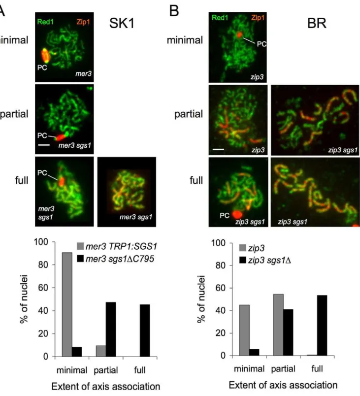
TRP1:SGS1 mer3D diploids displayed a severe synapsis
defect, with about 90% of nuclei showing separated homolog axes (as evidenced by Red1 staining) and little Zip1 local-ization (Figure 4). Homolog AA was partially restored in
mer3D sgs1DC795 mutants, with more than 90% of cells
displaying either some or all chromosomes with axes in close alignment along their lengths (Figure 4A). These fully aligned chromosomes displayed only low levels of discontinuous Zip1 staining, in contrast to the end-to-end continuous staining seen in sgs1 single mutants and in wild-type ([7,17,32] and unpublished data). Although about half of mer3Dsgs1DC795
nuclei displayed full pseudosynapsis of homologs, an equal number displayed partial pseudosynapsis, where only some homologs or parts of homologs appeared pseudosynapsed.
We also determined the effect of Sgs1 on synapsis inzip3D
BR strains, which display a relatively mild CO and pro-gression defect [28]. Only a small fraction ofzip3mutant cells displayed full homolog AA, and this occurred at relatively late times in meiosis. Despite this synapsis defect, about 75% of
zip3Dmutant cells progress to form mature asci (unpublished data). Deletion of SGS1 from zip3D mutants restored full
homolog AA to more than half of nuclei (Figure 4B). Of the chromosome pairs in which axes were closely juxtaposed, most were truly synapsed (i.e., displayed end-to-end Zip1 staining) and a minor fraction were pseudosynapsed (Figure 4B and unpublished data).
Discussion
In previous studies of BR strains, Rockmill et al. showed that the sgs1D and sgs1DC795 mutations increase crossing
over in allelic intervals (1.2- to 1.4-fold) and cause a corresponding increase (1.3- to 1.4-fold) in the number of Zip3 foci, which are thought to be cytological markers of CO sites [32]. In this study, we examined the effect of sgs1
mutations on meiotic recombination in SK1 strains, using assays that detected NCO and CO DNA molecules produced by ectopic recombination. We did not observe a statistically significant increase in CO molecules in two test intervals, although experiment-to-experiment variation would have obscured an increase of 30% or less. We also did not observe a consistent increase in CO recombination, measured by tetrad analysis in three genetic intervals on Chromosome III. We did observe an increase (by about 25%) in Zip3 foci in
sgs1DC795SK1 strains (relative toTRP1:SGS1). If Zip3 foci are
Figure 3.Sgs1 Prevents COs inzmmMutants
(A) Schematic representation of the ectopicURA3-ARG4interval. Symbols are as in Figure 1. EcoRI (E) and XhoI (X) restrictions sites are indicated. To detect COs and DSBs, DNA is digested with XhoI and probed withARG4sequences (ArgD; [16]). To detect NCOs and a CO product (CO19), DNA is digested with EcoRI and XhoI and probed withHIS4sequences (HisU; [16]).
(B) Southern blot of DNA isolated from a meiotic culture of MJL3035 (TRP1:SGS1) at the indicated time after initiation of sporulation, digested, and probed to detect COs and DSBs. mw, HinDIII digest of phagekDNA. DSBs occur inURA3-ARG4inserted at bothhis4andleu2,but are;7-fold stronger in thehis4insert than atleu2[16]. Palindrome cleavage, by unidentified activities, occurs at the same time as CO formation and results in a band about the size of DSB2 (T. Allers, L. Jessop, and M. Lichten, unpublished data).
(C) Southern blot of the same samples, digested and probed to detect NCO and CO19recombinants.HIS4,HIS4locus lacking an insert; DSB*, DSB1 product where resection has passed the EcoRI site inHIS4; mw, BstEII digest of phagekDNA.
accurate markers of CO sites, this finding would be consistent with a modest increase in crossing over on a genome-wide basis. In a separate study, Oh and Hunter have examined the effect ofsgs1DC795 on CO recombination in SK1 diploids,
using both genetic and molecular assays. Their data indicate that, in the SK1 background, loss ofSGS1function increases overall CO frequencies by no more than 20% (S. Oh and N. Hunter, personal communication).
Figure 4.Sgs1 Prevents AA inmer3Dandzip3DMutants
(A) Analysis of AAs in TRP1:SGS1mer3Dandmer3Dsgs1DC795SK1 strains. Nuclei from cells harvested 5 h after initiation of sporulation were surface spread and probed with anti-Zip (red) and anti-Red1 (green) antisera. Nuclei where chromosomes displayed linear Red1 were examined and classified as displaying minimal, partial, or full pseudosynapsis, as described in Materials and Methods. Nuclei with fully associated chromosomes displayed discontinuous Zip1 staining (right-hand example), Zip1 localization in polycomplexes (PC, left-hand example), or both. White bar: 2 microns. (B) Analysis of AAs inzip3Dandzip3Dsgs1DBR strains. Nuclei from cells harvested 18 h after the initiation of sporulation were spread, stained, and analyzed as former3strains. In most nuclei with full AA, at least some chromosome pairs displayed end-to-end Zip1 staining with the remainder being pseudosynapsed.
DOI: 10.1371/journal.pgen.0020155.g004
We also found that, in SK1 sgs1 mutants, NCO recombi-nants are recovered at frequencies similar to those seen in wild-type; similar results have been observed in BR strains [32]. Thus, the bulk of the genetic, DNA-based, and cytological assays suggest that, in wild-type cells, there must be proteins other than Sgs1 that prevent COs, and that promote NCO recombination.
Sgs1 Mediates the CO Defect inzmm Mutants
COs are required for proper chromosome segregation during meiosis, and both meiotic CO distributions and the time of their formation are tightly regulated in most organisms [9,50]. It is therefore likely that the anti-CO activity of the Sgs1/BLM helicase evident in mitotic cells [35–37,39–41] must be modulated during meiosis to allow high CO levels, either by reducing helicase activity or by preventing access to substrates. Our current study identi-fies SIC/ZMM proteins encoded by the budding yeast ZIP1, ZIP2, ZIP3, MER3, and MSH4 genes, as modulators of the impact of Sgs1. Although mutations in these genes reduce CO formation, the severity of this defect can vary widely. For example, under the sporulation conditions used in our study, this CO defect ranged from about a 4-fold reduction (in msh4D) to a virtual elimination (in mer3D).
These differences most likely reflect the different bio-chemical and structural roles played by the different ZMM proteins, either individually or as part of a larger complex. Nevertheless, the CO defect in these mutants was at least partially suppressed by the loss of Sgs1 activity. In molecular assays, CO restoration was not accompanied by a corresponding decrease in NCO recombinants, and COs were not restored to levels seen in wild-type or in the sgs1DC795 single mutant. Genetic assays, in both SK1
and BR backgrounds, also showed substantial restoration of COs to zmm mutants by sgs1 mutation. The sgs1DC795
mutation also has been found, in genetic and molecular assays, to restore crossing over to msh5 and mlh3 mutants in the SK1 background (S. Oh and N. Hunter, personal communication). These findings indicate that Sgs1 can act specifically to prevent CO formation during meiosis, but that this activity is primarily manifest in cells lacking intact SIC/ZMM protein function. Below, we briefly consider mechanisms by which Sgs1 might decrease COs in zmm mutants.
COs restored in zmm sgs1 double mutants come from NCOs. Allers and Lichten suggested that CO and NCO recombinants are the products of alternate processing of an early strand invasion intermediate [16]. Helicase-driven destabilization of this intermediate would produce NCOs via a process similar to synthesis-dependent strand annealing; stabilization would allow capture of the second break end, producing a dHJ intermediate that subsequently would be resolved as a CO. This model predicts that CO increases in
zmm sgs1 mutants should be accompanied by equivalent
decreases in NCOs. This prediction is not supported. There is no decrease in NCO levels inmsh4 sgs1andzip1 sgs1strains compared with msh4 and zip1 single mutants, respectively. The slight decrease in NCOs inmer3 sgs1andzip2 sgs1strains compared with themer3andzip2single mutants, respectively, cannot account completely for the restoration of COs (Figure 3). We therefore consider the alternate processing hypothesis to be unlikely.
COs insgs1 zmmdouble mutants depend on Mus81/Mms4.It has been suggested that, inS. cerevisiae,most meiotic COs are ZMM-dependent, with a minor fraction being produced by a ZMM-independent pathway that requires Mus81/Mms4 endo-nuclease activity to resolve recombination intermediates as COs [51,52]. One way to account for partial CO restoration in
sgs1 zmm double mutants would be to suggest that Sgs1
activity blocks this ZMM-independent pathway. If this putative second pathway were completely separate from ZMM-dependent processes, then allsgs1 zmmmutants should display a similar increase in COs, which we do not observe. However, our data do not exclude the possibility that, in the absence of Sgs1 activity, resolution of intermediates as COs requires Mus81/Mms4 activity. Experiments to test this possibility are ongoing.
ZMM proteins protect pre-CO intermediates from Sgs1.
ZMM proteins colocalize in foci whose number and distribu-tion are similar to those of meiotic COs, and it has been suggested that these foci correspond to the late recombina-tion nodules observed in higher eukaryotes that mark sites of crossing over [22]. One possible function for these large structures would be to promote progression of recombina-tion intermediates that are designated to produce COs [17], perhaps by shielding them from Sgs1 and other helicases, or by modifying Sgs1 so that it no longer has anti-CO activity. If this were the case, then the presence or absence of either ZMM proteins or fully functional Sgs1 should not substan-tially alter NCO levels. Our data are consistent with this suggestion, and thus provide further support for previous suggestions that NCOs and COs diverge at a very early step in meiotic recombination, and that only limited crosstalk occurs between the two pathways once the separation occurs [17,20]. What happens to the DSBs and recombination intermedi-ates that would have given rise to COs inzmmmutants? Some of these mutants display persistent unrepaired DSBs, which could come either from intermediates disassembled in vivo, or from intermediates, arrested at the strand invasion stage, that fall apart during DNA preparation. However, this is not universally true. In msh4D at 308C, COs are reduced significantly, yet DSBs do not visibly persist, and cells progress normally through both meiotic divisions. In this case, the DSBs that would have given rise to COs must have been repaired. Because COs are reduced, and NCOs are not increased in this mutant, it is likely that these ‘‘missing breaks’’ are repaired by sister-chromatid recombination, which would be undetected in our assays.
Sgs1 Inhibits Interhomolog AA
In zip1 single mutants, the cores of each pair of homologous chromosomes are connected at only a few sites, referred to as AAs [7,32]. The number of AAs is increased in
zip1 sgs1 double mutants to the point where homolog axes appear to be tightly associated along their lengths [32]. This
sgs1 effect appears to be a general phenomenon for zmm
mutants, as increased association between homolog axes occurs in mer3D sgs1DC795 and zip3 sgs1D double mutants
A previous study has reported that, in zip1 sgs1 double mutants, the increase in the number of AAs appears to be much greater than the increase in the number of COs [32]. Our cytological study of mer3D TRP1:SGS1 and mer3D sgs1DC795 provides further evidence for a discrepancy
between the number of COs and the number of AAs. In particular,mer3Dsgs1DC795 double mutants show complete
pseudosynapsis in about half of cells, but COs are restored to levels that are only 1.4-fold greater than those seen inzip1D
TRP1:SGS1 (Figure 3 and unpublished data), where axes
associate at only a few points per chromosome. It therefore appears likely that, when full Sgs1 activity is absent, AAs occur at more sites than the ones that give rise to COs.
The molecular nature of the interhomolog interactions at these association sites remains to be determined. However, it is likely that it involves recombination intermediates, since the formation of AAs depends on DSBs [53] and AAs are observed in SK1 strains at a time when most mature CO products have not yet appeared (Figure 3). Because the number of AAs seen inzip1single mutants approximates the number of COs seen in wild-type [29], it is unlikely that the additional AAs seen inzip1Dsgs1DC795or inmer3Dsgs1DC795
double mutants reflect interhomolog interactions that will eventually be processed to form COs. Instead, we suggest that they contain recombination intermediates that either are resolved as NCOs, or are disassembled and repaired by sister-chromatid recombination later in meiosis. Further study will be required to determine which of these suggestions is correct.
In summary, the data presented here point to a mutual antagonism between Sgs1 and functions that promote homologous chromosome colocalization and synapsis during meiosis. We suggest that, on one hand, the SIC/ZMM proteins prevent Sgs1 from disassembling nascent CO-designated intermediates; on the other hand, Sgs1 activity may limit stable, long-lived associations between homologous chromo-somes to sites that will be used for COs.
Materials and Methods
Strains and media.Strains used for molecular analyses (Table S1A) are all direct derivatives of SK1 [54]. TheURA3-ARG4recombination interval used has been described previously [16]. Strain construction details are given in Protocol S1. Deletions ofSGS1, MSH4, MER3,
ZIP1,and ZIP2 were made by replacing coding sequences with a
G418-resistance cassette [55]. Strains with sgs1DC795 contain this allele integrated atTRP1and the endogenousSGS1locus deleted; as controls, strains withSGS1atTRP1were used. The meiotic null allele
ofSGS1 (sgs1-mn)was made as described [56]. Thesesgs1-mnmutants
grow as well as wild-type in the presence of 0.012% MMS, which prevents growth ofsgs1-null mutants, indicating thatsgs1-mnretains normal mitotic function. Quantitative Western blots showed that the 3HA-Sgs1 protein expressed fromsgs1-mnis rapidly degraded during meiosis: 2 h after induction of sporulation, about 10% of the protein remains, and none remains after 4 h (Figure S1).
Zip3 foci were quantified in strains that were heterozygous for an insertion of pSA219 (ZIP3-GFP-URA3;[28]). Strains homozygous for this insert displayed greater than normal spore lethality, and were therefore not used.
Genetic and cytological analyses were also done with strains isogenic to BR1919-8B (Table S1B). Wild-type,sgs1::KAN, zip3::URA3,
andmsh4::ADE2strains have been described [22,32]. Genetic crosses
were used to make double mutants.
Genetic and molecular analyses.Yeast media and genetic proce-dures were as described [18]. Genetic distance determinations (map distance in cM and standard error of map distance) used the calculator at http://www.molbio.uoregon.edu/;fstahl. Only tetrads with four viable spores were considered.G-test analysis of tetrad class
distributions used to calculate map distances used a Microsoft Excel calculator kindly supplied by E. Hoffmann and R. Borts. Recombi-nation intermediates and recombiRecombi-nation products were detected and quantified as described [16,18].
Cytological analysis.Nuclear spreads were performed as described [29], with antibody staining and Zip3 focus quantification as described [22,28,32]. In mer3D and mer3D sgs1DC795 strains, Zip1 does not assemble properly into SC ([17] and this paper), so chromosome association was evaluated by examining nuclear spreads in which Red1 staining was continuous. AAs were scored as ‘‘minimal’’if chromosomes consisted of thin (i.e., single) axes with only a few points of association, as ‘‘full’’ if the majority of chromosome axes were thick (i.e., clearly doubled), and as‘‘partial’’ if they displayed a morphology intermediate between these two states (see Figure 4). 200 nuclei were scored for each mutant genotype.
Supporting Information
Figure S1.sgs1-mnIs a Meiosis-Specific Null Allele
(A) Western blot probed with anti-HA (top panel) to detect 3HA-Sgs1 expressed from theCLB2promoter, or anti-Tub2 (bottom panel) to detect Tub2 as a loading control. Protein was extracted from a synchronously sporulating culture of MJL3091 at the indicated times. * indicates cross-reacting protein that is present in all samples. (B) Graph of relative 3HA-Sgs1 levels, with 0 h sample levels set at 100%. This corresponds to between 1 and 1.5 times the level of Sgs1 seen in 0 h samples from wild-type cells (unpublished data). Found at DOI: 10.1371/journal.pgen.0020155.sg001 (1.0 MB TIF).
Figure S2.Effect ofsgs1DC795on CO Recombination in SK1 Strains CO recombination was measured as described in Figure 3B. Values reflect averages of 7 and 8 h samples from multiple blots of DNA from several independent cultures. Number of determinations were as follows: wild-type, 18 measurements, 5 cultures; TRP1::SGS1, 8 measurements, 3 cultures; sgs1DC795, 6 measurements, 3 cultures; sgs1-mn,5 measurements, two cultures.
Found at DOI: 10.1371/journal.pgen.0020155.sg002 (330 KB TIF).
Figure S3. sgs1DC795 Does Not Substantially Alter CO or NCO Recombination in a Second Interval
CO and NCO recombination were measured in a URA3-tel-ARG4 recombination reporter insert [8] at LEU2 and HIS4 on parental homologs.
(A) Structure of the insert and detection of recombinants. In this insert,URA3andARG4are in opposite orientations, and recombi-nation is initiated at a single DSB site, promoted by a 60 nucleotide insert containing telomere repeats(tel). CO1 and NCO recombinants were detected essentially as described in Figure 3, by digesting and probing as follows: CO1: XhoI digest, probe withARG4 sequences (black box); NCO: EcoRI/XhoI digest, probe withhis49sequences (blue box).
(B) Average CO and NCO product frequencies from 7 and 8 h samples for wild-type (MJL2984), TRP1::SGS1 (MJL3033), and sgsDC795 (MJL3034) strains. Bars indicate standard deviations for the following number of determinations: wild-type: CO 4, NCO 2;
TRP1::SGS1:CO 3, NCO 4;sgsDC795:CO 4, NCO 3.
Found at DOI: 10.1371/journal.pgen.0020155.sg003 (924 KB TIF).
Figure S4. An sgs1 Meiotic Null Mutant Restores COs to msh4D Mutants
Cultures of msh4D (MJL3120, red), sgs1-mn (MJL3091, black), and msh4Dsgs1-mn(MJL3124, blue) were sporulated, and samples taken at the indicated times were analyzed for nuclear divisions (MIþMII), DSBs, and CO and NCO recombinants (NCO and CO19) as in Figure 3C.
Found at DOI: 10.1371/journal.pgen.0020155.sg004 (719 KB TIF).
Protocol S1.Supplementary Online Methods
Found at DOI: 10.1371/journal.pgen.0020155.sd001 (37 KB DOC).
Table S1.Strain Genotypes
Found at DOI: 10.1371/journal.pgen.0020155.st001 (77 KB DOC).
Table S2. sgs1D Restores Crossovers to a zip1DMutant in the BR Strain Background
Accession Numbers
The UniProt (http://www.pir.uniprot.org) accession numbers for the proteins mentioned in this paper are BLM helicase (P54132), Cdc5 (P32562), Mer3/Hfm1 (P51979), Msh4 (P40965), Msh5 (Q12175), Ndt80 (P38830), Red1 (P14291), Sgs1 (P35187), Spo11 (P23179), TOP3 alpha (Q13472), Tub2 (P02557), Zip1 (P31111), Zip2 (P53061), Zip3/Cst9 (Q06032), and Zip4/Spo22 (P40511).
Acknowledgments
We thank E. Hoffmann, S. Brill, R. H. Borts, M. Basrai, and A. Amon for strains, reagents, and statistical tools, C. Mann for technical advice, and D. Chattoraj and Y. Rong for comments that improved
the manuscript. We also thank S. Oh and N. Hunter for communicat-ing results prior to publication.
Author contributions. LJ, BR, GSR, and ML conceived and designed the experiments. LJ, BR, and ML performed the experi-ments. LJ, BR, GSR, and ML analyzed the data, contributed reagents/ materials/analysis tools, and wrote the paper.
Funding. This work was supported in part by the Intramural Research Program of the NIH, National Cancer Institute, Center for Cancer Research (ML laboratory) and by the Howard Hughes Medical Institute (GSR laboratory).
Competing interests.The authors have declared that no competing interests exist.
References
1. Strathern JN, Shafer BK, McGill CB (1995) DNA synthesis errors associated with double-strand-break repair. Genetics 140: 965–972.
2. Paques F, Haber JE (1999) Multiple pathways of recombination induced by double-strand breaks inSaccharomyces cerevisiae. Microbiol Mol Biol Rev 63: 349–404.
3. Jones GH (1987) Chiasmata. In: Moens PB, editor. Meiosis. Orlando (Florida): Academic Press. pp. 213–244.
4. Nicklas RB (1997) How cells get the right chromosomes. Science 275: 632– 637.
5. Petronczki M, Siomos MF, Nasmyth K (2003) Un me´nage a` quatre: The molecular biology of chromosome segregation in meiosis. Cell 112: 423– 440.
6. Klapholz S, Waddell CS, Esposito RE (1985) The role of theSPO11gene in
meiotic recombination in yeast. Genetics 110: 187–216.
7. Sym M, Engebrecht JA, Roeder GS (1993)ZIP1is a synaptonemal complex
protein required for meiotic chromosome synapsis. Cell 72: 365–378. 8. Koehler KE, Hawley RS, Sherman S, Hassold T (1996) Recombination and
nondisjunction in humans and flies. Hum Mol Genet 5: 1495–1504. 9. Bishop DK, Zickler D (2004) Early decision; meiotic crossover interference
prior to stable strand exchange and synapsis. Cell 117: 9–15.
10. Alani E, Padmore R, Kleckner N (1990) Analysis of wild-type andrad50
mutants of yeast suggests an intimate relationship between meiotic chromosome synapsis and recombination. Cell 61: 419–436.
11. Baudat F, Manova K, Yuen JP, Jasin M, Keeney S (2000) Chromosome synapsis defects and sexually dimorphic meiotic progression in mice lacking Spo11. Mol Cell 6: 989–998.
12. Keeney S, Giroux CN, Kleckner N (1997) Meiosis-specific DNA double-strand breaks are catalyzed by Spo11, a member of a widely conserved protein family. Cell 88: 375–384.
13. Sun H, Treco D, Szostak JW (1991) Extensive 39-overhanging,
single-stranded DNA associated with the meiosis-specific double-strand breaks at
theARG4recombination initiation site. Cell 64: 1155–1161.
14. Hunter N, Kleckner N (2001) The single-end invasion: An asymmetric intermediate at the double-strand break to double-Holliday junction transition of meiotic recombination. Cell 106: 59–70.
15. Schwacha A, Kleckner N (1995) Identification of double Holliday junctions as intermediates in meiotic recombination. Cell 83: 783–791.
16. Allers T, Lichten M (2001) Differential timing and control of noncrossover and crossover recombination during meiosis. Cell 106: 47–57.
17. Bo¨rner GV, Kleckner N, Hunter N (2004) Crossover/noncrossover differ-entiation, synaptonemal complex formation, and regulatory surveillance at the leptotene/zygotene transition of meiosis. Cell 117: 29–45.
18. Jessop L, Allers T, Lichten M (2005) Infrequent co-conversion of markers flanking a meiotic recombination initiation site inSaccharomyces cerevisiae. Genetics 169: 1353–1367.
19. Clyne RK, Katis VL, Jessop L, Benjamin KR, Herskowitz I, et al. (2003) Polo-like kinase Cdc5 promotes chiasmata formation and cosegregation of sister centromeres at meiosis I. Nat Cell Biol 5: 480–485.
20. Storlazzi A, Xu L, Schwacha A, Kleckner N (1996) Synaptonemal complex (SC) component Zip1 plays a role in meiotic recombination independent of SC polymerization along the chromosomes. Proc Natl Acad Sci U S A 93: 9043–9048.
21. Xu L, Ajimura M, Padmore R, Klein C, Kleckner N (1995)NDT80, a
meiosis-specific gene required for exit from pachytene inSaccharomyces cerevisiae. Mol Cell Biol 15: 6572–6581.
22. Fung JC, Rockmill B, Odell M, Roeder GS (2004) Imposition of crossover interference through the nonrandom distribution of synapsis initiation complexes. Cell 116: 795–802.
23. Mazina OM, Mazin AV, Nakagawa T, Kolodner RD, Kowalczykowski SC (2004)Saccharomyces cerevisiaeMer3 helicase stimulates 39-59heteroduplex extension by Rad51; implications for crossover control in meiotic recombination. Cell 117: 47–56.
24. Snowden T, Acharya S, Butz C, Berardini M, Fishel R (2004) hMSH4-hMSH5 recognizes Holliday junctions and forms a meiosis-specific sliding clamp that embraces homologous chromosomes. Mol Cell 15: 437–451. 25. Perry J, Kleckner N, Borner GV (2005) Bioinformatic analyses implicate the
collaborating meiotic crossover/chiasma proteins Zip2, Zip3, and Spo22/ Zip4 in ubiquitin labeling. Proc Natl Acad Sci U S A 102: 17594–17599. 26. Hooker GW, Roeder GS (2006) A Role for SUMO in meiotic chromosome
synapsis. Curr Biol 16: 1238–1243.
27. Cheng CH, Lo YH, Liang SS, Ti SC, Lin FM, et al. (2006) SUMO modifications control assembly of synaptonemal complex and polycomplex in meiosis ofSaccharomyces cerevisiae. Genes Dev 20: 2067–2081.
28. Agarwal S, Roeder GS (2000) Zip3 provides a link between recombination enzymes and synaptonemal complex proteins. Cell 102: 245–255. 29. Chua PR, Roeder GS (1998) Zip2, a meiosis-specific protein required for
the initiation of chromosome synapsis. Cell 93: 349–359.
30. Novak JE, Ross-Macdonald PB, Roeder GS (2001) The budding yeast Msh4 protein functions in chromosome synapsis and the regulation of crossover distribution. Genetics 158: 1013–1025.
31. Ross-Macdonald P, Roeder GS (1994) Mutation of a meiosis-specific MutS homolog decreases crossing over but not mismatch correction. Cell 79: 1069–1080.
32. Rockmill B, Fung JC, Branda SS, Roeder GS (2003) The Sgs1 helicase regulates chromosome synapsis and meiotic crossing over. Curr Biol 13: 1954–1962.
33. Henderson KA, Keeney S (2004) Tying synaptonemal complex initiation to the formation and programmed repair of DNA double-strand breaks. Proc Natl Acad Sci U S A 101: 4519–4524.
34. Marcon E, Moens P (2003) MLH1p and MLH3p localize to precociously induced chiasmata of okadaic-acid-treated mouse spermatocytes. Genetics 165: 2283–2287.
35. Watt PM, Hickson ID, Borts RH, Louis EJ (1996)SGS1, a homologue of the
Bloom’s and Werner’s syndrome genes, is required for maintenance of genome stability inSaccharomyces cerevisiae. Genetics 144: 935–945. 36. Spell RM, Jinks-Robertson S (2004) Examination of the roles of Sgs1 and
Srs2 helicases in the enforcement of recombination fidelity inSaccharomyces cerevisiae. Genetics 168: 1855–1865.
37. Sugawara N, Goldfarb T, Studamire B, Alani E, Haber JE (2004) Heteroduplex rejection during single-strand annealing requires Sgs1 helicase and mismatch repair proteins Msh2 and Msh6 but not Pms1. Proc Natl Acad Sci U S A 101: 9315–9320.
38. Myung K, Datta A, Chen C, Kolodner RD (2001)SGS1, theSaccharomyces
cerevisiaehomologue of BLM and WRN, suppresses genome instability and homologous recombination. Nat Genet 27: 113–116.
39. Chaganti RS, Schonberg S, German J (1974) A manyfold increase in sister chromatid exchanges in Bloom’s syndrome lymphocytes. Proc Natl Acad Sci U S A 71: 4508–4512.
40. Wang W, Seki M, Narita Y, Sonoda E, Takeda S, et al. (2000) Possible association of BLM in decreasing DNA double strand breaks during DNA replication. EMBO J 19: 3428–3435.
41. Mankouri HW, Hickson ID (2004) Understanding the roles of RecQ helicases in the maintenance of genome integrity and suppression of tumorigenesis. Biochem Soc Trans 32: 957–958.
42. Wu L, Hickson ID (2003) The Bloom’s syndrome helicase suppresses crossing over during homologous recombination. Nature 426: 870–874. 43. Bennett RJ, Sharp JA, Wang JC (1998) Purification and characterization of
the Sgs1 DNA helicase activity ofSaccharomyces cerevisiae. J Biol Chem 273: 9644–9650.
44. Ira G, Malkova A, Liberi G, Foiani M, Haber JE (2003) Srs2 and Sgs1-Top3 suppress crossovers during double-strand break repair in yeast. Cell 115: 401–411.
45. Robert T, Dervins D, Fabre F, Gangloff S (2006) Mrc1 and Srs2 are major actors in the regulation of spontaneous crossover. EMBO J 25: 2837–2846. 46. Mullen JR, Kaliraman V, Brill SJ (2000) Bipartite structure of theSGS1DNA
helicase inSaccharomyces cerevisiae. Genetics 154: 1101–1114.
47. Nakagawa T, Ogawa H (1999) The Saccharomyces cerevisiae MER3 gene,
encoding a novel helicase-like protein, is required for crossover control in meiosis. EMBO J 18: 5714–5723.
48. Sym M, Roeder GS (1995) Zip1-induced changes in synaptonemal complex structure and polycomplex assembly. J Cell Biol 128: 455–466.
50. Zickler D, Kleckner N (1998) The leptotene-zygotene transition of meiosis. Annu Rev Genet 32: 619–697.
51. Argueso JL, Wanat J, Gemici Z, Alani E (2004) Competing crossover pathways act during meiosis inSaccharomyces cerevisiae. Genetics 168: 1805– 1816.
52. de los Santos T, Hunter N, Lee C, Larkin B, Loidl J, et al. (2003) The Mus81/ Mms4 endonuclease acts independently of double-Holliday junction resolution to promote a distinct subset of crossovers during meiosis in budding yeast. Genetics 164: 81–94.
53. Rockmill B, Sym M, Scherthan H, Roeder GS (1995) Roles for two RecA
homologs in promoting meiotic chromosome synapsis. Genes Dev 9: 2684– 2695.
54. Kane SM, Roth R (1974) Carbohydrate metabolism during ascospore development in yeast. J Bacteriol 118: 8–14.
55. Wach A, Brachat A, Pohlmann R, Philippsen P (1994) New heterologous
modules for classical or PCR-based gene disruptions in Saccharomyces
cerevisiae. Yeast 10: 1793–1808.
56. Lee BH, Amon A (2003) Role of Polo-like kinaseCDC5in programming
meiosis I chromosome segregation. Science 300: 482–486.