bc
Subunits
Jennie Garcia-Olivares1, Delany Torres-Salazar1, William A. Owens3, Tracy Baust1, David P. Siderovski4, Susan G. Amara1,2, Jun Zhu5, Lynette C. Daws3, Gonzalo E. Torres1,2*
1Department of Neurobiology, University of Pittsburgh School of Medicine, Pittsburgh, Pennsylvania, United States of America,2Department of Pharmacology and Chemical Biology, University of Pittsburgh School of Medicine, Pittsburgh, Pennsylvania, United States of America,3Department of Physiology, University of Texas Health Science Center at San Antonio, San Antonio, Texas, United States of America,4Department of Pharmacology and UNC Neuroscience Center, The University of North Carolina at Chapel Hill, Chapel Hill, North Carolina, United States of America,5Department of Pharmaceutical and Biomedical Sciences, South Carolina College of Pharmacy, University of South Carolina, Columbia, South Carolina, United States of America
Abstract
Uptake through the Dopamine Transporter (DAT) is the primary mechanism of terminating dopamine signaling within the brain, thus playing an essential role in neuronal homeostasis. Deregulation of DAT function has been linked to several neurological and psychiatric disorders including ADHD, schizophrenia, Parkinson’s disease, and drug addiction. Over the last 15 years, several studies have revealed a plethora of mechanisms influencing the activity and cellular distribution of DAT; suggesting that fine-tuning of dopamine homeostasis occurs via an elaborate interplay of multiple pathways. Here, we show for the first time that thebc subunits of G proteins regulate DAT activity. In heterologous cells and brain tissue, a physical association between Gbcsubunits and DAT was demonstrated by co-immunoprecipitation. Furthermore,in vitro pull-down assays using purified proteins established that this association occurs via a direct interaction between the intracellular carboxy-terminus of DAT and Gbc. Functional assays performed in the presence of the non-hydrolyzable GTP analog GTP-c-S, Gbcsubunit overexpression, or the Gbcactivator mSIRK all resulted in rapid inhibition of DAT activity in heterologous systems. Gbc activation by mSIRK also inhibited dopamine uptake in brain synaptosomes and dopamine clearance from mouse striatum as measured by high-speed chronoamperometry in vivo. Gbc subunits are intracellular signaling molecules that regulate a multitude of physiological processes through interactions with enzymes and ion channels. Our findings add neurotransmitter transporters to the growing list of molecules regulated by G-proteins and suggest a novel role for Gbcsignaling in the control of dopamine homeostasis.
Citation:Garcia-Olivares J, Torres-Salazar D, Owens WA, Baust T, Siderovski DP, et al. (2013) Inhibition of Dopamine Transporter Activity by G Proteinbc Subunits. PLoS ONE 8(3): e59788. doi:10.1371/journal.pone.0059788
Editor:Jean-Pierre Mothet, CNRS - Universite´ Aix Marseille, France
ReceivedOctober 25, 2012;AcceptedFebruary 18, 2013;PublishedMarch 26, 2013
Copyright:ß2013 Garcia-Olivares et al. This is an open-access article distributed under the terms of the Creative Commons Attribution License, which permits unrestricted use, distribution, and reproduction in any medium, provided the original author and source are credited.
Funding:This work was supported by the National Institute on Drug Abuse, National Institutes of Health (DA 016710-02 to G.E.T.). The funders had no role in study design, data collection and analysis, decision to publish, or preparation of the manuscript.
Competing Interests:The authors have declared that no competing interests exist. * E-mail: [email protected]
Introduction
Termination of dopamine (DA) neurotransmission is accom-plished primarily through a plasma membrane sodium-dependent re-uptake process mediated by the dopamine transporter (DAT). The physiological contribution of DAT to the control of DA homeostasis has been suggested by decades of pharmacological studies and further substantiated by genetic approaches [1]. DAT gene deletion in mice leads to profound neurochemical changes characterized by a 95% decrease in total DA levels [2]. Indeed, depletion of DA stores in DAT knock-out mice was observed despite the fact that the DA synthesis rate was elevated by two-fold, suggesting a major role for the transporter in both the termination of DA signaling and vesicular DA replenishment.
Because DAT plays a critical role in the control of DA homeostasis, much attention has been placed on mechanisms regulating DAT activity and trafficking. Signaling pathways involved in rapid regulation of DAT include the activation of G protein-coupled receptors, intracellular second messenger systems, and the effect of protein-protein interactions [3–6]. As a conse-quence, these multiples modes of regulation result in dynamic
changes in DAT function, which are expected to have profound consequences in DA transmission and DA-related behaviors.
We recently identified the synaptic vesicle protein synaptogyrin-3 as a DAT interacting protein [7]. The physical interaction between synaptogyrin-3 and DAT increases transporter activity and suggests a model in which the plasma membrane DA uptake process is physically and functionally coupled with vesicle mono-amine transporter 2 (VMAT2)-mediated synaptic vesicle refilling. In this scenario, a macromolecular complex involving DAT, synaptogyrin-3, and VMAT2 would ensure a rapid and efficient transport of DA from the extracellular space into synaptic vesicles. Given the functional interaction between these two transporter systems, it is then tempting to speculate that DAT and VMAT2 might be regulated through similar signaling pathways.
VMAT2-mediated DA transport into synaptic vesicles has been shown to be regulated by G proteins [8]. Specifically, the synaptic vesicle-associated G protein Gao2 decreases VMAT2-mediated
by G proteins was dependent on the vesicular content of monoamines [9]. Site-directed mutagenesis identified the first luminal domain of VMAT2 as being responsible for the G protein down-regulation, leading the authors to propose this domain as a monoamine level sensor regulated by Gao2subunits. Because G
subunits are associated with synaptic vesicles [10], this mode of regulation appears to be G protein-coupled receptor-independent. Motivated by these provocative findings describing a functional interaction between VMAT2 and synaptic vesicle-associated G proteins, we examined the possibility that DAT-mediated uptake might also be regulated by G proteins. In the present study, we identify a novel interaction between DAT and Gbcsubunits that result in modulation of DAT activity.
Materials and Methods
Cell Culture and Transfections
HEK293 cells were obtained from the American Type Culture Collection. HEK293 cells were cultured in MEM supplemented with 10% fetal bovine serum (FBS), 1 mM glutamine, and 50mg/ ml each penicillin and streptomycin at 37uC in a humidified, 5% CO2incubator. MN9D cells were provided by Dr. Alfred Heller
(University of Chicago, Chicago, IL) and maintained in DMEM high glucose, supplemented with 10% FBS, at 37uC in a humid-ified, 5% CO2 incubator. The human DAT cDNA was cloned
into pcDNA3.1(+) usingKpnI andXbaI sites and used to transfect HEK293 or MN9D cells with Lipofectamine 2000 (Invitrogen) combined with a CombiTag Magneto transfection reagent (OZ Biosciences). DAT-expressing single clones (HEK293-DAT or MN9D-DAT cells) were selected with G418 (Gibco), verified by DAT immunoblot and immunofluorescence, and maintained in appropriate media containing 0.5 mg/ml G418. Cells were transiently transfected using Lipofectamine 2000 (Invitrogen).
Immunoprecipitations and Western Blot Analysis C57BL/6 mice were purchased from Jackson Laboratories, whereas DAT knockout mice tissue was a generous gift from Dr. Marc Caron (Duke University). All procedures were carried out in accordance with the National Institute of Health’s Guide to the Care and Use of Laboratory Animals, and were approved by the University of Pittsburgh’s Institutional Animal Care and Use Committee (IACUC) (Protocol number: IC-IS00000171-1) and in the case of DAT knockout mice brain samples, procedures were approved by the Duke University IACUC (Protocol number: A183-10-07). Striatum and cerebellum from wild-type and DAT knockout mice, HEK293-DAT, MN9D-DAT, or control cells were lysed in buffer containing (in mM): 20 HEPES, 125 NaCl, 1 EDTA, 1 EGTA and 10% glycerol containing protease inhibitors. After homogenization, 1% Triton X-100 was added, incubated for 1 h at 4uC, and centrifuged at 120006g for 15 min at 4uC to
remove cellular debris. Protein concentration was determined using the Dc Protein Assay kit (Bio-Rad Laboratories). Immuno-precipitations were carried out using 1 mg of total protein as described previously [7]. Two different DAT antibodies were used for immunoprecipitations; DAT1(Mab369, Millipore), DAT2 (H-80, Santa Cruz). Immunoblotting was performed with a third DAT antibody, DAT3(c-20, Santa Cruz) and a Gbpan-antibody (T-20, Santa Cruz).
GST Fusion Protein Pull-down Assays, Western Blot, and Immuno-Far Western Blot
cDNA fragments coding for intracellular domains of DAT were amplified by PCR and subcloned into the pGEX4T-1 vector. Plasmids construction, sequencing, and characterization have
been described previously [11]. We generated three Gluthatione-S-Transferase (GST) fusion proteins: (1) GST-DATN(amino acids
1 to 60); (2) GST-DATL, (amino acids 119–139); and (3)
GST-DATC (amino acids 582–620) of DAT. For pull-down
experi-ments, 5–20mg of GST fusion proteins were incubated with striatum lysates for 30 min at 22uC. GST fusion proteins and subsequent interacting proteins were isolated with 40ml of Gluthatione-resin and samples were analyzed by SDS-PAGE and Western blotting. Direct interactions were examined by Immuno-Far Western. Briefly, 5mg of GST fusion proteins were loaded into SDS-PAGE gels and electro-transferred to nitrocellu-lose membranes. Membranes were then washed, blocked over-night with PBS containing 1% BSA and 0.05% Tween-20, and incubated with purified Gbc protein from bovine brain (EMD Biosciences) for 30 min. After washing membranes with 50 mM Tris, 150 mM NaCl, pH 8.0 four times, Western Blot was performed with specified antibodies.
[3H]-DA and [3H]-Glutamate Uptake inXenopus laevis
Oocytes
Capped RNAs (cRNA) encoding human DAT or human excitatory amino acid transporter 1 (EAAT1) were synthesized from SmalI-linearized pOTV-hDAT or pOTV-hEAAT1 using a MESSAGE machine kit (Ambion). Synthesized cRNA was resuspended in 10ml of water and stored in 2ml aliquots at
280uC until use. 50 nl of cRNA was injected into Xenopus laevis oocytes using a nanoliter injector (nanoliter 2000, World Precision Instruments), and oocytes were kept at 18uC in ND-96 buffer (in mM: 96 NaCl, 4 KCl, 0.3 CaCl2, 1 MgCl2 and 5 Hepes, pH 7.4) supplemented with 2.5 mM sodium pyruvate and 100mg/ml gentamycin sulfate. Experiments were performed 2–3 days after cRNA injection. Intracellular injections of 50 nl of 100mM
GDP-b-S or GTP-c-S (Sigma-Aldrich) were performed 15 min before the uptake experiment. Control experiments were performed with intracellular injections of H2O. After treatment with GTP analogs,
oocytes were incubated for 10 min in 1 ml of ND-96 buffer containing 0.2mM of [3H]-DA and 9.8mM of DA or 0.2mM of [3H]-Glutamate (PerkinElmer). Oocytes were transferred to
non-radioactive ND-96 buffer, and washed three more times with ice-cold stop solution. Individual oocytes were lysed with 1 ml of 1% SDS for at least 1 h before adding the scintillation counting solution. For experiments with the mSIRK peptide, oocytes expressing DAT or EAAT1 were incubated for 30 min with ND-96 solution containing 10mM of the peptide or 0.1% DMSO as control.
[3H]-DA Uptake Assay in Cell Lines
The conditions to examine DAT-mediated uptake in cultured cells have been described previously [12]. Briefly, 72–96 h after transfections, medium was removed, and DAT-mediated uptake was measured after incubation of cells for 5 min with 250ml of uptake buffer (in mM: 5 Tris base, 7.5 HEPES, 120 NaCl, 5.4 KCl, 1.2 CaCl2, 1.2 MgSO4, 1 ascorbic acid, and 5 glucose,
pH 7.4). For HEK293-DAT cells, 20 nM of [3H]DA (3,4-[7-3H] dihydroxyphenylethylamine) (34.8 Ci/mmol; PerkinElmer) and increasing concentrations of cold DA ranging from 0.1mM to 30mM were used. After rinsing with 1 ml of NaCl-free uptake buffer, cells were solubilized in 0.5 ml of 1% SDS and the radioactivity incorporated into the cells was measured by liquid scintillation counting. Nonspecific uptake was determined in the presence of 300mM cold DA. Data are presented as the mean6
modifications. Briefly, cells were incubated for 15 min at 37uC in Hank’s Balance Salt solution (HBSS) (in mM: 137 NaCl, 5.4 KCl, 0.25 Na2HPO4, 0.44 KH2PO4, 1.0 MgSO4, 4.2 NaHCO3, 5
Glucose, and 30 HEPES, pH 7.2). Cells were incubated for 20 min at 37uC with 100 ng/ml SLO containing 50mM GTP-c-S and 1 mM Dithiotrietol, followed by incubation with ice-cold HBSS containing 1.4 mM CaCl2and 30 mM HEPES for 2 h. In
other experiments, cells were incubated with the mSIRK peptide (myr-SIRKALNILGYPDYD) (EMD Chemicals) or the scramble version (scb-mSIRK) (myr-SLYRLISLAPRGDYD) (Neo-BioScience) prior to uptake. In these experiments, control cells were incubated with 0.1% DMSO. Uptake was normalized to protein concentrations determined using the Dcprotein assay kit.
Biotinylation Assay
Transfected cells were washed three times with PBS and then incubated with gentle agitation for 30 min at 4uC with 1 ml of 1.5 mg/ml sulfo-NHS-SS-biotin prepared in Biotinylation buffer (in mM: 150 NaCl, 2 CaCl2, 10 triethanolamine, pH 7.8). The
reaction was quenched by incubating the cells for an additional 10 min with 50 mM glycine in PBS. Cells were then washed with PBS and incubated in radioimmune precipitation assay buffer (RIPA) (in mM, 10 Tris, 150 NaCl, 1 EDTA, 0.1% SDS, 1% Triton X-100, and 1% sodium deoxycholate, pH 7.4) at 4uC for 1 h. Each sample was divided into two aliquots. One aliquot was used for isolation of biotinylated proteins with ultralink-immobi-lized avidin beads (Pierce). The second aliquot was used to determine total DAT levels. Samples were analyzed by SDS-PAGE and Western blotting with the anti-DAT antibody (MAB369, Millipore) and an HRP-conjugated secondary antibody (Jackson Immunoresearch Lab). Densitometry analysis of bands was performed with ImageJ software (U.S. National Institutes of Health).
[3H]-DA Uptake in Synaptosomes
Synaptosomes were prepared from rat striata as described [14]. Striatal synaptosomes were preincubated with various concentra-tions of mSIRK or scb-mSIRK (0.01mM-100mM) at 34uC for 10 min followed by the addition of [3H]-DA (final concentration, 0.1mM) for 8 min. Data are expressed as percentage of control values (2020562065 dpm). Nonspecific [3H]-DA uptake was
determined in the presence of 10mM nomifensine. Kinetic analysis of the synaptosomal [3H]-DA uptake was determined in the absence (control) or presence of 5mM mSIRK. Synaptosomes were preincubated with or without mSIRK at 34uC for 10 min followed by the addition of one of eight mixed concentrations of the [3H]-DA. In parallel, nonspecific uptake at each concentration of [3H]-DA in the presence of 10mM nomifensine, was subtracted from total uptake to calculate DAT-mediated uptake.
In vivoElectrochemical Recordings of Striatal DA Clearance
Clearance of exogenously applied DA from the striatum of anesthetized male C57Bl/6 mice was measured by high-speed chronoamperometry using the FAST-12 system (Quanteon) as previously described [15]. All procedures involving the use of mice were approved by the University of Texas Health Science Center at San Antonio IACUC (Protocol number 020146). Carbon fiber electrodes were coated with NafionH(Aldrich Chemical Compa-ny). Neither peptide (mSIRK nor scrambled) elicited an electro-chemical signal in vitro or in vivo. The center-to-center distance between the microelectrode and the micropipette ejector was
,200mm. The micropipette was filled with DA (200mM),
mSIRK, or scrambled peptide (both 100mM). The electrode/ micropipette assembly was lowered into the striatum (in mm from bregma: A/P,+1.1; M/L,61.4; D/V,22.25). Drug application was accomplished using a Picospritzer II (Parker Hannifin Corporation) in an ejection volume of,20 nl for DA to deliver ,4 pmol and attain signals at the recording electrode in the range
of 0.5 to 1.0mM, and 50 nl for peptides to deliver 5 pmol (5– 25 psi for 0.25–3 s) with an estimated concentration of peptide reaching the recording electrode in the range of 0.5–10mM [16]. Oxidation potentials consisting of 100 ms pulses of 550 mV each separated by a 1 s interval during which the resting potential was maintained at 0 mV were applied to the microelectrode with respect to an Ag/AgCl reference electrode implanted into the contralateral superficial cortex. Oxidation and reduction currents were digitally integrated during the last 80 ms of each 100 ms voltage pulse. For each recording session, DA was pressure-ejected at 5 min intervals until reproducible signals were obtained.
Data Analysis
Functional experiments and densitometry data were analyzed with SigmaPlot (Systat Software Incorporation). Data are pre-sented as the mean of at least three independent experiments with the standard error of the mean (S.E.M.) value. For uptake experiments, the Vmaxand Kmvalues were estimated by fitting the
data to the Michaelis-Menten equation and represent the means from three independent experiments 6 S.E.M. Statistics for uptake experiments were performed using a non-paired t-test with an accepted significance level at p,0.05. In vivo clearance was analyzed with ANOVA followed by Bonferroni or Newman-Keuls post-hoc comparisons.
Results
Physical Interaction between DAT and GbcSubunits To begin to investigate the possibility that G proteins regulate DAT activity, we first examined whether there is a physical association between the transporter and the subunits of G proteins. We generated three lines of evidence supporting a physical interaction between DAT and G protein bc subunits. First, immunoprecipitations with two antibodies directed against differ-ent DAT epitopes resulted in co-precipitation of Gbc subunits from mouse striatum, while no bands were detected in samples precipitated using the corresponding control IgGs (Figure 1A). In mouse cerebellum or striatal tissue from DAT knockout mice; where DAT expression is not detectable, immunoprecipitation with DAT antibodies failed to co-precipitate Gbc subunits (Figure 1B), confirming the specificity of our DAT antibodies. Furthermore, the DAT-Gbc interaction was recapitulated in heterologous cells lines (MN9D and HEK293) stably expressing human DAT (Figure 1C–D). We failed to detect any interaction with Ga using a pan-antibody in co-immunoprecipitation experiments (data not shown). While these results cannot rule out the possibility of an interaction between DAT and Ga
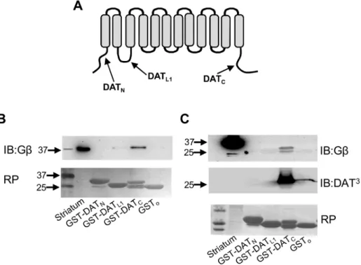
subunits, they strongly support a physical association between the transporter and Gbc subunits. Secondly, to identify the DAT domains involved in the interaction with Gbc subunits, we generated and purified three GST fusion proteins containing the amino-terminus, the first intracellular loop, or the carboxy-terminus of DAT (Figure 2A) and performed pull-down assays. Only the carboxy-terminus of DAT was able to pull down Gbc
these results demonstrate a direct physical interaction between DAT and Gbcsubunits and identify the carboxy terminus of the transporter as the site of the interaction.
Activation of G Proteins by GTP-c-S Inhibit DAT Activity in Heterologous Systems
According to the classical model, G proteins are activated when GTP binds to the Ga subunit of the Gabc trimeric complex, resulting in the dissociation of the Ga from the Gbc dimer. Subsequent hydrolysis of GTP into GDP allows the Gaand Gbc
subunits to re-associate, consequently inactivating the Gabc
complex [17]. Based on this model, we employed various pharmacological approaches to examine the effects of G protein activation on DA uptake. The non-hydrolyzable GTP analog GTP-c-S binds to Gaand activates G proteins by dissociating Ga
subunits from Gbcdimers. Because GTP-c-S is cell impermeable, we employedXenopus laevisoocytes expressing human DAT where the GTP analog can be injected directly into the cytoplasm. Injection of GTP-c-S (10mM) resulted in 30.566.1% reduction of [H3]-DA uptake inXenopus laevisoocytes expressing human DAT (Figure 3A). In contrast, intracellular injection of GDP-b-S (10mM), which prevents Gabc complex dissociation, failed to alter DAT uptake (Figure 3A). We repeated these experiments in HEK293-DAT cells permeabilized with streptolysin prior to incubation with the GTP analog GTP-c-S. As shown in Figure 3B, GTP-c-S incubation produced a significant decrease in DAT uptake activity, consistent with the results obtained in oocytes. Thus, these findings suggest that the activation of endogenous G proteins in both, Xenopus oocytes and HEK293 cells expressing DAT results in inhibition of transporter activity.
Overexpression of GbcSubunits Inhibit DAT Activity in Heterologous Cells
As a second functional approach, we examined the effect of overexpressing Gbc subunits on DAT activity in HEK293 cells stably transfected with DAT. Overexpression of Gb1c2, the most common Gbcdimer expressed in brain, resulted in a 37.1613.8% reduction in [H3]-DA uptake (Figure 4A). In an effort to show that the inhibitory effect mediated by Gbcsubunits is reversible, we repeated the experiments in the presence of a Ga subunit. Overexpression of Gai2 failed to alter DAT activity, but more
importantly prevented the inhibitory effect of Gb1c2 on DAT-mediated uptake (Figure 4A). The changes in DAT function by Gb1c2 overexpression were the result of a reduction in the maximal velocity (Vmax), but not in the affinity (Km) of the
transporter for DA (Figure 4B). There are five distinct Gb
isoforms, all expressed in brain. Therefore, to investigate the possibility that the inhibitory effect observed by Gb1c2 is isoform specific, we repeated the experiments using the most divergent Gb4 or Gb5 subunits. Overexpression of Gb4 or Gb5 either alone or in combination withc2 subunits produced similar decreases in DAT activity in HEK293 cells (Figure 4C) suggesting that the effect is not isoform specific. Biotinylation experiments revealed that Gb5, Gb4, or Gb1c2 overexpression resulted in no differences in cell surface levels of the transporter compared to control cells (Figure 4D and 4E). Thus, these results indicate that the decrease in DAT uptake activity by Gbc subunits is not a consequence of decreased plasma membrane levels of the transporter.
Figure 1. Co-immunoprecipitation of DAT and Gbcsubunits.(A, B) Immunoprecipitation of DAT with two DAT antibodies directed against different DAT epitopes results in the co-precipitation of Gbsubunits from mouse striatum. Control experiments include immunoprecipitations with nonspecific rat or rabbit IgGs or immunoprecipitations from DAT knockout striatum or mouse cerebellum. (C, D) Immunoprecipitation of DAT resulted in the co-immunoprecipitation of Gbsubunits from MN9D-DAT cells or HEK293-DAT cells. Control experiments include immunoprecipita-tions from MN9D or HEK293 mock-transfected cells. Three different DAT antibodies were used; DAT1(Mab369, Millipore), DAT2(H-80, Santa Cruz), and DAT3(c-20, Santa Cruz). Immunoblotting was performed with a Gbpan-antibody (T-20, Santa Cruz). Cereb = cerebellum, St = striatum, DAT-KO = DAT knockout.
Sequestration of Endogenous GbcSubunits Increase DAT activity in Heterologous Cells
Next, we examined the involvement of endogenous Gbc
subunits on DAT modulation by transfecting HEK293-DAT with either Gatransducin (Gat) or the carboxy-terminal domain of G
protein–coupled receptor kinase 2 (GRK2ct), which are both known scavengers of Gbc subunits [18–19]. Overexpression of Gat or
GRK2ct significantly increased DAT activity (126.3%69.1% and 123.2%66.7%, respectively) (Figure 4C). These results suggest that DAT is tonically inhibited by Gbc subunits expressed in HEK293 cells.
Activation of Endogenous GbcSubunits with mSIRK Inhibit DAT Activity in Heterologous Systems
As an additional approach to assess the role of Gbc on DAT function, we tested the effect of mSIRK, a cell-permeable myristoylated peptide that specifically activates Gbc subunits [20–21]. InXenopus laevisoocytes expressing DAT, incubation with 5mM or 25mM of mSIRK resulted in a 34.068.0% and 57.366.3% decrease in DAT activity, respectively (Figure 5A). To examine specificity, we tested the effect of mSIRK on glutamate uptake in oocytes expressing the excitatory amino acid transporter 1 (EAAT1). Here, glutamate uptake was not affected by mSIRK under the same conditions (Figure 5A). Next, we
Figure 2. Direct interaction between Gbcsubunits and the carboxy terminus of DAT.(A) Schematic representation of DAT depicting cytoplasmic segments used in GST pull-down assays. (B) The carboxy terminus of DAT (GST-DATc) precipitated Gbcsubunits from mouse striata. (C) Immuno-far-western assay showing a direct interaction between GST-DATcand purified Gbcfrom bovine brain. RP = Ponceau Red.
doi:10.1371/journal.pone.0059788.g002
Figure 3. Activation of G proteins with GTP-c-S inhibits DAT function in heterologous systems.(A) [3H]-DA uptake assays inXenopus laevisoocytes expressing DAT after intracellular injection with water (white bar, n = 8), 10mM GTP-c-S (black bar, n = 8), or 10mM GDP-b-S (gray bar, n = 8). (B) Specific [3H]-DA uptake and kinetic analysis for HEK293-DAT permeabilized with streptolysin-O (SLO) and dialyzed with 50
mM GTP-c-S
repeated these experiments in HEK293-DAT cells. mSIRK pre-incubation for 5 min produced a dose-dependent inhibition of [H3]-DA uptake with an IC50 of 10.662.6mM; whereas,
a scramble myristoylated peptide (scb-mSIRK) failed to alter DA uptake (Figure 5B). Longer incubation times with mSIRK did not augment the inhibitory effect on DAT function (IC50= 13.261.2mM). Kinetic analysis revealed that mSIRK
incubation resulted in a reduction of Vmaxwithout changes in the
Kmof DAT (Figure 5C).To confirm that the effect of mSIRK is mediated by Gbc subunits, we repeated these experiments in HEK293-DAT cells transfected with the Gbcscavenger GRK2ct. Consistent with our previous results (Figure 4C), overexpression of GRK2ct in HEK293-DAT cells increased DAT activity. More importantly, the inhibitory effect of mSIRK was significantly attenuated in the presence of GRK2ct (Figure 5D). Thus, these results are consistent with those obtained with GTP-c-S and Gbc
subunit overexpression and further support the contention that activation of Gbcsubunits results in inhibition of DAT activity.
Activation of Endogenous GbcSubunits with mSIRK Inhibit DAT Activity in Striatal Synaptosomes
To examine the Gbc-mediated regulation of DAT in native systems, we first assessed the effect of mSIRK on DAT activity in striatal synaptosomes. Pre-incubation of synaptosomes for 5 min with mSIRK, but not with scb-mSIRK, resulted in a dose-dependent inhibition of DAT activity with an IC50of 5.460.9mM
(Figure 6A), which is consistent with reported IC50 values for
mSIRK in other systems [20–22]. As observed with heterologous
cells, the effect of mSIRK on DAT function reflected changes in Vmax(37.764.6% reduction) without changes in Km(Figure 6B).
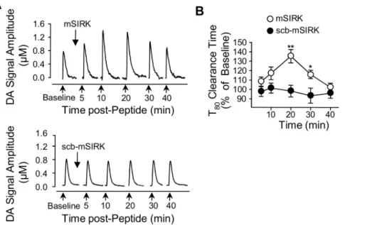
Activation of Endogenous GbcSubunits with mSIRK Inhibit DAT ActivityIn Vivo
Finally, we performed high-speed chronoamperometry to examine thein vivoeffect of the mSIRK peptide on DA clearance in striatum. In these experiments, a recording microelectrode was placed in the striatum and a glass multi-barrel micropipette was positioned adjacent to the electrode to locally deliver DA, mSIRK, or scrambled peptide. For each recording session, DA was pressure-ejected at 5 min intervals before and after mSIRK or scrambled peptide. mSIRK application, but not scrambled peptide produced an increase in both the amplitude of the DA signal (Figure 7A) and the clearance time of extracellular DA (Figure 7B). This pattern is consistent with an inhibition of DAT uptake activity, which is typically observed with known inhibitors of DA transport [15]. Taken together, our findings from synaptosomal preparations and intact animals provide compelling evidence that G proteinbcsubunits modulate DAT activity within a physiolog-ical context.
Discussion
Here, we have used a combination of biochemical and functional approaches in heterologous systems, brain synapto-somes, andin vivoto demonstrate that Gbcsubunits regulate DAT activity. Biochemically, we provide evidence for a direct in-teraction between DAT and Gbc through
co-immunoprecipita-Figure 4. Overexpression of Gbcsubunits results in decreased DAT uptake activity in HEK-293-DAT cells.(A, B) Specific [3H]-DA uptake and kinetic analysis for HEK293-DAT control cells (white bar, n = 6) or transfected with Gb1c2 (n = 3), Gai2 (n = 3), or Gai2Gb1c2 (n = 3). (C) Overexpression of Gb4 (n = 5) or Gb5 (n = 7) (gray bars), decreased [3H]-DA uptake in HEK293-DAT cells when compared to control (white bar, n = 19). Overexpression of the Gbcscavengers (black bars), Gat(n = 6), or GRK2ct (n = 4) increased [3H]-DA uptake. (D, E) Overexpression of Gb1c2 (n = 4), Gb4 (n = 5), or Gb5 (n = 7) in HEK-DAT cells did not alter the plasma membrane levels of DAT as measured by biotinylation. T = total fraction, B = biotinylated fractions, **p,0.01.
tion and pull-down assays with purified proteins. Functionally, Gbcsubunit activation by multiple approaches resulted in a rapid inhibition of transporter activity without alterations in levels of the transporter at the cell membrane. To our knowledge, no data are available describing an interaction between plasma membrane neurotransmitter transporters and G proteins and thus, this is the first study to identify a Gbc-mediated modulation of a neurotrans-mitter transporter.
According to the traditional view, upon binding of an extracellular agonist to a G-protein coupled receptor (GPCR), G proteins dissociate into active Ga and Gbc subunits. Originally, the Ga subunits were believed to transduce cellular responses, while the Gbc subunits were regarded to function solely as negative regulators of Ga-mediated signaling. Investigations over the last 20 years have revealed that the activation and function of G proteins is far more complex than previously anticipated. There
Figure 5. Activation of Gbcwith mSIRK resulted in an inhibition of DAT activity in heterologous systems.(A) Oocytes expressing DAT (white bars) or EAAT1 (gray bars) were incubated for 30 min with 0.5% DMSO (DAT/n = 3; EAAT1/n = 25), 5mM mSIRK (DAT/n = 20, EAAT1/n = 23), or 25mM mSIRK (DAT/n = 15, EAAT1/n = 15) before uptake assays with [H3]-DA or [3H]-Glutamate, respectively. (B) Dose-dependent inhibition of [3H]-DA
uptake in HEK293-DAT cells by mSIRK (n = 4, white circle). (C) 5 min pre-incubation of HEK293-DAT cells with 10mM mSIRK produced a reduction of Vmaxwith no changes in Km(n = 3). (D) In HEK293-DAT, 10mM mSIRK reduced uptake (control, black bar, n = 32; mSIRK, white bar, n = 15). The
inhibitory effect of 10mM mSIRK was attenuated in HEK-DAT cells expressing GRK2ct (control, n = 16; mSIRK, n = 16). **p,0.01 or *p,0.05. doi:10.1371/journal.pone.0059788.g005
Figure 6. Activation of Gbcsubunits reduces DAT activity in brain synaptosomes(A)mSIRK reduces [3H]-DA uptake in rat striatal synaptosomes.Samples were pre-incubated for 10 min with various concentrations of mSIRK (open circles) or scb-mSIRK (filled circles) at 34uC, followed by the addition of [3H]-DA (final concentration, 0.1mM) for 8 min, n = 3. Nonspecific [3H]-DA uptake was determined in the presence of 10mM nomifensine. (B) Kinetic analysis of the synaptosomal [3H]-DA uptake was determined in the absence (vehicle) or presence of 5mM mSIRK,
n = 3. **p,0.01.
is now evidence for both, receptor-dependent and -independent Ga- and Gbc-mediated responses [23]. In the case of VMAT2, the Ga-mediated inhibition of the transporter appears to be receptor-independent as Gasubunits are associated with synaptic vesicles [8], [10]. Our findings from immunoprecipitation experiments and those using Gbc subunits scavengers suggest a receptor-independent regulation, however, we can’t rule out a receptor-mediated regulation of DAT activity through Gbc
subunits. Potential GPCRs expressed in DA neurons, which could modulate DAT activity include receptors for glutamate, norepi-nephrine, serotonin, ATP, and DA. Indeed, several studies have documented changes in DAT activity as a result of the activation of D2 DA receptors (D2R) [24–25]. Interestingly, recent pro-vocative evidence suggests that the dopamine D2 receptor (D2R) regulates the trafficking of DAT to the plasma membrane through a direct protein-protein interaction [26]. Together, the available data suggest up-regulation of DAT through two mechanisms involving the activation of second messenger systems through D2R [25], as well as a direct protein-protein interaction between D2R and DAT [26]. Thus, it is unlikely that the Gbc-dependent decrease in DAT activity is related to activation of D2R. Future studies will be required to examine whether the Gbc-mediated inhibition of DAT described in this report is related to the activation of additional GPCRs or represents a receptor-in-dependent mechanism.
Additionally, evidence has accumulated indicating that Gbc
subunits also transduce cellular signals. In fact, Gbc subunits have been reported to directly regulate a diverse array of effector molecules including ion channels, enzymes, and in-tracellular regulators [27]. These interactions provide a role for Gbc in important physiological functions such as cardiac membrane potential, heart rate, inflammation, pain modulation, and neurotransmitter release [17], [28–29]. Of the many identified Gbc effectors, the Gbc-mediated inhibition of voltage-dependent calcium channels has been one of the most widely studied and best characterized [30]. Gbc directly binds
and inhibits calcium channel activity. Likewise, Gbc has also been reported to interact with syntaxin 1A [31], which is part of a SNARE protein complex that links the calcium channel to the vesicular release machinery. These findings suggest a com-plex interplay between the channel, G proteins, and SNARE proteins that modulate neurotransmitter release. Based on this model, we speculate that a similar network may exist between DAT, Gbc, and synaptic vesicles. Indeed, we previously reported that DAT is also physically and functionally coupled to synaptic vesicles via an interaction with synaptogyrin-3 [7]. Further supporting this idea, syntaxin 1A has also been reported to interact and modulate DAT [32–34]. Future studies will be needed to explore this possibility.
Despite the fact that there is much to learn regarding the modulation of DAT by Gbc subunits, the identification of DAT as a novel Gbc effector will open new avenues to our understanding of DA homeostasis regulation. DAT plays a crucial role in the control of DA homeostasis and has been implicated in a variety of psychiatry disorders and drug addiction [35]. Our findings suggest a novel mechanism for the control of DA homeostasis and may introduce a novel target for treatment of DA-related disorders. It remains to be seen whether additional plasma membrane transporters such as the norepinephrine, serotonin, GABA, or glutamate transporters are also modulated by G proteins.
Acknowledgments
We are grateful to the Torres and Amara laboratories for helpful discussions. We also thank Dr. Marc Caron and Ms. Wendy Roberts (Duke University) for providing DAT knockout tissue and Dr. Luis Aguayo (Universidad de Concepcion, Chile) for providing cDNAS expressing bsubunits and GRK2ct.
Figure 7. Activation of Gbcsubunits reduces DAT activityin vivo.(A) Clearance of exogenously applied DA from the striatum of anesthetized mice. Once a baseline for DA clearance was established, mSIRK or scr-mSIRK was applied, and then 5 min later, DA was applied and again, 10, 20, 30 and 40 min after the peptides were introduced (arrows). The amplitude of the DA signal augment as observed in a representative traces of DA signal after intrastriatal injection of mSIRK (upper panel) or scb-mSIRK (bottom panel). (B) Clearance time (T80) of exogenous DA was increased in the presence of mSIRK treatment. **p,0.01, *p,0.05.
Author Contributions
Conceived and designed the experiments: JG-O LD GET. Performed the experiments: JG-O DT-S TB WAO JZ. Analyzed the data: JG-O LCD
GET. Contributed reagents/materials/analysis tools: DPS SGA. Wrote the paper: JG-O TB GET.
References
1. Gainetdinov RR, Sotnikova TD, Caron MG (2002) Monoamine transporter pharmacology and mutant mice. Trends Pharmacol Sci 23: 367–373. 2. Jones SR, Gainetdinov RR, Jaber M, Giros B, Wightman RM, et al. (1998)
Profound neuronal plasticity in response to inactivation of the dopamine transporter. Proc Natl Acad Sci USA 95: 4029–4034.
3. Zahniser NR, Sorkin A (2004) Rapid regulation of the dopamine transporter: role in stimulant addiction. Neuropharmacology 47 Suppl 1: 80–91. 4. Foster JD, Cervinski MA, Gorentla BK, Vaughan RA (2006) Regulation of the
dopamine transporter by phosphorylation. Handb Exp Pharmacol 175: 197– 214.
5. Eriksen J, Jorgensen TN, Gether U (2011) Regulation of dopamine transporter function by protein-protein interactions: new discoveries and methodological challenges. J Neurochem 113: 27–41.
6. Sager JJ, Torres GE (2011) Proteins interacting with monoamine transporters: current state and future challenges. Biochemistry 50: 7295–7310.
7. Egan˜a LA, Cuevas RA, Baust TB, Parra LA, Leak RK, et al. (2009) Physical and functional interaction between the dopamine transporter and the synaptic vesicle protein synaptogyrin-3. J Neurosci 29: 4592–4604.
8. Ahnert-Hilger G, Nurnberg B, Exner T, Schafer T, Jahn R (1998) The heterotrimeric G protein Go2 regulates catecholamine uptake by secretory vesicles. EMBO J 17: 406–413.
9. Brunk I, Blex C, Rachakonda S, Holtje M, Winter S, et al. (2006) The first luminal domain of vesicular monoamine transporters mediates G-protein-dependent regulation of transmitter uptake. J Biol Chem 281: 33373–33385. 10. Ahnert-Hilger G, Schafer T, Spicher K, Grund C, Schultz G, et al. (1994)
Detection of G-protein heterotrimers on large dense core and small synaptic vesicles of neuroendocrine and neuronal cells. Eur J Cell Biol 65: 26–28. 11. Carneiro AM, Ingram SL, Beaulieu JM, Sweeney A, Amara SG, et al. (2002)
The multiple LIM domain-containing adaptor protein Hic-5 synaptically colocalizes and interacts with the dopamine transporter. J Neurosci 22: 7045– 7054.
12. Torres GE, Carneiro A, Seamans K, Fiorentini C, Sweeney A, et al. (2003) Oligomerization and trafficking of the human dopamine transporter. Mutational analysis identifies critical domains important for the functional expression of the transporter. J Biol Chem 278: 2731–2739.
13. Walev I, Bhakdi SC, Hofmann F, Djonder N, Valeva etal. (2001) Delivery of proteins into living cells by reversible membrane permeabilization with streptolysin-O. Proc Natl Acad Sci USA 98: 3185–3190.
14. Zhu J, Mactutus CF, Wallace DR, Booze RM (2009) HIV-1 Tat protein-induced rapid and reversible decrease in [3H]dopamine uptake: dissociation of [3H]dopamine uptake and [3H]2beta-carbomethoxy-3-beta-(4-fluorophenyl)-tropane (WIN 35,428) binding in rat striatal synaptosomes. J Pharmacol Exp Ther 329: 1071–1083.
15. Zahniser NR, Larson GA, Gerhardt GA (1999) In vivo dopamine clearance rate in rat striatum: regulation by extracellular dopamine concentration and dopamine transporter inhibitors. J Pharmacol Exp Ther 289: 266–277. 16. Callaghan PD, Irvine RJ, Daws LC (2005) Differences in the in vivo dynamics of
neurotransmitter release and serotonin uptake after acute para-methoxyamphe-tamine and 3,4-methylenedioxymethamphepara-methoxyamphe-tamine revealed by chronoampero-metry. Neurochem Int 47: 350–361.
17. Clapham DE, Neer EJ (1997) G protein beta gamma subunits. Ann Rev Pharmacol 37: 167–203.
18. Ford CE, Skiba NP, Bae H, Daaka Y, Reuveny E, et al. (1998) Molecular basis for interactions of G protein betagamma subunits with effectors. Science 280: 1271–1274.
19. Yevenes GE, Peoples RW, Tapia JC, Parodi J, Soto X, et al. (2003) Modulation of glycine-activated ion channel function by G-protein betagamma subunits. Nat Neurosci 6: 819–824.
20. Goubaeva F, Ghosh M, Malik S, Yang J, Hinkle PM, et al. (2003) Stimulation of cellular signaling and G protein subunit dissociation by G protein betagamma subunit-binding peptides. J Biol Chem 278: 19634–19641.
21. Smrcka AV (2008) G protein betagamma subunits: central mediators of G protein-coupled receptor signaling. Cell Mol Life Sci 65: 2191–2214. 22. Zhao Y, Fang Q, Straub SG, Lindau M, Sharp GW (2010) Noradrenaline
inhibits exocytosis via the G protein betagamma subunit and refilling of the readily releasable granule pool via the alpha(i1/2) subunit. J Physiol 588: 3485– 3498.
23. Blumer JB, Smrcka AV, Lanier SM (2007) Mechanistic pathways and biological roles for receptor-independent activators of G-protein signaling. Pharmacol Therapeut 113: 488–506.
24. Cass WA, Gerhardt GA (1994) Direct in vivo evidence that D2 dopamine receptors can modulate dopamine uptake. Neurosci Lett 176: 259–263. 25. Bolan EA, Kivell B, Jaligam V, Oz M, Jayanthi LD, et al. (2007) D2 receptors
regulate dopamine transporter function via an extracellular signal-regulated kinases 1 and 2-dependent and phosphoinositide 3 kinase-independent mechanism. Mol Pharmacol 71: 1222–1232.
26. Lee FJ, Pei L, Moszczynska A, Vukusic B, Fletcher PJ, et al. (2007) Dopamine transporter cell surface localization facilitated by a direct interaction with the dopamine D2 receptor. EMBO J 26: 2127–2136.
27. Smrcka AV, Lehmann DM, Dessal AL (2008) G protein betagamma subunits as targets for small molecule therapeutic development. Comb Chem High Throughput Screen 11: 382–395.
28. Zhang XL, Upreti C, Stanton PK (2011) Gbetagamma and the C Terminus of SNAP-25 Are Necessary for Long-Term Depression of Transmitter Release. PLoS One 6: e20500.
29. McCudden CR, Hains MD, Kimple RJ, Siderovski DP, Willard FS (2005) G-protein signaling: back to the future. Cell Mol Life Sci 62: 551–577. 30. Zamponi GW (2001) Determinants of G protein inhibition of presynaptic
calcium channels. Cell Biochem Biophys 34: 79–94.
31. Jarvis SE, Magga JM, Beedle AM, Braun JE, Zamponi GW (2000) G protein modulation of N-type calcium channels is facilitated by physical interactions between syntaxin 1A and Gbetagamma. J Biol Chem 275: 6388–6394. 32. Binda F, Dipace C, Bowton E, Robertson SD, Lute BJ, et al. (2008) Syntaxin 1A
interaction with the dopamine transporter promotes amphetamine-induced dopamine efflux. Mol Pharmacol 74: 1101–1108.
33. Carvelli L, Blakely RD, DeFelice LJ (2008) Dopamine transporter/syntaxin 1A interactions regulate transporter channel activity and dopaminergic synaptic transmission. Proc Natl Acad Sci USA 105: 14192–14197.
34. Cervinski MA, Foster JD, Vaughan RA (2010) Syntaxin 1A regulates dopamine transporter activity, phosphorylation and surface expression. Neuroscience 170: 408–416.

![Figure 6. Activation of Gbc subunits reduces DAT activity in brain synaptosomes (A) mSIRK reduces [ 3 H]-DA uptake in rat striatal synaptosomes](https://thumb-eu.123doks.com/thumbv2/123dok_br/17245160.245346/7.918.86.626.89.495/activation-subunits-reduces-activity-synaptosomes-reduces-striatal-synaptosomes.webp)