J of Evolution of Med and Dent Sci/ eISSN- 2278-4802, pISSN- 2278-4748/ Vol. 3/ Issue 57/Oct 30, 2014 Page 12989
CLINICAL ANALYSIS OF FIFTY CASES OF NEOVASCULAR GLAUCOMA
K. Stephen
1, C. Shankar
2, V. Velayutham
3, R. Pandurangan
4, C. Charanya
5, R. Adnan
6HOW TO CITE THIS ARTICLE:
K. Stephen, C. Shankar, V. Velayutham, R. Pandurangan, C. Charanya, R. Adnan. Clinical Analysis of Fifty Cases of Neovascular Glaucoma . Journal of Evolution of Medical and Dental Sciences 2014; Vol. 3, Issue 57, October 30; Page: 12989-12996, DOI: 10.14260/jemds/2014/3724
ABSTRACT: AIM: to clinically analyze fifty cases of neo vascular glaucoma in a tertiary care eye hospital. MATERIALS AND METHOS: a two year prospective study was done in patients with neo vascular glaucoma attending the glaucoma clinic. Detailed history with ocular and systemic examination was done in all patients. Fundus fluorescein angiogram and B scan was done in selected patients. Gonioscopic evaluation was done and neovascularization was graded. RESULTS: mean age of presentation of NVG was 52.06 years with male preponderance (2.06:1). Post traumatic NVG presented at early age. Proliferative diabetic retinopathy(PDR) accounted for 15 cases(30%) followed by recurrent anterior uveitis in 12 cases(24%), post central retinal vein occlusion and post cataract surgery accounted for 6 cases each (12%) and post traumatic and post chronic retinal detachment accounted for 5 cases each (10%). Patients with NVG secondary to CRVO presented earlier than other groups and all patients had ischemic type of CRVO. Hyphaema was present in 16 cases. (32%) corneal edema and corneal decompensation was universal and it contributed to poor visual acuity. Mean IOP range was 51.14mmHg ranged between (28.2-81.7mmHg). Mean intra ocular pressure in neo vascular glaucoma due to chronic retinal detachment was 33.2mmHg which is comparatively less than other group. In most patients neovascularization of iris was in advanced grading (C & D). CONCLUSION: In all patients the NVG was unilateral. PDR was the most common cause followed by recurrent uveitis, Ischemic CRVO, post cataract surgery, post traumatic and post retinal detachment. Patients with neo vascular glaucoma secondary to CRVO presented earlier than 6 months when compared to other groups which presented after 6 months. Mean IOP secondary to long standing RD was lower when compared to other groups. Most patients with NVG presented with defective vision weeks to months before the onset of redness and pain.
KEYWORDS: neo vascular glaucoma, etiology, clinical features.
INTRODUCTION: Neovascular glaucoma is defined as increased intraocular pressure associated with iris and/or angle neovascularization.[1] It is mainly secondary to posterior segment ischemia.[2] It is
also known as hemorrhagic glaucoma (referring to hyphema that is present in some patients), thrombotic glaucoma (underlying vascular thrombotic etiology) rubeotic glaucoma.[3]Clinical course
is characterized by open angle glaucoma caused by invasion of anterior chamber angle and later on angle closure due to secondary synechiae formation
OCULAR: Vascular disease such as central retinal artery occlusion (CRVO), central retinal artery occlusion, proliferative diabetic retinopathy (PDR), Ocular ischemic syndrome, Ocular neoplasms, chronic uveitis, Fuchsiridocyclitis.
SYSTEMIC: Carotid artery occlusion, Giant cell arteritis and Aortic arch syndrome.[3,4] Diabetes and
J of Evolution of Med and Dent Sci/ eISSN- 2278-4802, pISSN- 2278-4748/ Vol. 3/ Issue 57/Oct 30, 2014 Page 12990 proliferative diabetic retinopathy.[5] PDR accounts for 32-43% of NVG patients.[6,7] Retinal hypoxia
causes increased vascular endothelial growth factor(VEGF) which stimulates the neovascularization of iris and angle of anterior chamber. In patients with NVG without precipitating fundus condition, carotid occlusive disease should be suspected.[8]
PROPHYLAXIS: Key stone for prevention of NVG is retinal ablation by photocoagulation or cryotherapy in which retinal tissue is destroyed leading to inhibition of vasoproliferation in anterior segment.[9] If ocular media is hazy then retinal cryoabalation can be effective in anterior segment
vaso proliferation.
Goniophotocoagulation can also prevent the development of NVG by interrupting critical tissue response to angiogenic stimulus, while retinal photocoagulation reduces the stimulus itself. Prophylactic methods are performed only if anterior segment neovascularization develops.
VARIOUS TREATMENT MODALITIES INCLUDE: Medical treatment is with cycloplegics. Panretinal photocoagulation should be done even if IOP is medically controlled as it reverses the pathogenesis. It may not reverse the fibrous component infibrovascular proliferation.[10] Pan retinal cryotherapy is
done if media is hazy and photocoagulation cannot be done.[11]
Filtering surgery with mitomycin C and glaucoma shunt procedures can be done in established cases. Anti VEGF’s are beneficial and are used as adjuvant in treatment of NVG due to its antiangiogenic properties. They are used for both prophylaxis and treatment of NVG.[12] Intravitreal
antiVEGF injection is not very effective once peripheral anterior synechial angle closure develops.[13]
Retrobulbar injection of alcohol and cyclocryotherapy may be done to relieve the pain
MATERIALS AND METHODS: This prospective two-year study has been conducted on patients with Neo Vascular Glaucoma (NVG) attending the glaucoma clinic in Regional Institute of Ophthalmology, Chennai. A detailed history regarding onset, duration, presenting complaints, ocular and systemic predisposing factors like diabetes, hypertension, vascular diseases, and chronic uveitis were recorded.A thorough evaluation of patients, which included general and ocular examination were performed.
Anterior segment examination by oblique illumination, slit lamp biomicroscopy, tonometry, gonioscopy, visual acuity recording, fundus examination, fundus flurorescein angiography in selected cases, ultrasound B scan with vector A scan examination was done in cases with suspected posterior segment involvement in which fundus examination showed hazy media. For suspected cases of diabetes and patients with family history of diabetes post prandial blood sugar, urine for albumin and sugar was done.
GONIOSCOPY: angle evaluated with goldmann single mirror indirect gonioprism and following factors noted:
1. Grading of anterior chamber angle(SHAFFERS). 2. Presence of new vessels.
J of Evolution of Med and Dent Sci/ eISSN- 2278-4802, pISSN- 2278-4748/ Vol. 3/ Issue 57/Oct 30, 2014 Page 12991 Grading of neo vascularization was done based on extent of neo vascularization, presence of peripheral anterior synechiae, and the number of quadrants involved. (JoesphTauba et al). A B C – represent extent of neo vascularization.
A- At pupillary margin. B- Mid stromal iris. C- Angle.
D- PAS.
NUMBER 1,2,3,4 – indicate number of quadrants involved. IOP greater than 21mmHg is denoted by adding a + sign.
INCLUSION CRITERIA:
1. All patients with neovascular glaucoma of varied etiology.
2. Patients with neovascularization of iris and raised intraocular pressure.
EXCLUSION CRITERIA:
3. Patients with anterior segment neovascularization alone without glaucoma. 4. Patients who have evidence of treatment for primary glaucoma.
5. Patients with dilated vessels alone in iris without evidence of new vessels.
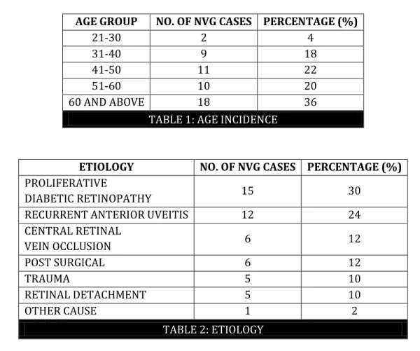
RESULTS: This study was conducted in fifty established cases of neovascular glaucoma attending glaucoma clinic. In all cases detailed history regarding mode of onset, duration of illness, patient’s symptoms and detailed general and ocular examination was done to find out incidence, mode of presentation, etiological factors and associated systemic risk factors related to development of NVG. The mean age of presentation was 52.06 years with male preponderance (2.06:1). In all cases the neo vascular glaucoma was unilateral. Post traumatic NVG presented at early age than other groups. (TABLE 1).
The patients were divided into seven groups based on etiology of NVG of which PDR accounted for 15 cases. Recurrent chronic anterior uveitis accounted for 12 cases, CRVO and post cataract surgery NVG 6 cases, post traumatic and post retinal detachment NVG formed 5 cases each. PDR accounted for major cause followed by anterior uveitis and CRVO. In all the RD cases causing NVG, the RD was long standing. (TABLE 2 AND 3).
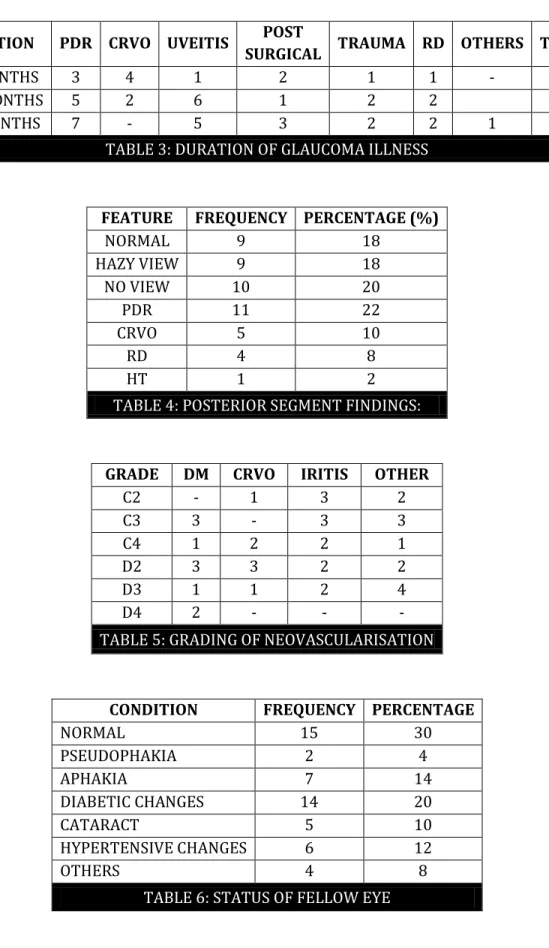
In all cases of neo vascular glaucoma due to CRVO the cause of neovascularization was due to ischemic type CRVO. Patients with NVG due to PDR and uveitis usually presented after 6 months. In contrast to patients with CRVO who presented within the first 6 months.
ANTERIOR SEGMENT FINDINGS: Corneal decompensation and edema was found in almost all the cases reflecting advanced stage of presentation and high IOP. Other findings were corneal Keratic precipitates, Bullae, Vascularization, new vessels on iris extending to the angle, with synechiae angle closure. Hyphaema was present in 16 cases (32%).
J of Evolution of Med and Dent Sci/ eISSN- 2278-4802, pISSN- 2278-4748/ Vol. 3/ Issue 57/Oct 30, 2014 Page 12992
GRADING OF NEOVASCULARIZATION: Out of 41 patients where grading was done, 21 patients had grade C and 20 had grade D signifying the advanced stage of disease. In 9 cases grading was not possible due to advanced corneal decompensation. (TABLE 5).
FELLOW EYE: Fellow eyes in patients with NVG secondary to vascular causes such as diabetes and CRVO showed diabetic retinopathic changes and hypertensive retinopathy changes respectively. (TABLE 6).
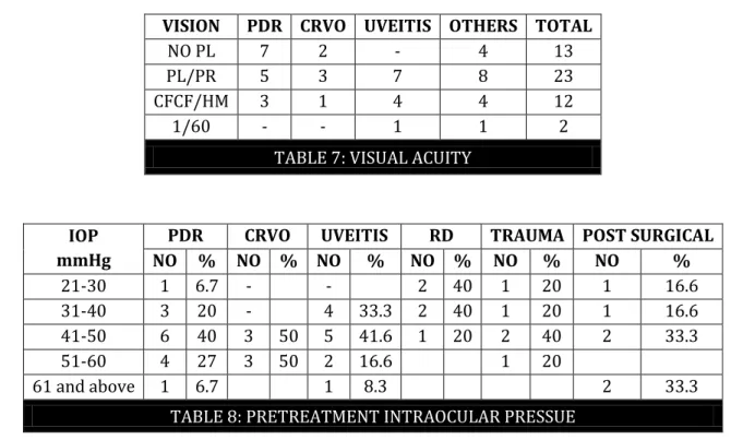
VISUAL ACUITY: All cases had visual acuity 1/60 or less with visual acuity in range of PL/PR constituted majority of cases. Corneal decompensation was the most common cause of defective vision in most of the cases. (TABLE 7)
INTRAOCULAR PRESSURE:
Mean IOP range was 51.14mmHg ranged between (28.2-81.7mmHg). Mean intra ocular pressure in neo vascular glaucoma due to chronic retinal detachment was 33.2mmHg which is comparatively less than other group. (TABLE 8).
DISCUSSION: Total incidence of neo vascular glaucoma was maximum in age group more than 60 years. Maximum age of presentation was 80 years and minimum age was 21 years. Mean age of neo vascular glaucoma due to central retinal vein occlusion was 64.33 years which correlates with Ohrt et al study in which the mean age of NVG secondary to CRVO was 66.4 _+10.9 years.[14]
In this study percentage of male patients with neo vascular glaucoma secondary to central retinal vein occlusion was 83.3%. Male sex is significant risk factor for retinal vein occlusion according to Hayreh et al study.[15] In all 6 CRVO cases the cause of neovascularization was due to
ischemic type CRVO which was similar to Hayreh et all retinal vein occlusion study which showed 50% of ischemic CRVO to develop NVG.[16]
More risk of anterior segment neovascularization and NVG in PDR. Most patients with diabetic neovascularization (80%) presented after 6 months and 91.6% of patients with NVG due to recurrent uveitis also presented after 6 months. This may be due to less virulent nature of new vessels.[17] Most patients of CRVO presented within first 6 months which may be due to rapid
development of neovascularization process which correlates with study of Jocsons study.[18]
Neovascularization could not be demonstrated in one patient in iris. However new vessels was seen in the angle. This may be due to regression of new vessels. This was similar to Madsen et al study.[19] All cases had visual acuity 1/60 or less. No PL accounted for 13 cases (26%) PL/PR
accounted for 23 cases. The poor visual acuity in these cases was due to primary posterior segment pathology, the glaucoma process, late stage of presentation, advanced corneal decompensation. Advanced corneal decompensation was the most common cause of defective vision.
The mean pre-treatment IOP was 51.14mmHg. The mean IOP in diabetic group was 50.24 mmHg, CRVO group was 43.00mmHg, and uveitic group was 43.00mmHg. Mean IOP in NVG due to chronic RD is 33.2 which are comparatively less than other groups. IOP is usually >40mmHg in NVG. In rare cases of NVG due to chronic RD, IOP is comparatively lower than other group or even normal according to Brown et al study.[20]
post-J of Evolution of Med and Dent Sci/ eISSN- 2278-4802, pISSN- 2278-4748/ Vol. 3/ Issue 57/Oct 30, 2014 Page 12993 traumatic etiology and retinal detachment. PDR and recurrent anterior uveitis constituted to about 54%.
The mean age of presentation was 52.06 years with post traumatic neo vascular glaucoma presented at early age. Male to female ratio is 2.06:1.
Patients with neo vascular glaucoma secondary to CRVO presented relatively earlier than other groups.
In all cases of neo vascular glaucoma due to CRVO the neovascularization was due to ischemic type of CRVO.
Almost all cases had corneal and iris involvement with new vessels extending to the angle with or without synechial angle closure.
In all cases of NVG secondary to RD the detachment was long standing.
Fellow eyes in patients with NVG secondary to diabetes and CRVO showed diabetic retinopathy and hypertensive retinopathy changes.
In most cases visual acuity was PL/PR and advanced corneal decompensation was the cause for defective vision.
Mean intraocular pressure in NVG secondary to long standing RD was lowest among all groups. Hyphaema was significant sign of anterior segment neovascularization accounting to 32% of cases.
In many patients depending on underlying disease patients presented with defective vision weeks to months before onset of pain and redness.
AGE GROUP NO. OF NVG CASES PERCENTAGE (%)
21-30 2 4
31-40 9 18
41-50 11 22
51-60 10 20
60 AND ABOVE 18 36
TABLE 1: AGE INCIDENCE
ETIOLOGY NO. OF NVG CASES PERCENTAGE (%)
PROLIFERATIVE
DIABETIC RETINOPATHY 15 30
RECURRENT ANTERIOR UVEITIS 12 24
CENTRAL RETINAL
VEIN OCCLUSION 6 12
POST SURGICAL 6 12
TRAUMA 5 10
RETINAL DETACHMENT 5 10
OTHER CAUSE 1 2
J of Evolution of Med and Dent Sci/ eISSN- 2278-4802, pISSN- 2278-4748/ Vol. 3/ Issue 57/Oct 30, 2014 Page 12994
DURATION PDR CRVO UVEITIS POST
SURGICAL TRAUMA RD OTHERS TOTAL
0-6 MONTHS 3 4 1 2 1 1 - 12
6-12 MONTHS 5 2 6 1 2 2 18
>12 MONTHS 7 - 5 3 2 2 1 20
TABLE 3: DURATION OF GLAUCOMA ILLNESS
FEATURE FREQUENCY PERCENTAGE (%)
NORMAL 9 18
HAZY VIEW 9 18
NO VIEW 10 20
PDR 11 22
CRVO 5 10
RD 4 8
HT 1 2
TABLE 4: POSTERIOR SEGMENT FINDINGS:
GRADE DM CRVO IRITIS OTHER
C2 - 1 3 2
C3 3 - 3 3
C4 1 2 2 1
D2 3 3 2 2
D3 1 1 2 4
D4 2 - - -
TABLE 5: GRADING OF NEOVASCULARISATION
CONDITION FREQUENCY PERCENTAGE
NORMAL 15 30
PSEUDOPHAKIA 2 4
APHAKIA 7 14
DIABETIC CHANGES 14 20
CATARACT 5 10
HYPERTENSIVE CHANGES 6 12
OTHERS 4 8
J of Evolution of Med and Dent Sci/ eISSN- 2278-4802, pISSN- 2278-4748/ Vol. 3/ Issue 57/Oct 30, 2014 Page 12995
VISION PDR CRVO UVEITIS OTHERS TOTAL
NO PL 7 2 - 4 13
PL/PR 5 3 7 8 23
CFCF/HM 3 1 4 4 12
1/60 - - 1 1 2
TABLE 7: VISUAL ACUITY
IOP mmHg
PDR CRVO UVEITIS RD TRAUMA POST SURGICAL NO % NO % NO % NO % NO % NO %
21-30 1 6.7 - - 2 40 1 20 1 16.6
31-40 3 20 - 4 33.3 2 40 1 20 1 16.6
41-50 6 40 3 50 5 41.6 1 20 2 40 2 33.3
51-60 4 27 3 50 2 16.6 1 20
61 and above 1 6.7 1 8.3 2 33.3
TABLE 8: PRETREATMENT INTRAOCULAR PRESSUE
REFERENCES:
1. Călugăru D, Călugăru M.Neovascular glaucoma--etipathogeny and diagnosis. Oftalmologia. 2012; 56 (2): 3-14.
2. Shazly TA, Latina MA. Neovascular glaucoma: etiology, diagnosis and prognosis. SeminOphthalmol. 2009; 24: 113–121.
3. Mocanu C, Barascu D, Marinescu F. Neovascular glaucoma-Retrospective study. Ophthalmologica 2005; 49: 58-65.
4. Ashaye AO1, Adeoti CO. Neovascular glaucoma in a Nigerian African population. East Afr Med J.
2006 Oct; 83 (10): 559-64.
5. Schertzer RM, Wang D, Bartholomew LR: Diabetes mellitus and glaucoma. Int Ophthalmol Clin 1998; 38: 69–87.
6. Madsen PH. Experiences in surgical treatment of haemorrhagic glaucoma. A follow-up study.
Acta Ophthalmol Suppl. 1973; 120: 88-91.
7. Brown, G.C, Magargal, L.E, Schachat, A, and Shah, H. Neovascular glaucoma. Etiologic considerations. Ophthalmology. 1984; 91: 315–320.
8. Coppeto, JR, Wand, M, Bear, L, and Sciarra, R. Neovascular glaucoma and carotid artery obstructive disease. Am J Ophthalmol. 1985; 99: 567–570.
9. Laatikainen L. Preliminary report on effect of retinal panphotocoagulation on rubeosisiridis and neovascular glaucoma. Br J Ophthalmol. 1977 Apr; 61 (4): 278-84.
10.Little HL, Rosenthal AR, Dellaporta A, and Jacobson DR. The effect of pan-retinal photocoagulation on rubeosisiridis. Am J Ophthalmol. 1976; 81: 804–809
J of Evolution of Med and Dent Sci/ eISSN- 2278-4802, pISSN- 2278-4748/ Vol. 3/ Issue 57/Oct 30, 2014 Page 12996 12.Luke J, Nassar K, Luke M. Ranibizumab as adjunct in treatment of rubeosisiridis. Grafes arch
clin. Exp Ophthalmol 2013, Oct: 251(10) 2403.
13.Jack.J.kanski.Brad Bowling clinical ophthalmology. 7th ed. Page 361.
14.Ohrt V. The frequency of rubeosisiridis in diabetic patients. Acta Ophthalmol (Copenh) 1971; 49 (2): 301–307.
15.Hayreh SS1, Zimmerman MB. Ocular neovascularization associated with central and hemicentral retinal vein occlusion. Retina. 2012 Sep; 32 (8): 1553-65.
16.Hayreh, S.S, Klugman, M.R, Podhajsky, P et al. Argon laser panretinal photocoagulation in ischemic central retinal vein occlusion. A 10-year prospective study. Graefes Arch Clin Exp Ophthalmol. 1990; 228: 281–296 (I).
17.Blinder, K.J, Friedman, S.M, and Mames, R.N. Diabetic iris neovascularization. Am J Ophthalmol. 1995; 120: 393–395 (II).
18.Jocson VL .Microvascular injection studies in rubeosisiridis and neovascular glaucoma. Am J Ophthalmol. 1977 Apr; 83 (4): 508-17.
19.P H Madsen. Rubeosis of the iris and haemorrhagic glaucoma in patients with proliferative diabetic retinopathy. Br J Ophthalmol. Jun 1971; 55 (6): 368–371.
20.Brown, G.C and Margargal, L.E. Central retinal artery obstruction and visual acuity. Ophthalmology. 1982; 89: 14–19.
AUTHORS:
1. K. Stephen 2. C. Shankar 3. V. Velayutham 4. R. Pandurangan 5. C. Charanya 6. R. Adnan
PARTICULARS OF CONTRIBUTORS:
1. Associate Professor, Department of Ophthalmology, Chetthinad Hospital and Research Institute.
2. Assistant Professor, Department of Ophthalmology, Chetthinad Hospital and Research Institute.
3. Professor, Department of Ophthalmology, Chetthinad Hospital and Research Institute.
4. Professor & HOD, Department of
Ophthalmology, Chetthinad Hospital and Research Institute.
5. Post Graduate, Department of
Ophthalmology, Chetthinad Hospital and Research Institute.
6. Post Graduate, Department of
Ophthalmology, Chetthinad Hospital and Research Institute.
NAME ADDRESS EMAIL ID OF THE CORRESPONDING AUTHOR:
Dr. C. Charanya
Urbanville Apartments, Velachery Main Road, Velachery,
Chennai-600042.
Email: charanyachendilnathan@gmail.com