63
ABSTRACT
A case of a pseudoneoplastic lesion of the breast clinically and sonographically suggestive of a fibroadenoma is reported. Excisional biopsy revealed the nodule was an inflammatory process consequent to infection by Schistosoma mansoni.
Key-words : Breast. Schistosomiasis. Pseudoneoplastic lesion. Schistosoma mansoni.
RESUMO
Relata-se um caso de uma lesão pseudoneoplásica da mama clinicamente e ultrasonograficamente sugestiva de um fibroadenoma. A biópsia excisional revelou que o nódulo tratava-se de um processo inflamatório conseqüente à infecção pelo Schistosoma mansoni.
Palavras-chaves : Mama. Esquistossomose. Lesão pseudoneoplásica. Schistosoma mansoni.
1. Department of Oncology, Hospital João Alves, Aracaju, SE, Brazil. 2. Department of Infectious Diseases, Hospital João Alves, Aracaju, SE, Brazil. 3. Laboratório de Patologia e Citologia, Aracaju, SE, Brazil.
Address to: Dr. Carlos Anselmo Lima. Av. Sizino Martins Fontes 84/102, Farolândia, 49032-510 Aracaju, SE, Brazil. Telefax: 55 79 243-1713.
e-mail: [email protected] Recebido em 28/7/2003 Aceito em 23/9/2003
RELATO DE CASO/CASE REPORT
Revista da Sociedade Brasileira de Medicina Tropical 37:63-64, jan-fev, 2004
Pseudoneoplastic lesion of the breast caused
by
Schistosoma mansoni
Pseudoneoplasma da mama causado
pelo
Schistosoma mansoni
Carlos Anselmo Lima
1, Aécio Costa Cavalcanti
1,
Márcia Maria Macêdo Lima
2and Nestor Piva
3Schistosomiasis is an important health problem in the world, mainly in developing countries where it is responsible for more than 200 million infected people. The disease is caused by the schistosome and, although various species are found worldwide, only five cause disease in man: S. mansoni, S. japonicum, S. haematobium, S. mekongi, and S. intercalatum2. In South America, only S. mansoni is known.
There are several clinical presentations of schistosomiasis, defined by manifestations mainly of the gut, urinary tract, portal system, and lungs. Also, ectopic granulomas can occur in every organ. The breast is a very rare site of disease manifestation. Only six cases of this localization were found in the world literature1 3 4 5 6 7. Thus, we report what seems to be the seventh case of a breast lesion caused by S. mansoni, with a clinical and sonographic appearance of fibroadenoma.
CASE REPORT
This 23-year-old woman sought medical attendance because of a painless breast nodule noted six months before admission. Physical examination revealed a non tender, elastic, mobile 2.5 x 2cm mass
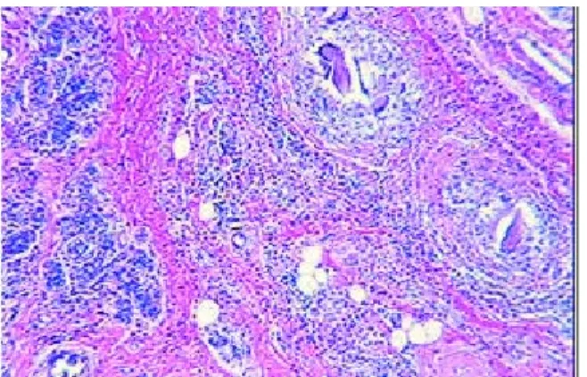
located between the lower quadrants, near the right nipple. A sonogram revealed a hypoechogenic nodule suggestive of a fibroadenoma. No mammogram was done because of the age of the patient. Excisional biopsy showed a dense fibrous brown mass. Microscopically, two features were outstanding: several granulomas with either viable ova or remnants of ova in the center (Figures 1 and 2) and couples of adult worms inside veins amongst mammary lobules, surrounded by an intense eosinophil-rich inflammatory process (Figure 3). These formed a pseudoneoplastic breast lesion cause by S. mansoni. Since there were viable worms, treatment with oxamniquine was prescribed.
DISCUSSION
64
Figure 1 - Granulomas centered by a viable S. mansoni egg. Figure 2 - Granulomas centered by eggshells.
enabled the diagnostic confirmation of a mammary nodule caused byS. mansoni. This finding was considered conclusive and so no other diagnostic methods were necessary. Treatment with Oxamniquine was initiated because viable ova were observed. A fecal test revealed no ova soon after treatment. In older women, such a clinical and mammographic appearance would not rule out the possibility of cancer. Thus, either an excision or needle biopsy is necessary for differential diagnosis as well as providing the only form of diagnosing pseudoneoplastic lesions caused by schistosome.
The rarity of the mammary presentation should be emphasized and the six cases reported in the world literature were due to S. japonicum1 5 6 7 andS. haematobium3 4.
To the best of our knowledge, this is the first case to be reported in the world literature, with Schistosoma mansoni as the etiologic agent.
Lima CA et al
Figure 3 - Couples of adult worms inside veins.
REFERENCES
1. Gorman JD, Champaign JL, Sumida FK, Canavan L. Schistosomiasis involving the breast. Radiology 185:423-424, 1992.
2. Mahmoud AA. Trematodes (Schistosomiasis) and other flukes. In: Mandell GL, Bennet JE, Dolin R (eds) Principles and Practice of Infectious Diseases, 5th Edition,
Churchill-Livingstone, Philadelphia, p. 2950-2956, 2000.
3. Nkanza NK. Schistosomal ova in a female breast. Tropical and Geographic Medicine 41:365-367, 1989.
4. Peyromaure M, Antoine M, Gadonneix P, Villet R. La bilharziose: une cause exceptionelle de microcalcifications mammaires. Journal of Gynecology, Obstetrics, Biology and Reproduction 29:790-792, 2000.
5. Sloan BS, Rickman LS, Blan EM, Davis CE. Schistosomiasis masquerading as carcinoma of the breast. Southern Medical Journal 89:345-347, 1996. 6. Varin CR, Eisenberg BL, Ladd W. Mammographic microcalcifications associated
with schistosomiasis. Southern Medical Journal 82:1060-1061, 1989. 7. Wu DM. A case of mammary schistosomiasis complicated with breast cancer.