Cop
yright
© ABE&M t
odos os dir
eit
os r
eser
vados
.
Thyroglobulin measurements in
washout of ine needle aspirates in
cervical lymph nodes for detection of
papillary thyroid cancer metastases
Determinação da tireoglobulina no lavado da agulha da punção aspirativa de linfonodos cervicais para detecção de metástases do câncer papilar de tireoide
André B. Zanella1, Erika L. Souza Meyer1, Letícia Balzan1, Antônio C. Silva1, Joíza Camargo1, Alceu Migliavacca1, José Ricardo Guimarães1,
Ana Luiza Maia1
ABSTRACT
Objective: The aim of this study was to evaluate the accuracy of the measurement of thyroglo-bulin in washout needle aspiration biopsy (FNAB-Tg) to detect papillary thyroid cancer (PTC) metastases. Subjects and methods: Forty-three patients (51.4 ± 14.6 years) with PTC diagno-sis and evidence of enlarged cervical lymph nodes (LN) were included. An ultrasound-guided ine-needle aspiration of suspicious LN was performed, for both cytological examination and measurement of FNAB-Tg. Results: The median values of FNAB-Tg in patients with metastatic LN (n = 5) was 3,419 ng/mL (11.1-25,538), while patients without LN metastasis (n = 38) showed levels of 3.7 ng/mL (0.8-7.4). Considering a 10 ng/mL cutoff value for FNAB-Tg, the sensitivity and speciicity was 100%. There were no differences on the median of FNAB-Tg measurements between those on (TSH 0.07 mUI/mL) or off levothyroxine (TSH 97.4 mUI/mL) therapy (3.3 vs. 3.8 ng/mL, respectively; P = 0.2). Conclusion: The results show that evaluation of FNAB-Tg in cervical LN is a valuable diagnostic tool for PTC metastases that can be used independent of the thyroid status. Arq Bras Endocrinol Metab. 2010;54(6):550-4
Keywords
Papillary thyroid carcinoma; metastases in cervical lymph nodes; thyroglobulin; TSH
RESUMO
Objetivo: O objetivo deste estudo foi avaliar a acurácia da dosagem de tireoglobulina no lavado da agulha da punção aspirativa (PAAF-Tg) de linfonodos (LN) cervicais para detecção de me-tástases do câncer papilar de tireoide (CPT). Sujeitos e métodos: Foram incluídos 43 pacientes (51,4 ± 14,6 anos) com diagnóstico de CPT e evidência de LN cervicais aumentados. Os LN suspeitos foram submetidos à punção aspiração com agulha ina guiada por ecograia para análise citológica e dosagem de tireoglobulina (PAAF-Tg). Resultados: A mediana dos valores de PAAF-Tg nos LN metastáticos (n = 5) foi 3.419,0 ng/mL (11,1-25.538), enquanto nos LN não metastáticos (n= 38) a mediana foi de 3,7 ng/mL (0,8-7,4). Utilizando-se o nível de 10 ng/mL como ponto de corte, observaram-se sensibilidade e especiicidade de 100%. Os níveis de TSH sérico não interferiram na dosagem de PAAF-Tg (3,3 e 3,8 ng/mL nos grupos com TSH supresso (TSH 0,07 mUI/mL) e hipotireoidismo (TSH 97,4 mUI/mL), respectivamente, P = 0,2). Conclusão: Os resultados demonstram que a dosagem de PAAF-Tg é uma ferramenta importante no diag-nóstico de metástases do CPT, podendo ser utilizada independente do “status” tireoidiano. Arq Bras Endocrinol Metab. 2010;54(6):550-4
Descritores
Câncer papilar de tireoide; metástases em linfonodos cervicais; tireoglobulina; TSH
1 Seção de Tireoide, Divisão de Endocrinologia, Hospital de Clínicas de Porto Alegre, Universidade Federal do Rio Grande do Sul (HC-UFRGS), Porto Alegre, RS, Brazil
Correspondence to:
Ana Luiza Maia
Seção de Tireoide, Divisão de Endocrinologia, Hospital de Clínicas de Porto Alegre Rua Ramiro Barcelos, 2.350 90035-003 − Porto Alegre, RS, Brazil
Cop
yright
© ABE&M t
odos os dir
eit
os r
eser
vados
.
InTRODUCTIOn
T
hyroid cancer is the most common malignant neo-plasm of the endocrine system, mostly affecting women and individuals between 25 to 65 years (1). Differentiated thyroid cancer (DTC) is responsible for 90% of the malignant thyroid gland neoplasias (2). Ac-cording to current guidelines, the initial therapeutic approach to DTC consists of total thyroidectomy and cervical lymph node (LN) dissection for the papillary type (3,4). The treatment is completed with a thera-peutic dose of radioactive iodine and TSH suppression by the use of levothyroxine (3).The disease has a generally good prognosis, espe-cially among patients younger than 45 years of age. However, recurrence of the tumor in cervical LN oc-curs in up to 15% of patients after initial therapy (5). Therefore, monitoring of recurrence is mandatory. Three main diagnostic tools can be used in detecting residual or recurrent disease: serum thyroglobulin (sTg) basal and stimulated by endogenous or recom-binant TSH, cervical ultrasound (US) and whole body scan with radioactive iodine in selected cases (6). Cur-rently, it is a general consensus that the most sensitive method to detect DTC recurrence is the sTg measu-rement (7). Nevertheless, this method is not entirely accurate because 20% of patients with metastatic disease have undetectable sTg when in use of levothyroxine. In addition, sTg can suffer interference with the presence of antithyroglobulin antibodies, present in 25% of the patients with DTC (7).
Because recurrent disease usually occurs in the neck, cervical US has been advocated as an important tool in the follow-up of low risk thyroid cancer patients (8). Despite the presence of speciic characteristics of ma-lignancy, such as cystic appearance, hyperechoic punc-tuations, loss of hilum and peripheral vascularization (9), the diagnosis of cervical LN metastases of DTC can be frequently complex, because inlammatory lym-phadenopathies are extremely frequent in this region and, furthermore, metastasis in cervical LN from non-thyroid cancers is also relatively common (10). There-fore, US alone is not enough to distinguish a metastatic nodule from reaction hyperplasia (7).
Studies have shown that adding ine-needle aspira-tion biopsy to US increased the sensitivity and speciici-ty in detecting metastases of DTC up to 87% and 76%, respectively (11,12). Consequently, most suspicious LN undergo ine needle aspiration biopsy for cytological evaluation. Nevertheless, inadequate cellularity or
non-representative sampling precludes diagnosis in up to 20% of specimens, depending on the cytopathologist’s experience and skill (11,12).
In an attempt to improve the accuracy of detection of DTC metastases, the measurement of thyroglobu-lin in the wash-out (FNAB-Tg) from the same needle used in aspiration biopsy has been proposed (13). Inde-ed, previous studies have demonstrated a sensitivity of 84%-100% and speciicity of 85%-95% for this method (13,14). However, there is still uncertainty related to the cutoff values, particularly for the latest generation of highly sensitive thyroglobulin assays. Moreover, it is not known whether changes in thyrotropin (TSH) le-vels interfere in FNAB-Tg lele-vels.
Here we have evaluated the accuracy of the FNAB-Tg measurement in detecting metastases of PTC in a series of patients attending the Endocrine Outpa-tient Clinics at Hospital de Clínicas de Porto Alegre (HCPA).
SUBJECTS AnD METHODS
Patients
Between November 2007 and May 2009, 43 consecu-tive patients with thyroid cancer and enlarged cervical LN (≥ 1 cm), detected by palpation or cervical US, attending the Endocrine Division at our Institution were invited to participate in the study. According to our current protocol, all patients with DTC un-derwent total thyroidectomy followed by a therapeu-tic dose of radioactive iodine and suppressive thyroxi-ne therapy.
Patients underwent a physical examination and blood was obtained for measurement of TSH, sTg and antithyroglobulin antibodies. Neck US was performed on all patients by the same operator using a 7.5 MHz linear transducer. An ultrasound-guided ine-needle as-piration (FNA) of suspicious LN was performed, allo-wing for both cytological examination and FNAB-Tg. FNA was done with 22-25 G needles. The cells were spread on a glass slide and 1 mL normal saline (0.9% NaCl) was aspirated through the needle with a syringe from a test tube (2 mL-eppendorf) and the washout was stored in test tubes in -20°C until the analyses were performed. At the time of FNA, 10 patients were off thyroxine replacement therapy for sTg stimulation.
non-diag-Cop
yright
© ABE&M t
odos os dir
eit
os r
eser
vados
.
nostic: presence of blood cells without lymphocytes, plasma cells, histiocytes and epithelial cells; ii) negati-ve cytology: presence of lymphocytes and occasional plasma cells without malignant epithelial cells; and iii) positive cytology for DTC metastases: presence of epi-thelial cells with malignant cytological characteristics. According to the sTG measurements and cytological reports, patients were referred for surgery by the at-tending physician. Histology was considered the gold standard to determine the accuracy of FNAB-Tg. For patients who did not undergo surgery, we have used the negative cytological result and follow-up for at least one year. The following criteria were used to consider patients free of metastatic neck disease: cervical US wi-thout enlargement of LN or presence of LN wiwi-thout malignant characteristics (hyperechoic punctuations, loss of hilum and peripheral vascularization) and unde-tectable stimulated sTg. The study was approved by the Ethics Committee of the Hospital and all patients gave informed consent.
Laboratory measurements
Serum TSH, sTG and FNAB-Tg were measured by eletrochemiluminescent method, using a commer-cially available kit (Modular E-170 Roche). The TSH reference range was 0.4 to 4.2 mIU/L. The referen-ce range for thyroglobulin measurements (serum and washout) was 1.4-7.8, with a sensitivity of 1 ng/mL. The antithyroglobulin antibodies were measured by the passive agglutination method (Serodia – ATG, Bayer Diagnostica).
Statistical analysis
Results are expressed as frequencies, mean ± standard deviation (SD) or median (range). Clinical and labora-tory data were compared using the unpaired Student’s t test, Mann-Whitney U test, or χ2, as appropriate.
A two-tailed P < 0.05 was considered statistically signi-icant. All analyses were performed by Statistical Packa-ge for Social Science professional software version 15.0 (SPSS, Chicago, IL, USA).
RESULTS
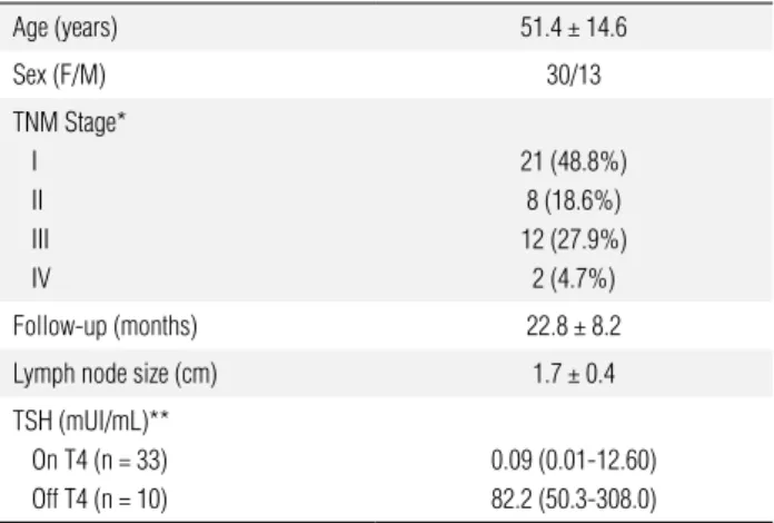
Table 1 shows the clinical and laboratory characteris-tics of the studied patients. Forty-three patients with suspect LN were included (30 women and 13 men) with a mean age of 51.4 ± 14.6 years. According to
TNM staging for DTC (15), patients were distributed as follows: stage I (48.8%), II (18.6%), III (27.9%) and IV (4.7%). Thirty-three patients were receiving levo-thyroxine TSH-suppressive therapy, while 10 patients were in hypothyroidism (levothyroxine withdrawn). Mean size of LN was 1.7 ± 0.4 cm and mean follow-up was 22.8 ± 8.2 months. The median (range) values of sTg and FNAB-Tg in patients on T4 and off T4 were as follows: 1.0 ng/mL (1.0-4,283.0) and 4.0 ng/mL (0.9-3,742.0); 2.3 ng/mL (1.0-191.8) and 3.9 ng/ mL (0.8-25,538), respectively. Five patients presented positive antithyroglobulin antibodies.
Table 1. Clinical and laboratory characteristics of the 43 patients with papillary thyroid cancer and enlarged cervical lymph nodes
Age (years) 51.4 ± 14.6
Sex (F/M) 30/13
TNM Stage* I II III IV
21 (48.8%) 8 (18.6%) 12 (27.9%) 2 (4.7%)
Follow-up (months) 22.8 ± 8.2
Lymph node size (cm) 1.7 ± 0.4
TSH (mUI/mL)** On T4 (n = 33) Off T4 (n = 10)
0.09 (0.01-12.60) 82.2 (50.3-308.0)
* TNM staging for differentiated thyroid cancer; ** Median (range).
Four patients presented positive cytological results and were referred for surgery. Three of them had their results conirmed by histology. The FNAB-Tg values of these patients were 3,419 (TSH 0.49 mUI/mL; sTg 732.6 ng/mL), 25,538 (TSH 62.08 mUI/mL; sTg 191.8 ng/mL) and 3,742 (TSH 0.1 mUI/mL; sTg 4,283 ng/mL). For the patient who presented a negative histology, the FNAB-Tg value was 3.1ng/mL.
Cop
yright
© ABE&M t
odos os dir
eit
os r
eser
vados
.
differ between patients with negative (3.6 ng/mL) or unsatisfactory cytology (6.2 ng/mL; P = 0.07).
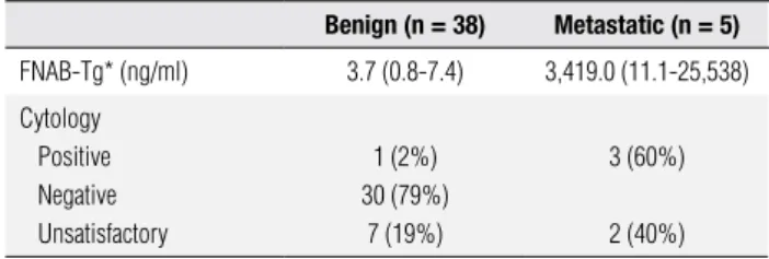
Considering a cutoff value of 10 ng/mL, based on previous reports (7,16), all metastatic LN (n = 5) had higher FNAB-Tg values (median of 3,419.0 ng/mL, range 11.1-25,538.0). In all patients without LN disea-se (n = 38), conirmed by negative cytology and at least 1-year follow-up, the FNAB-Tg was below 10 ng/mL with a median of 3.7 ng/mL (0.8-7.4) (Table 2).
DISCUSSIOn
In the present work we have further demonstrated that the measurement of FNAB-Tg is an excellent tool for de-tecting PTC recurrence, improving the evaluation of sus-picious LN in these patients. Furthermore, we showed that the serum TSH does not seem to interfere in the detectable FNAB-Tg levels observed in negative cases.
Previous studies have already demonstrated the accuracy of FNAB-Tg in suspicious LN metastases of DTC (14,17,18), with a sensitivity and speciicity ran-ging from 84% to 100% and 85% to 95.4%, respective-ly. Cutoffs used in previous studies have varied widely, without a consensus about the most appropriate level (6,19,20). However, a recent study with 168 patients that analyzed 4 different levels of cutoffs (1, 10, 100 ng/mL and FNAB-Tg mean ± SD) indicated that the best level for diagnosis of DTC persistence/recurrence was < 10 ng/mL (16). Here, considering this cutoff, the sensitivity and speciicity of FNAB-Tg was 100% and all cases were distinguishable as positive or negative based on FNAB-Tg. In two patients with LN metas-tasis showed FNAB-Tg levels close to the cutoff value (11.1 and 13.5 ng/mL). In these cases, the likely ex-planation for the low levels of FNAB-Tg might be the small amount of cells (unsatisfactory sample) since the histology had conirmed classical PTC. Nevertheless, low FNAB-Tg levels may also occur in LN metastases of poorly differentiated PTC that are unable to synthe-size/secrete quantiiable amounts of thyroglobulin. It is interesting to note that the diagnostic performance of FNAB-Tg compared favorably with cytology, allo-wing the diagnosis in 3 patients in whom cytological results were false-positive or nondiagnostic. These ca-ses further illustrate the importance of this method to improve the accuracy of detection of DTC metastases.
In agreement with previous studies (7,13,16), we observed that some patients without LN metas-tasis presented detectable FNAB-Tg levels. In these patients, the levels of FNAB-Tg ranged between the method detection limit (< 1 ng/mL) and the cut-off value (10 ng/mL) and, in some cases were even su-perior to sTg. The reasons for these FNAB-Tg values are unclear. A possible contamination by circulating sTg can be theoretically ruled out based on the results of Borel and cols. (21) that demonstrated that sTg in-terferes with FNAB-Tg measurement only when high sTg levels are present. These authors, based on recent studies (19,20), suggest that in all patients who do not have high levels of sTg, a detectable level of FNAB-Tg
Table 2. FNAB-Tg* values and cytological results according to the lymph node histological examination
Benign (n = 38) Metastatic (n = 5)
FNAB-Tg* (ng/ml) 3.7 (0.8-7.4) 3,419.0 (11.1-25,538)
Cytology Positive Negative Unsatisfactory
1 (2%) 30 (79%)
7 (19%)
3 (60%)
2 (40%)
* Thyroglobulin in washout of fine-needle aspiration.
TSH effect on FnAB-Tg
Next we evaluated the role of serum TSH on FNAB-Tg levels in patients without evidence of LN metasta-ses by cytology. Twenty-eight patients were receiving levothyroxine suppressive therapy (TSH = 0.07 mUI/ mL) and 10 patients were on hypothyroidism for a sti-mulated sTg measurement (TSH = 82.2 mUI/mL). Interestingly, we found no signiicant difference in the median values of FNAB-Tg between the groups 3.3 ng/mL (0.9-6.8) vs. 3.8 ng/mL (0.8-7.4; P = 0.2),
indicating that serum TSH has no effect on FNAB-Tg levels (Figure 1).
8
6
4
2
0
FNAB-Tg (ng/mL)
Supressed
Serum TSH
Estimulated
Cop
yright
© ABE&M t
odos os dir
eit
os r
eser
vados
.
in a cervical mass is related to the presence of thyroid cells. Nevertheless, low levels of FNAB-Tg have been observed in control patients (7,13) and can be explai-ned by presence of ‘matrix effects’ artifacts depending on the antibodies and medium used (21). To further support this hypothesis, we demonstrated here that the FNAB-Tg level does not change according to the se-rum TSH in patients without LN disease.
A possible limitation of our study is that none of the 30 patients with a benign cytology underwent LN surgery. In these patients, we cannot provide evidence of benign histology. Consequently, false negatives in cytology associated with negative FNAB-Tg cannot be excluded. To overcome this limitation, we have used stimulated sTg measurement after 1 year follow-up considering sTg values < 1 ng/mL as indicative of di-sease-free. The small number of patients with lymph node metastases could also be a limiting factor to the conclusions of this study.
In conclusion, our results showed that US-guided FNAB-Tg should be performed adjunct to cytology in patients with suspicious cervical LN. This method proved to be a useful exam in the follow-up of patients with PTC and can contribute to diminish the number of unnecessary surgeries, reducing costs and patient morbidity.
Acknowledgments: we thank Conselho Nacional de Desenvol-vimento Cientíico e Tecnológico (CNPq), Fundação de Apoio à Pesquisa do Estado do Rio Grande do Sul (FAPERGS), and Fundo de Incentivo a Pesquisa e Eventos, Hospital de Clínicas de Porto Alegre (FIPE), Brazil, for Grant support.
Disclosure: all authors declare that there is no conlict of interest that would impair the impartiality of this scientiic study.
REFEREnCES
1. Instituto Nacional do Câncer. Câncer da tireóide. Rev Bras Cance-rol. 2002;48(2):181-85.
2. Takashi U, Akira M, Kazuo S, Chisato T, Yuuki T, Yasuhiro I, et al. Usefulness of thyroglobulin measurement in ine-needle aspira-tion biopsy specimens for diagnosing cervical lymph node me-tastasis in patients with papillary thyroid cancer. World J Surg. 2005;29:483-85.
3. Maia AL, Ward LS, Carvalho GA, Graf H, Maciel RMB, Maciel LMZ, et al. Nódulos de tireóide e câncer diferenciado de tireóide: con-senso brasileiro. Arq Bras Endocrinol Metab. 2007;51(5):867-93. 4. Cooper D, Doherty G, Haugen B, Kloos R, Lee S, Mandel S, et
al. Management guidelines for patients with thyroid nodules and differentiated thyroid cancer. Thyroid. 2006;16(2):109-42. 5. Grant CS, Hay ID, Gough IR, et al. Local recurrence in papillary
thyroid carcinoma: is extent of surgical resection important? Sur-gery. 1988;104:954-62.
6. Boi F, Baghino G, Atzeni F, Lai ML, Faa G, Mariotti S. The diag-nostic value for differentiated thyroid carcinoma metastases of thyroglobulin (Tg) measurement in washout luid from ine-needle aspiration biopsy of neck lymph nodes is maintained in the presence of circulating anti-Tg antibodies. J Clin Endocrinol Metab. 2006;91(4):1364-9.
7. Baskin HJ. Detection of recurrent papillary thyroid carcinoma by thyroglobulin assessment in the needle washout by ine-needle aspiration of suspicious lymph nodes. Thyroid. 2004;14:959-63. 8. Boland G, Lee M, Mueller P, Mayo-Smith W, Dawson S,
Sime-one J. Eficacy of sonographically guided biopsy of thyroid masses and cervical lymph nodes. AJR Am J Roentgenol. 1993;161:1053-6.
9. Leboullex S, Girard E, Rose M, Travagli J, Sabbah N, Caillou B, et al. Ultrasound criteria of malignancy for cervical lymph nodes in patients followed up for differentiated thyroid cancer. J Clin Endocrinol Metab. 2007;92(9):3590-4.
10. Cignareli M, Ambrosi A, Marino A, Lamacchia O, Campo M, Picca G, et al. Diagnostic utility of thyroglobulin detection in ine-needle aspiration of cervical cystic metastatic lymph nodes from papilla-ry thyroid cancer with negative cytology. Thyroid. 2003;13:1163-7. 11. Leonard N, Melcher DH. To operate or not operate? The value
of ine needle aspiration cytology in the assessment of thyroid swellings. J Clin Pathol. 1997;50(11):941-3.
12. Cáp J, Ryska A, Rehorková P, Hovorková E, Kerekes Z, Pohneta-lová D. Sensitivity and speciicity of the ine needle aspiration biopsy of the thyroid: clinical point of view. Clin Endocrinol (Oxf). 1999;51:509-15.
13. Pacini F, Fugazzola L, Lippi F, Ceccareli C, Centoni R, Miccoli P, et al. Detection of thyroglobulin in ine the needle aspirates of non-thyroidal neck masses: a clue to the diagnosis of metastatic diffe-rentiated thyroid cancer. J Clin Endocrinol Metab. 1992;74:1401-4. 14. Frasoldati A, Toschi E, Zini M, Flora M, Caroggio A, Dotti C, et
al. Role of thyroglobulin measurement in ine-needle aspiration biopsies of cervical lymph nodes in patients with differentiated thyroid cancer. Thyroid. 1999;9:105-11.
15. Greene FL, Page DL, Fleming ID, et al. AJCC Cancer Staging Han-dbook. 6th ed. New York: Springer-Verlag, 2002. p. 89-98. 16. Kim MJ, Kim EK, Kim BM, Kwak JY, Lee EJ, Park CS, et al.
Thyro-globulin measurement in ine-needle aspirate washouts: the cri-teria for neck node dissection for patients with thyroid cancer. Clin Endocrinol (Oxf). 2009;1:145-51.
17. Lee MJ, Ross DS, Mueller PR, Daniels GH, Dawson SL, Simeone JF. Fine-needle biopsy of cervical lymph nodes in patients with thyroid cancer: a prospective comparison of cytopathology and tissue marker analyses. Radiology. 1993;187:851-4.
18. Biscolla RPM, Ikejiri ES, Mamone MC, Nakabashi CCD, Andrade VP, Kasamatsu T, et al. Diagnóstico de metástases de carcinoma papi-lífero de tireóide através da dosagem de tireoglobulina no líquido obtido da lavagem da agulha utilizada na punção aspirativa. Arq Bras Endocrinol Metab. 2007;51(3):419-25.
19. Snozek C, Chambers EP, Reading CC, Sebo TJ, Sistrunk JW, Singh RJ, et al. Serum thyroglobulin, high-resolution ultrasound and lymph node thyroglobulin in diagnosis of differentiated thyroid carcino-ma nodal metastases. J Clin Endocrinol Metab. 2007;92(11):4278-81.
20. Cunha N, Rodrigues F, Curado F, Ilhéu O, Cruz C, Naidenov P, et al. Thyroglobulin detection in ine-needle aspirates of cervical lym-ph nodes: a technique for the diagnosis of metastatic differentia-ted thyroid cancer. Eur J Endocrinol. 2007;157:101-7.