ABSTRACT
ORIGINAL AR
Eddie Fernando Candido Murta
from patients with risk factors for
cervical cancer
Instituto de Pesquisa em Oncologia (IPON), Discipline of Gynecology
and Obstetrics, Universidade Federal do Triângulo Mineiro (UFTM),
Uberaba, Minas Gerais, Brazil
CONTEXT AND OBJECTIVE: Pap smears are the most common and inexpensive screening method for cervical cancer. We analyzed micronucleus prevalence in exfoliated cervical mucosa cells, to investigate associations between increased numbers of micronuclei and risk factors for cervical cancer.
DESIGN AND SETTING: Analytical cross-sectional study, at Instituto de Pesquisa em Oncologia (IPON).
METHODS: Exfoliated cervical cells were ob-tained from 101 patients between September 2004 and November 2005. Patients’ ages, habits (passive or active smoking, alcoholism and numbers of sexual partners), age at fi rst sexual intercourse, contraceptive methods used, histories of sexually transmitted diseases, use of hormone replacement therapy, numbers of pregnancies and abortions, infl ammatory cytol-ogy and cervical intraepithelial neoplasia (CIN) were obtained. Cells were collected using Ayre spatulas, transferred to vials containing 0.9% saline solution for micronucleus tests and ana-lyzed at 1000x magnifi cation. The number of micronuclei in 1,000 epithelial cells per patient sample was counted.
RESULTS: Comparisons between groups with active (7.9 ±7.8) and passive (7.2 ± 10.6) smoking versus no smoking (3.7 ± 5.1); with/ without alcoholism (7.8 ±1.4 and 6.9 ± 10.1); with/without infl ammatory cytology (10.7 ± 10.5 and 1.3 ± 1.7); and with CIN I, II and III and no CIN (respectively 4.3 ±4.3, 10.6 ± 5.3, 22.7 ±
11.9 and 1.3 ± 1.4) found elevated micronucleus prevalence (P < 0.05).
CONCLUSIONS: We concluded that the preva-lence of micronuclei in exfoliated uterine cervical cells was greater in patients with one or more risk factors for uterine cervical cancer than in patients without risk factors.
KEY WORDS: Cells. Cervix uteri. Risk factors. Neoplasms. Micronucleus test.
INTRODUCTION Cervical cancer is one of the most frequent female cancers. The estimated worldwide incidence of cervical cancer is approximately 500,000 new cases per year, and the overall fi ve-year survival rate is in the range of 44 to 66% for all clinical stages. Pap smears are the most common and inexpensive method of screening for cervical cancer.1,2
Since most cancers arise in epithelial tis-sues, exfoliated epithelial cells may be par-ticularly useful for monitoring patients who are exposed to risk factors.3 Epidemiological
evidence indicates that in most cervical cancer patients, squamous cell carcinoma is the pre-dominant histological type. This carcinoma results from progression of preinvasive cervical intraepithelial neoplasia (CIN) grade I to CIN III.4 The evolution of CIN I to III is
accompa-nied by increased genetic instability or muta-bility, such as losses or gains of chromosomes or fragments of chromosomes.5,6 Progression
to advanced-stage cervical carcinoma is charac-terized by a recurrent pattern of chromosomal rearrangements. The pattern of abnormalities varies greatly between malignancies, ranging from simple balanced rearrangements to com-plex abnormalities affecting both chromosome structure and number.7
In addition to genetic factors, various environmental factors have also been impli-cated in the neoplastic process. Among these, human papillomavirus (HPV) infection and smoking have been cited. HPV infection is one of the most common sexually transmit-ted diseases and is associatransmit-ted with a higher risk of cervical cancer.8 Extensive screening
programs and the development of safe and effective vaccines against HPV would dimin-ish mortality and morbidity from this disease, which has been reported to affect poor women disproportionately.8,9
Behavioral risk factors such as smoking indirectly infl uence the manifestation of
cer-vical cancer and thereby accelerate the tumor progression induced by HPV. Smoking may contribute towards the development of cervical cancer through direct exposure of the DNA of epithelial cells to nicotine and cotinine, or through reactions with the metabolic products from the smoke, such as aromatic polycyclic hy-drocarbons and aromatic amines.10,11
Accord-ing to Weiderpass et al.,12 alcohol consumption
may have an indirect infl uence on the develop-ment of cervical cancer, by triggering malignant transformation of HPV lesions.
Carcinogens affect cells by altering genetic material and thus causing instability. Chro-mosomal instability manifested by increased aneuploidy and structural chromosomal aber-rations is believed to play a critical role in the intermediate to late stages of the development of cervical malignancies.13 Chromosomes or
chromosome segments that fail to be incorpo-rated into nuclei during cell division confi gure micronuclei. Thus, micronuclei represent a measure of both chromosome breakage and chromosome loss, and can function as a sensi-tive indicator of genetic damage.14
OBJECTIVE The purpose of this study was to analyze the prevalence of micronuclei in exfoliated cells from the cervical mucosa in order to in-vestigate associations between increased num-bers of micronuclei and risk factors for cervical cancer, including smoking, large numbers of sexual partners,histories of sexually transmit-ted diseases (STDs), presence of infectious agent for vaginitis, and diagnoses of cervical intraepithelial neoplasia (CIN).
MATERIALS AND METHODS
Patient and control characteristics
Gy-necology and Obstetrics outpatient service of the teaching hospital of Universidade Federal do Triângulo Mineiro (UFTM), Uberaba, Mi-nas Gerais, Brazil, between September 2004 and November 2005. All these patients had previously undergone the Papanicolaou test and colposcopy, respectively to detect likely infectious agents for vaginitis and preinvasive or invasive malignant lesions of the uterine cervix. All the patients underwent video col-poscopy, and biopsies were performed in the cases where test abnormalities were present, in accordance with the Rome recommenda-tions.15 The patients’ ages ranged from 36 to
82 years, with a mean of 58.2 ± 11.5 years (mean ± standard deviation, SD).
Out of the total of 101 patients, 12 did not present any of the risk factors and were therefore considered to be controls. The other patients were divided into groups according to the factors considered in this study: num-bers of partners, pregnancies and abortions, smoking, alcoholism and presence of agents for vaginitis infection. A Specific ELISA (en-zyme linked immunosorbent assay) was used to investigate the serological presence of the human immunodeficiency virus (HIV), and positive findings were confirmed by western blot and indirect immunofluorescence.
After the patients signed an informed consent form, cells were collected. The study was approved by the Research Ethics Com-mittee of UFTM (protocol no. 528/2005). All the participants answered personal
ques-tions regarding their lifestyles. Information on the patients’ ages, habits (smoking, drug use and numbers of sexual partners), con-traceptive methods used, histories of STDs and use of hormone replacement therapy was obtained using a modified version of the questionnaire of the Commission for Protection against Environmental Mutagens and Carcinogens.16
Cervical cell collection and preparation
Exfoliated cervical cells were collected using Ayre spatulas and were transferred to vials containing 0.9% physiological serum for micronucleus tests. The material was cen-trifuged and the supernatant was discarded, leaving the exfoliated cells in the pellet. The cells were fixed for 20 minutes using 1 ml of a methanol-acetic acid solution (3:1). Drops of the material were placed on cold damp slides and allowed to dry. Samples were stained using 4% Giemsa for 12 minutes.
The cytological criteria were used as previously described.17-19 The clue cells were
squamous cells covered with coccobacilli that presented smudged cytoplasmic borders.
Candida sp. was diagnosed through the pres-ence of pseudohyphae that stained weakly with hematoxylin-eosin and/or small spores (diameters of 2-4 mm) that stained pale pink. The cytological diagnosis for possible HPV infection was based on the morphological criteria of Schneider et al.20
Cytogenetic analysis of micronuclei
The slides were analyzed using an opti-cal microscope at a magnification of 1000 x (objective = 100 x with eyepiece = 10 x), and 1,000 epithelial cells were counted per microscope field. Within the samples, only cells that were separate, without overlapping or folds, were analyzed. Micronuclei were counted if the structures had a regular border and were located inside the cytoplasm, with an intensity of staining less than or equal to that of the main nucleus and a size less than two-thirds of the size of the main nucleus (Figure 1). The frequency of micronuclei was obtained as the ratio between the number of micronuclei and the total number of cells analyzed, multiplied by 100.
Statistical analysis
Statistical analyses were computed using GraphPad InStat 3 (GraphPad Software, Inc). All variables were subjected to the Kol-mogorov-Smirnov test to determine whether there was normal distribution. The data with normal distribution were then analyzed using the parametric Kruskal-Wallis method and the Dunn multiple comparisons test. The data that did not present normal distribution were analyzed using the nonparametric Mann-Whitney method. The significance level con-sidered for all the tests was P < 0.05.
of micronuclei, an analysis comparing the nonsmoking women in the control group with those in the active and passive smoking group was performed.
The presence of cytological signs of HPV infection increased the frequency of micronu-clei among the infected women (Table 2). The patient samples were divided into two groups (with and without cytological signs of HPV infection), and a stratified analysis in relation to the other risk factors for cervical cancer was performed. The risk factors combined with HPV infection resulted in statistically sig-nificant differences in micronucleus frequency between the group in question and the groups that did not present HPV. Furthermore, vagi-nal and cervical inflammation was associated with very significantly increased frequency of micronuclei. Even when inflammation intensity (mild, moderate or severe) was taken into account in the analysis, the frequency of micronuclei was significantly greater in subjects with inflammation than in controls without inflammation (Table 3).
We observed (Table 4) that CIN corre-lated with increasing numbers of micronuclei. During the process of chromosomal damage, more than one micronucleus may be found within cells. Most of the cells observed in this study had one micronucleus. Cells with two micronuclei were most frequently observed in women who had had more than three pregnancies. The groups with more than three pregnancies, CIN, cytological signs of HPV infection and inflammation were the only subjects that presented more than three micronuclei per cell.
DISCUSSION In this study, we observed that 88% of the women had some type of risk factor for cervical cancer. Of these, 64% showed in-creased micronucleus frequency in exfoliated cervical cells. Young adult women were more vulnerable to risk factors, since they present a cervical transformation zone located in the ectocervix.21 Nonetheless, our results did not
demonstrate any influence from age, age at first sexual intercourse or number of partners, with regard to micronucleus formation. How-ever, the number of pregnancies and abor-tions increased the frequency of micronuclei. Prolonged exposure to hormones has been reported to be a strong risk factor for progres-sion of intraepithelial neoplasia, since steroid hormones may facilitate the transforming activity of HPV.22 The use of oral
contracep-tives seems to increase the transforming activ-ity of HPV oncogenes and interfere with the
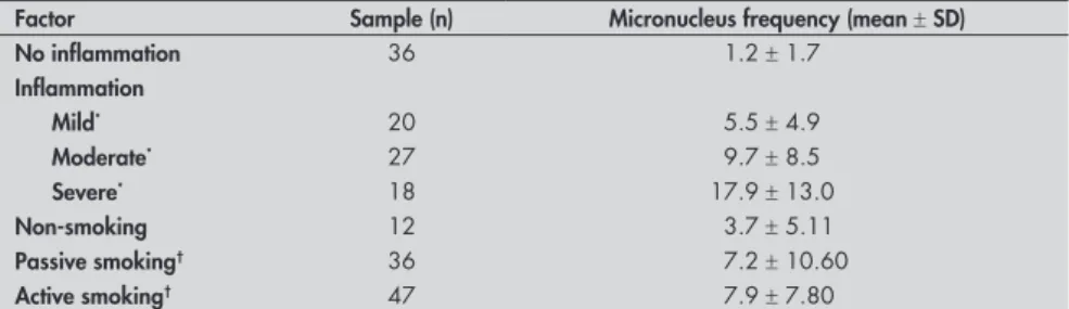
Table 3. Comparison of the mean frequencies of micronuclei in the cytology with inflam-mation and smoking groups compared to controls
Factor Sample (n) Micronucleus frequency (mean ± SD)
No inflammation 36 1.2 ± 1.7
Inflammation
Mild* 20 5.5 ± 4.9
Moderate* 27 9.7 ± 8.5
Severe* 18 17.9 ± 13.0
Non-smoking 12 3.7 ± 5.11
Passive smoking† 36 7.2 ± 10.60
Active smoking† 47 7.9 ± 7.80
*P < 0.001 versus no inflammation; †P < 0.05 versus non-smoking; SD = standard deviation.
Table 2. Comparison of mean frequencies of micronuclei in women with and without HPV infection, in relation to risk factors for cervical cancer
Factor
HPV No HPV
n
Micronucleus frequency (mean ± SD)
n
Micronucleus frequency (mean ± SD)
Alcoholic* 25 9.6 ± 8.1 6 2.5 ± 1.1
Smoker* 32 10.0 ± 7.8 15 1.7 ± 1.8
Infectious agents for vaginitis* 22 9.7 ± 8.2 9 4.0 ± 4.4
Cytology with inflammation 47 13.2 ± 10.7 18 10.7 ± 1.05
CIN* 53 14.2 ± 18.9 3 4.6 ± 4.9
Oral contraceptive* 30 9.8 ± 9.6 25 1.7 ± 2.2
*P < 0.0001, for all data in comparison with no HPV; HPV = human papillomavirus; CIN = cervical intraepithelial neoplasia.
Table 1. Micronucleus frequency in relation to risk factors for cervical cancer
Factor Groups defined Sample (n) Micronucleus frequency (mean ± SD) P-value
Control 12 3.70 ± 5.1
HPV* 57 11.01 ± 1.08 0.04
Age (years)* ≤ 35 years 59 6.80 ± 8.80 0.486
> 35 years 42 8.00 ± 10.30
Age at first sexual intercourse* ≤ 16 years 56 6.70 ± 9.70 0.299
> 16 years 45 8.10 ± 9.20
Partner*,† 1 19 5.40 ± 8.20 0.328
> 1 82 7.70 ± 9.70
Oral contraceptive Yes 55 6.10 ± 8.30 0.109
No 46 8.70 ± 10.60
Pregnancy 0 23 4.40 ± 6.70 0.093
1 to 3 55 7.20 ± 8.90
> 3 23 10.40 ± 12.10
Abortion* Yes 31 9.70 ± 10.80 0.090
No 70 6.20 ± 8.70
Active smoker*,† Yes 47 7.90 ± 7.80 0.006
No 54 5.10 ± 10.00
Passive Smoker*,† Yes 36 7.20 ± 10.60 0.038
No 65 5.90 ± 4.80
Alcoholic*,† Yes 31 7.80 ± 1.40 0.012
No 70 6.90 ± 10.10
Cytology with inflammation*,† Yes 65 10.70 ± 10.50 0.0001
No 36 1.20 ± 1.70
CIN*,† Yes 56 11.00 ± 10.40 0.0001
No 35 1.30 ± 1.40
STD* Yes 51 6.70 ± 8.90 0.508
No 50 7.90 ± 10.60
*P < 0.05, compared with control; †P < 0.05, compared within the same group; STD = sexually transmitted disease; CIN =
efficient resolution of lesions caused by this virus in the cervix among young women.23
Although use of oral contraceptives did not affect the expression of HPV activity, women who were both HPV-infected and oral contra-ceptive users presented significantly increased frequency of micronuclei, thus suggesting that the use of these steroid hormones might boost the oncogenicity of HPV infection.24,25
In the present study, we observed greater numbers of micronuclei in specimens from women who were active or passive smokers, relative to control specimens. These results corroborate those of Cerqueira et al.,26 who
found greater numbers of micronuclei in ex-foliated cervical cells in women who smoked than in those who did not. Among patients without lesions (both smokers and nonsmok-ers), the frequency of micronuclei was lower than among those who had some type of abnormal pathological condition. Among nonsmokers, the frequency of micronuclei was higher in patients with lesions than in those who were cytologically normal. Among smokers, the frequency of micronuclei was high, even in women with a low degree of inflammation. In an epidemiological study, Matsumoto et al.27 showed that smoking and
Chlamydia infection were cofactors for CIN progression. Passive smokers were found to have the same risk of developing CIN as did active smokers.28 Two main mechanisms have
been suggested through which smoking may contribute towards cervical carcinogenesis: one involves direct exposure of the deoxyri-bonucleic acid (DNA) in cervical epithelial cells to nicotine and cotinine, and the other involves exposure to metabolic products re-sulting from the reactions of other compo-nents of cigarettes such as aromatic polycyclic hydrocarbons and aromatic amines.11,29 Other
mechanisms that may explain smoking-related carcinogenesis include abnormalities in the peripheral immune system of smokers, such as elevated numbers of cytotoxic/suppressor T lymphocytes, diminished numbers of helper T lymphocytes, suppression of T lymphocyte
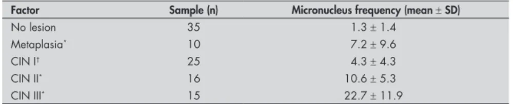
Table 4. Comparison between women without cervical lesions and women with dif-ferent degrees of cervical intraepithelial neoplasia (CIN), in relation to the frequency of micronuclei
Factor Sample (n) Micronucleus frequency (mean ± SD)
No lesion 35 1.3 ± 1.4
Metaplasia* 10 7.2 ± 9.6
CIN I† 25 4.3 ± 4.3
CIN II* 16 10.6 ± 5.3
CIN III* 15 22.7 ± 11.9
*P < 0.001 and †P < 0.05 versus no lesion; SD = standard deviation.
activity, significantly decreased numbers of natural killer lymphocytes and low levels of immunoglobulins other than immunoglobu-lin E (IgE).30 These effects may result from
decreased numbers of Langerhans cells in the cervix of women who smoke.31
Women who consume alcohol are con-sidered to present a high and progressive risk of developing in situ and invasive cervical and vaginal cancer. Epidemiological data have sug-gested a direct link between alcoholism and lifestyle factors such as promiscuity, smoking, use of hormonal contraceptives and dietary deficiencies.12 In the data obtained from the
present study, significantly increased frequency of micronuclei was observed among the women who consumed alcohol.
Our results showed that women present-ing inflammation had significantly greater numbers of micronuclei than did those with-out inflammation or those in the control group. Another important result was that the progressive increase in inflammation severity was directly proportional to the observed in-crease in micronucleus numbers. We observed a strong association between high numbers of micronuclei and the presence of inflam-mation with concomitant HPV infection, in relation to those without HPV infection. This observation may suggest that the presence of inflammation among women with HPV infection increases the genetic damage in cervical epithelial cells. The observation that only a small minority of the lesions resulting from HPV infection progressed to invasive cancer led Nishimura et al.32 to suggest that
additional events were necessary for malignant cellular transformation.
In evaluating micronuclei in relation to the presence of CIN, we observed that greater numbers of micronuclei were seen in women with progressive increases in the severity of CIN (CIN I < CIN II < CIN III), compared with controls. This evidence corroborates the importance of the micronucleus test as a biomarker for malignancy. A study by Guzmán et al.4 showed an association between
lesion severity and micronucleus frequency in epithelial cells, which contributes towards validating micronucleus frequency as a pos-sible biomarker for cancer risk.
Women with STDs presented signifi-cantly higher frequencies of micronuclei than did the control group. Fischer observed that Chlamydia infection could cause cervical hypertrophy in women with or without CIN or carcinoma.33 However, concomitant
infec-tion by Chlamydia and HPV increased the expression of Ki67 in the epithelium. More-over, Chlamydia infection has been shown to increase HPV-16 activity, possibly explaining variations in the HPV mechanism for cervical carcinogenesis.33,34 In a study based solely on
cytological criteria, there was an association between G. vaginalis and HPV infection.35
Two other studies addressed the matter.36,37
One of them36 showed that, compared with
the group of pregnant women without HPV infection, those with HPV infection had a significantly higher percentages of bacterial vaginosis (BV) (53.8 versus 15.4%; P = 0.007) and Chlamydia trachomatis (34.6 versus 7.7%; P = 0.039). No cases of Neisseria gonorrhoeae
were diagnosed. All cases of C. trachomatis and BV had high-risk HPV.36 The other
manu-script showed that there were higher frequen-cies of BV and HPV in patients with atypical squamous cells of undetermined significance than in patients with normal cytology.37
By most accounts, the oncogenic mecha-nism of HPV is well understood. Following infection by HPV, oncoproteins from the virus integrate with the tumor suppressor proteins p53 and pRb, which in turn alter the function of oncoproteins, thereby resulting in uncon-trolled transcription activity, abnormal DNA replication and cell division, hence leading to tumor formation.38 Our results showed
that the women with HPV infection had an elevated frequency of micronuclei in relation to the control group. In our study, HPV status was compared with a variety of risk factors for cervical cancer and the results were concordant with the literature.
There are three mechanisms that may contribute towards the formation of micronu-clei: metabolic stress caused by tumor growth, clastogenic products released from tumor cells and the presence of HPV.39 Chromosomal
in-stability, particularly in chromosomes 1, 3, 5, 11 and 17, is associated with the development of cervical carcinoma.40 The results presented
1. Waggoner SE. Cervical cancer. Lancet. 2003;361(9376):2217-25.
2. Valdespino VM, Valdespino VE. Cervical cancer screening: state of the art. Curr Opin Obstet Gynecol. 2006;18(1):35-40. 3. Gonsebatt ME, Guzmán P, Blas J. Cytogenetic and cytotoxic
damage in exfoliated cells as indicators of effects in humans. In: Biomonitors and biomarkers as indicators of environmental change. Butterworth F, Gunatilaka A, Gonsebatt ME, editors. New York: Kluwer/Plenum Press; 2000. p. 317-32. 4. Guzmán P, Sotelo-Regil RC, Mohar A, Gonsebatt ME.
Posi-tive correlation between the frequency of micronucleated cells and dysplasia in Papanicolaou smears. Environ Mol Mutagen. 2003;41(5):339-43.
5. Giannoudis A, Evans MF, Southern SA, Herrington CS. Basal keratinocyte tetrasomy in low-grade squamous intra-epithelial lesions of the cervix is restricted to high and interme-diate risk HPV infection but is not type-specific. Br J Cancer. 2000;82(2):424-8.
6. Loeb LA. A mutator phenotype in cancer. Cancer Res. 2001;61(8):3230-9.
7. Gisselsson D. Refined characterisation of chromosome aber-rations in tumours by multicolour banding and electronic mapping resources. Methods Cell Sci. 2001;23(1-3):23-8. 8. Masumoto N, Fujii T, Ishikawa M, et al. Dominant human
papillomavirus 16 infection in cervical neoplasia in young Japanese women; study of 881 outpatients. Gynecol Oncol. 2004;94(2):509-14.
9. Oliveira LH, Rosa ML, Pereira CR, et al. Human papillomavirus status and cervical abnormalities in women from public and private health care in Rio de Janeiro State, Brazil. Rev Inst Med Trop Sao Paulo. 2006;48(5):279-85.
10. Hellberg D, Nilsson S, Haley NJ, Hoffman D, Wynder E. Smoking and cervical intraepithelial neoplasia: nicotine
and cotinine in serum and cervical mucus in smokers and nonsmokers. Am J Obstet Gynecol. 1988;158(4):910-3.
11. Simons AM, Philips DH, Coleman DV. Damage to DNA in cervical epithelium related to smoking tobacco. BMJ.
1993;306(6890):1444-8.
12. Weiderpass E, Ye W, Tamimi R, et al. Alcoholism and risk
for cancer of the cervix uteri, vagina, and vulva. Cancer Epidemiol Biomarkers Prev. 2001;10(8):899-901.
13. Olaharskil AJ, Sotelo R, Solorza-Luna G, et al. Tetraploidy and chromosomal instability are early events during cervical carcinogenesis. Carcinogenesis. 2006;27(2):337-43. 14. Fenech M, Holland N, Chang WP, Zeiger E, Bonassi S. The
HUman MicroNucleus Project--An international collaborative study on the use of the micronucleus technique for measuring
DNA damage in humans. Mutat Res. 1999;428(1-2):271-83. 15. Stafl A, Wilbanks GD. An international terminology of
colposcopy: report of the Nomenclature Committee of the International Federation of Cervical Pathology and Colposcopy. Obstet Gynecol. 1991;77(2):313-4.
16. Carrano AV, Natarajan AT. International Commission for Protection Against Environmental Mutagens and Carcinogens. ICPEMC publication no. 14. Considerations for popula-tion monitoring using cytogenetic techniques
.
Mutat Res. 1988;204(3):379-406.17. Gupta PK. Microbiology, inflammation, and viral infections. In: Bibbo M, editor. Comprehensive cytopathology. Philadelphia: WB Saunders Company; 1997. p. 125-60.
18. Solomon D. The Bethesda system for cervicovaginal cytopa-thology. In: Bibbo M, editor. Comprehensive Cytopacytopa-thology. Philadelphia: WB Saunders Company; 1997. p. 93-100. 19. Adad SJ, de Lima RV, Sawan ZT, et al. Frequency of
Trichomo-nas vaginalis, Candida sp and Gardnerella vaginalis in cervical-vaginal smears in four different decades. Sao Paulo Med J.
2001;119(6):200-5.
20. Schneider A, Meinhardt G, De-Villiers EM, Gissmann L. Sensitivity of the cytologic diagnosis of cervical condyloma in comparison with HPV-DNA hybridization studies. Diagn
Cytopathol. 1987;3(3):250-5.
21. Mangan SA, Legano LA, Rosen CM, et al. Increased prevalence of abnormal Papanicolaou smears in urban adolescents. Arch Pediatr Adolesc Med. 1997;151(5):481-4.
22. Park JS, Rhyu JW, Kim CJ, et al. Neoplastic change of squamo-columnar junction in uterine cervix and vaginal epithelium by exogenous estrogen in hpv-18 URR E6/E7 transgenic mice. Gynecol Oncol. 2003;89(3):360-8.
23. Shapiro S, Rosenberg L, Hoffman M, et al. Risk of invasive cancer of the cervix in relation to the use injectable proges-togen contraceptives and combined estrogen/progesproges-togen oral contraceptives (South Africa). Cancer Causes Control. 2003;14(5):485-95.
24. Dziubinska-Parol I, Gasowska U, Rzymowska J, Kwasniewska A. Wplyw fizjologicznych stezen 17 beta-estradiolu na ek-spresje in vitro genu E6 wirusa brodawczaka ludzkiego typ 18. [Influence of physiologic 17 beta-estradiol concentrations on gene E6 expression in HPV type 18 in vitro]. Ginekol Pol. 2003;74(9):710-3.
25. Shields TS, Falk RT, Herrero R, et al. A case-control study of endogenous hormones and cervical cancer. Br J Cancer. 2004;90(1):146-52.
26. Cerqueira EM, Santoro CL, Donozo NF, et al. Genetic damage in exfoliated cells of the uterine cervix. Association and
interac-tion between cigarette smoking and progression to malignant transformation?Acta Cytol. 1998;42(3):639-49. 27. Matsumoto K, Yasugi T, Oki A, et al. Are smoking and
Chlamydia infection risk factors for CIN? Different results after adjustment for HPV DNA and antibodies. Br J Cancer. 2003;89(5):831-3.
28. Tay SK, Tay KJ. Passive cigarette smoking is a risk factor in cervical neoplasia. Gynecol Oncol. 2004;93(1):116-20. 29. Hellberg D, Nilsson S, Haley NJ, Hoffman D, Wynder E.
Smok-ing and cervical intraepithelial neoplasia: nicotine and cotinine in serum and cervical mucus in smokers and nonsmokers.Am J Obstet Gynecol. 1988;158(4):910-3.
30. Johnson JD, Houchens D, Kluwe WM, Craig DK, Fisher GL. Effects of mainstream and environmental tobacco smoke on the immune system in animals and humans: a review. Crit Rev Toxicol. 1990;20(5):369-95.
31. Poppe WA, Drijkoningen M, Ide PS, Lauweryns JM, Van Assche FA. Langerhans’ cells and L1 antigen expression in normal and abnormal squamous epithelium of the cervical transformation zone. Gynecol Obstet Invest. 1996;41(3):207-13. 32. Nishimura M, Furumoto H, Kato T, Kamada M, Aono T.
Microsatellite instability is a late event in the carcinogenesis of uterine cervical cancer. Gynecol Oncol. 2000;79(2):201-6. 33. Fischer N. Chlamydia trachomatis infection in cervical
intraepi-thelial neoplasia and invasive carcinoma. Eur J Gynaecol Oncol. 2002;23(3):247-50.
34. Smith JS, Muñoz N, Herrero R, et al. Evidence for Chlamydia trachomatis as a human papillomavirus cofactor in the etiology of invasive cervical cancer in Brazil and the Philippines. J Infect Dis. 2002;185(3):324-31.
35. Murta EF, Souza MA, Araújo Júnior E, Adad SJ. Incidence of Gardnerella vaginalis, Candida sp and human papilloma virus in cytological smears. Sao Paulo Med J. 2000;118(4):105-8. 36. da Silva CS, Adad SJ, Hazarabedian de Souza MA, Macêdo
Barcelos AC, Sarreta Terra AP, Murta EF. Increased frequency of bacterial vaginosis and Chlamydia trachomatis in pregnant women with human papillomavirus infection. Gynecol Obstet Invest. 2004;58(4):189-93.
37. Barcelos AC, Adad SJ, Michelin MA, Murta EF. Atypical squamous cells of undetermined significance: analysis of mi-crobiology, cytological criteria and clinical conduct. Tumori. 2006;92(3):213-8.
38. Deluca GD, Lucero RH, Martin de Civetta MT, et al. Human papillomavirus genotypes in women with cervical cytological abnormalities from an area with high incidence of cervical cancer. Rev Inst Med Trop Sao Paulo. 2004;46(1);9-12. 39. Leal-Garza CH, Cerda-Flores RM, Leal-Elizondo E,
Cortés-REFERENCES Micronuclei are indicative of numerical and/
or structural chromosome aberrations during cell mitosis. Other authors have used the micronucleus test as a biomarker for chromo-some instability and malignancy, observing higher frequencies of micronucleated cells among cancer patients than among healthy individuals.41,42 The presence of micronuclei
has been considered to be a very useful bio-marker for detecting malignant cervical uter-ine carcinomas.39 According to Bonassi et al.,43
several studies have confirmed the presence of micronuclei in different cell types, thus sug-gesting that micronuclei may be a morphologi-cal marker that may be useful for predicting several types of cancer risk. Furthermore, the ease and low cost of this method may allow
further development of the micronucleus test as a prognostic indicator during the plan-ning and validation of programs for cancer monitoring and prevention. Furthermore, the micronucleus test has been considered to be a very useful biomarker for detecting malignancy in the uterine cervix, with regard to other factors like surgical margins and the numbers of mitoses and methylated genes, for predicting recurrence of CIN III.44-46
The number of micronuclei correlates with the severity of genetic damage. Cells containing several micronuclei present greater genetic damage than do cells that present only one micronucleus. The data obtained in this study suggest that factors such as HPV infection, number of pregnancies, CIN and
inflammation are more clastogenic because they increase the frequency of cells contain-ing more than three micronuclei. This could explain the number of micronuclei found in metaplasia. Taken together, comparison in our study between patients with and without risk factors for cervical cancer showed that there was a significant difference, thus suggesting that micronuclei may be a valid biomarker for cancer risk.
Gutiérrez EI. Micronuclei in cervical smears and peripheral blood lymphocytes from women with and without cervical uterine cancer. Mutat Res. 2002;515(1-2):57-62. 40. Paz-y-Miño C, Ocampo L, Narváez R, Narváez L. Chromosome
fragility in lymphocytes of women with cervical uterine lesions produced by human papillomavirus. Cancer Genet Cytogenet. 1992;59(2):173-6.
41. Kamboj M, Mahajan S. Micronucleus--an upcoming marker of genotoxic damage. Clin Oral Investig. 2007;11(2):121-6. 42. Lou J, He J, Zheng W, et al. Investigating the genetic
instabil-ity in the peripheral lymphocytes of 36 untreated lung cancer patients with comet assay and micronucleus assay. Mutat Res. 2007;617(1-2):104-10.
43. Bonassi S, Znaor A, Ceppi M, et al. An increased micronucleus
frequency in peripheral blood lymphocytes predicts the risk of cancer in humans. Carcinogenesis. 2007;28(3):625-31. 44. Murta EF, Resende AV, Souza MA, Adad SJ, Salum R.
Importance of surgical margins in conization for cervical intraepithelial neoplasia grade III. Arch Gynecol Obstet. 1999;263(1-2):42-4.
45. Maluf PJ, Adad SJ, Murta EF. Outcome after conization for cervical intraepithelial neoplasia grade III: relation with surgical margins, extension to the crypts and mitoses. Tumori. 2004;90(5):473-7.
46. Terra AP, Murta EF, Maluf PJ, Caballero OL, Brait M, Adad SJ. Aberrant promoter methylation can be useful as a marker of recurrent disease in patients with cervical intraepithelial neoplasia grade III. Tumori. 2007;93(6):572-9.
Acknowledgements: The authors are grateful to Universidade
Federal do Triângulo Mineiro (UFTM), Coordenação de Aperfeiçoamento de Pessoal de Nível Superior (Capes), for funding; to Cláudio Fidalgo for performing the statistical analysis; and to Ricardo Manoel da Cruz for providing technical assistance
Sources of funding: Conselho Nacional de
Desenvolvi-mento Científico e Tecnológico (CNPq) (Grant number 303949/2006-6) and Fundação de Amparo à Pes-quisa do Estado de Minas Gerais (Fapemig) (Grant number 1965/05)
Conflict of interest: None
Date of first submission: November 21, 2007
Last received: November 4, 2008
Accepted: November 4, 2008
AUTHOR INFORMATION
Lízia Maria Franco dos Reis Campos, MSc. Postgraduate
student, Department of Biological Sciences, Universidade Federal do Triângulo Mineiro (UFTM), Uberaba, Minas Gerais, Brazil.
Francisca da Luz Dias, PhD. Visiting professor, Department
of Biological Sciences, Universidade Federal do Triângulo Mineiro (UFTM), Uberaba, Minas Gerais, Brazil.
Lusânia Maria Greggi Antunes, PhD. Associate professor,
Department of Clinical, Toxicological and Bromatologi-cal Analyses, Faculdade de Ciências Farmacêuticas de Ribeirão Preto (FCFRP), Universidade de São Paulo (USP), Ribeirão Preto, São Paulo, Brazil.
Eddie Fernando Candido Murta, MD, PhD. Titular professor,
Instituto de Pesquisa em Oncologia (IPON), Discipline of Gynecology and Obstetrics, Universidade Federal do Triân-gulo Mineiro (UFTM), Uberaba, Minas Gerais, Brazil.
Address for correspondence:
Eddie Fernando Candido Murta
Instituto de Pesquisa em Oncologia (IPON), Disciplina de Ginecologia e Obstetrícia, Universidade Federal do Triângulo Mineiro (UFTM)
Av. Getúlio Guarita, s/no
Uberaba (MG) — Brasil — CEP 38025-440 Tel. (+55 34) 3318-5326
Fax. (+55 34) 3318-5342 E-mail: [email protected] E-mail: [email protected]
Copyright © 2008, Associação Paulista de Medicina
RESUMO
Prevalência de micronúcleos em células esfoliativas do colo uterino de pacientes com fatores de risco para o câncer de colo uterino
CONTEXTO E OBJETIVO: O câncer do colo uterino é uma das mais freqüentes neoplasias na mulher. O exame de Papanicolaou é o método mais comum e econômico para rastreamento. As células esfoliativas epiteliais podem ser úteis para o monitoramento de pacientes expostas a fatores de risco para o câncer. O objetivo foi analisar a prevalência de micronúcleos em células esfoliativas da mucosa cervical uterina e associar com fatores de risco para o câncer de colo uterino.
TIPO DE ESTUDO E LOCAL: Estudo transversal analítico, no Instituto de Pesquisa em Oncologia (IPON).
MÉTODOS: Células esfoliativas do colo uterino foram obtidas de 101 pacientes ambulatoriais entre setembro/2004 e novembro/2005. As células foram coletadas usando espátula de Ayre e transferidas para um tubo de ensaio com soro fisiológico 0,9% para o teste do micronúcleo. Informações obtidas das pacientes foram: idade, hábitos (fumo e número de parceiros sexuais), métodos contraceptivos, história de doença sexualmente transmissível e uso de terapia hormonal. Células foram analisadas com magnificação de 1000 X e os micronúcleos contados em 1.000 células epiteliais por paciente.
RESULTADOS: A comparação do grupo de pacientes fumantes ativas (7,9 ±7,8) e passivas (7,2 ± 10,6) versus não fumantes (3,7 ± 5,1); alcoolismo e não alcoolismo (7,8 ±1,4 e 6,9 ± 10,1); citologia infla-matória e citologia normal (10,7 ± 10,5 e 1,3 ± 1,7); neoplasia intraepitelial cervical (NIC) I, II e III e a ausência de NIC, respectivamente, (4,3 ±4,3; 10,6 ± 5,3; 22,7 ± 11,9 e 1.3 ± 1.4) mostrou maior prevalência de micronúcleos (P < 0,05).
CONCLUSÕES: A prevalência de micronúcleo nas células esfoliativas do colo uterino foi maior no grupo de pacientes com pelo menos um dos fatores de risco para câncer do colo uterino do que no grupo controle (sem fatores de risco).