ABSTRACT
ORIGINAL AR
INTRODUCTION
Computed tomography guided biopsy hastomography guided biopsy hasomography guided biopsy hasy guided biopsy has guided biopsy has been widely accepted as an effective and safeas an effective and safe effective and safe procedure with high accuracy for diagnosis in with high accuracy for diagnosis incuracy for diagnosis inuracy for diagnosis iny for diagnosis in for diagnosis inis in in many clinical settings.1-6 It is a simple way to
obtain diagnoses in deep-seated lesions ands in deep-seated lesions and in deep-seated lesions and guide therapeutic decisions, thus avoiding surgical biopsies in many instances. Fine--needle aspiration biopsy and, more recently,on biopsy and, more recently,and, more recently, core biopsy have become the main choices forbiopsy have become the main choices for investigating many conditions.1-6 ComputedComputedomputed
tomography guidance is very versatile and is very versatile andis very versatile and can be used for various organs, from super-used for various organs, from super-various organs, from super-organs, from super-, from super-ficial soft-tissue lesions to deep-seated lesions in the thoracic or abdominal cavities. The sensitivity, specificity and accuracy also vary,ty and accuracy also vary, and accuracy also vary,curacy also vary,uracy also vary,y also vary, also vary, depending on the target organ and the typetarget organ and the typeorgan and the type of needle used. Some previous studies haveSome previous studies have studies havestudies have have demonstrated advantages in using cuttinged advantages in using cutting advantages in using cutting needles instead of fine needles, especially for of fine needles, especially for fine needles, especially for diagnosing benign lesions.ing benign lesions.ng benign lesions.5,7-11 The choice ofThe choice ofhe choice of
needle will depend on the patient’s coagulation state, the characteristics of each lesion, the access route and the experience of both the radiologist and the pathologist.1-12
The complications depend on patient-re-depend on patient-re- patient-re-lated conditions, target organ, type of needletarget organ, type of needle organ, type of needle, type of needletype of needle used and access route. Minor complications such as local discrete hemorrhage, pain and vasovagal reaction are not rare but are easy to deal with; mild complications such as pneu-mothorax are not rare in lung lesion biopsies but usually do not require drainage; major complications such as severe hemorrhage are rare and may even lead to death. Proper patient selection and technique can reduce the complication rates and, overall, percutaneous biopsy has a high benefit/risk ratio.13 The
objective of this study is to present the resultsis to present the results to present the results from a large series of computed tomography guided biopsies performed by the same radi-ologist, or by radiology residents under his supervision, at the Department of Radiology
of Hospital do Câncer A.C. Camargo, an oncology center in São Paulo, Brazil. Ourur specific results for different sites and needles used are reported.
METHODS
We retrospectively analyzed 1,300 con- retrospectively analyzed 1,300 con-ly analyzed 1,300 con- analyzed 1,300 con-secutive computed tomography guided needle biopsies performed from July 1994 to February 2000 in 1,174 patients. All the February 2000 in 1,174 patients. All the biopsies accomplished during this period wereaccomplished during this period were during this period were evaluated. 728 (56%) of the 1,300 lesions were. 728 (56%) of the 1,300 lesions were biopsied in male patients and 572 (47%) in female patients. The patients’ ages ranged from 0.3 to 98.0 years (mean: 59 years).mean: 59 years).: 59 years).
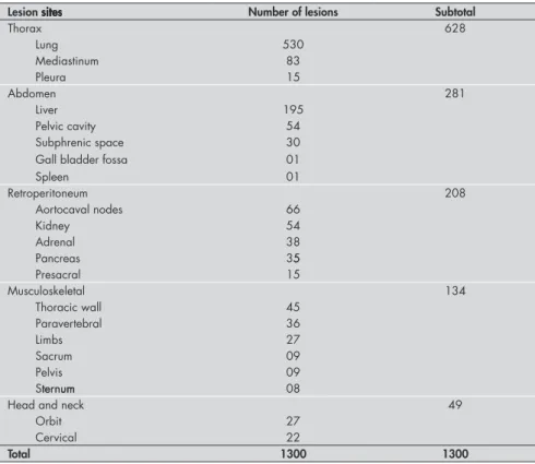
The biopsy sites were the following: 628sites were the following: 628 were the following: 628 cases in the thoracic cavity (530 in the lung, 83 in the mediastinum and 15 in the pleura); 281 cases in the abdomen (195 in the liver, 54 in the pelvis and 32 in other abdominalis and 32 in other abdominal and 32 in other abdominal sites); 208 cases in the retroperitoneum (66 aortocaval nodes, 54 in the kidney, 38 in the adrenals, 35 in the pancreas and 15 in thes, 35 in the pancreas and 15 in the presacral space); 134 in the musculoskeletal system (45 in the thoracic wall, 36 in the para-vertebral area, 27 in the limbs, 9 in the sacrum, area, 27 in the limbs, 9 in the sacrum,area, 27 in the limbs, 9 in the sacrum, 8 in the sternum and 9 in the superficial pelvicsternum and 9 in the superficial pelvicum and 9 in the superficial pelvic bones or muscles); 49 in the head and neck (27 in the orbit and 22 in the cervical area)orbit and 22 in the cervical area) and 22 in the cervical area) (Table 1). The musculoskeletal lesions were all described together in one group because of their similar characteristics, such as being superficial and extracavitary. The clinical in-dication for the procedures was a focal lesion, nodule or mass that in all cases was suspected to be malignant (primary malignancy in 845primary malignancy in 845malignancy in 845 cases (65%) or metastatic lesion of a known cancer in 455 (35%).
The patients’ preparation and lesion local-ization were the same for both types of needle used. The patients were admitted following six hours of fasting in preparation for the use of anesthetics and, if needed, intravenous contrast.
Alessandro Amorim Aita
Fernando Augusto Soares
1,300 procedures
Departments of Radiology and Pathology,
Hospital do Câncer A. C. Camargo, São Paulo, Brazil
CONTEXT AND OBJECTIVE: C o m p u t e d tomography (CT) guided biopsy is widely ac-omography (CT) guided biopsy is widely ac-y (CT) guided biopsy is widely ac- guided biopsy is widely ac-cepted as effective and safe for diagnosis inas effective and safe for diagnosis ineffective and safe for diagnosis inis in in many settings. Accuracy depends on targetcuracy depends on targeturacy depends on targety depends on target depends on targettarget organ and needle type. Cutting needles present advantages over fine needles. This study presents experience from CT guided biopsies performed at an oncology center.
DESIGN AND SETTING: Retrospective study at Hospital do Câncer A. C. Camargo, São Paulo. METHODS: 1,300 consecutive CT guided biop-sies performed between July 1994 and February 2000 were analyzed. Nodules or masses were suspected as primary malignancy in 845 casesprimary malignancy in 845 casesmalignancy in 845 cases (65%) or metastatic lesion in 455 (35%). 628 lesions were thoracic, 281 abdominal, 208 retroperitoneal, 134 musculoskeletal and 49 head/neck. All biopsies were performed by one radiologist or under his supervision: 765 (59%) with 22-gauge fine-needle/aspiration technique and 535 (41%) with automated 16 or 18-gauge cutting-needle biopsy.
RESULTS: Adequate samples were obtained insamples were obtained ines were obtained in 70-92% of fine-needle and 93-100% of cutting-needle biopsies. The specific diagnosis rates were 54-67% for fine-needle and 82-100% for cutting-needle biopsies, according to biopsy site.site.. For any site, sample adequacy and specific diag-nosis rate were always better for cutting-needle biopsy. Among 530 lung biopsies, there were 84 pneumothorax (16%) and two hemothoraxhorax (16%) and two hemothoraxorax (16%) and two hemothorax (0.3%) cases, with thoracic drainage in 24 (4.9%). Among abdominal and retroperitoneal biopsies, there were two cases of major bleeding and one of peritonitis.
Computed tomography scans were used to localize the lesion and to guide the insertion of the needle. Most procedures were performed under local anesthesia, but some younger and uncooperative patients required general anes-thesia. The patients were positioned to allow the most direct access to the lesion: when the lesions were located in the thorax, passage throughlocated in the thorax, passage throughthe thorax, passage through the aerated lung was minimized to reduce theaerated lung was minimized to reduce the was minimized to reduce the risk of pneumothorax; when the lesions wereof pneumothorax; when the lesions werepneumothorax; when the lesions werehorax; when the lesions wereorax; when the lesions were mediastinal or abdominal, passage throughl or abdominal, passage through or abdominal, passage through large vessels and intestinal loops was avoided.vessels and intestinal loops was avoided. Whenever necessary, intravenous contrast injec-intravenous contrast injec-venous contrast injec-us contrast injec- contrast injec-tion or changes in the patient’s posiinjec-tion werechanges in the patient’s position weres in the patient’s position were in the patient’s position werein the patient’s position werepatient’s position were’s position weres position were wereere performed. Lesions were localized by means of contiguous scans of 5 mm in thickness. The best path was displayed on the monitor and electronic cursors were used to measure the distance from the skin and from a body marker. This information enabled calculation of the needle entry point, the degree of inclination and the distance from the entry point to the lesion. The entry point was localized on the patient’s skin by correlation with the light beam from the computed tomography scanner. After positioning the needle, computed tomography scanning was done to confirm the position of the needle in relation to the lesion. The mate-rial was then collected and, after the procedure, all patients underwent computed tomography scans to evaluate the presence of possible com- to evaluate the presence of possible com-plications (such as pneumothorax, hemorrhagehorax, hemorrhageorax, hemorrhage and perforation of other structures).
In the cases of fine-needle biopsies, the-needle biopsies, theneedle biopsies, the biopsies, the 22-gauge “Chiba” needle was used with the as-piration technique, applying some firm back-firm back- back-and-forth movements and then withdrawingovements and then withdrawing and then withdrawingand then withdrawingthen withdrawing the needle under low negative pressure (toneedle under low negative pressure (to(toto keep the collected material inside the needle and to avoid excessive hemorrhage). The slidesive hemorrhage). The slideshemorrhage). The slides). The slides. The slides The slides obtained were then fixed in alcohol.
In the cases of cutting-needle biopsies, automated 16-gauge or 18-gauge side-cut needles were used and the automatic device was fired to advance the cutting edge. Theto advance the cutting edge. The advance the cutting edge. Theadvance the cutting edge. Thethe cutting edge. Thecutting edge. The. The fragments obtained were then preserved inpreserved in 10% formalin solution.alin solution.lin solution.in solution. solution.
In either case, the material obtained was examined by the pathology department staff atby the pathology department staff atthe pathology department staff at atat our institution, where they were routinely clas-, where they were routinely clas-sified as adequate or inadequate for analysis and, when adequate, according to whether or not they provided a specific pathological diagnosis.
We reviewed the pathology reports and the patients’ records for all the procedures. All the reported information was collected, such as the indication for the biopsy, results and complica-tions from the procedures. Finally, the success rate for diagnoses in different anatomical areas,
Table 1. Biopsy site distribution among 1,174 patients studiedsite distribution among 1,174 patients studied distribution among 1,174 patients studied
Lesion sites sitessitesites Number of lesions Subtotal
Thorax 628
Lung 530
Mediastinum 83
Pleura 15
Abdomen 281
Liver 195
Pelvic cavity 54
Subphrenic space 30
Gall bladder fossa 01
Spleen 01
Retroperitoneum 208
Aortocaval nodes 66
Kidney 54
Adrenal 38
Pancreas 355
Presacral 15
Musculoskeletal 134
Thoracic wall 45
Paravertebral 36
Limbs 27
Sacrum 09
Pelvis 09
Sternumternumum 08
Head and neck 49
Orbit 27
Cervical 22
Total 1300 1300
in relation to the different types of needles and the biopsy complications detected, were described. The results from the different needles were compared by means of the Fisher and χ2
(chi-squared) frequency tests, and differences were considered significant when p < 0.05.
RESULTS
The anatomical sites where the biopsies were performed are shown in Table 1. 765 procedures (59%) were performed using the 22-gauge fine-needle aspiration technique and 535 (41%) using automated 16-gauge or 18-gauge cutting-needle biopsy.
Out of the 765 fine-needle aspiration biopsies, 663 (87%) obtained material con-sidered adequate for pathological analysis, while out of 535 cutting-needle biopsies, 503 (94%) obtained material considered adequate for analysis. Adequate samples were obtainedsamples were obtainedes were obtained from 70 to 92% of the fine-needle biopsies and from 93 to 100% of the cutting-needle biopsies. The specific diagnosis rate ranged from 54 to 67% for fine-needle and from 82 to 100% for cutting-needle biopsies, depending on the biopsy site.site..
The adequacy of the material and specific diagnosis rates obtained at different sites are
Table 2. Thoracic biopsies guided by computed tomography according to the needleaccording to the needlethe needle type, sample adequacy rate and specific diagnosis rate and specific diagnosis rateand specific diagnosis rate
Needle n Adequacy (%) Specific diagnosis (%)is (%) (%)
Lung
Fine 448 (85%) 392 (88%)8%)%) 263 (67%)
Cutting 82 (15%) 78 (95%)
p = 0.045
64 (82%)
p = 0.001
Total 530 470 (89%) 327 (70%)
Thorax (except lung)
Fine 38 (39%) 30 (79%) 14 (47%)
Cutting 60 (61%) 56 (93%)
p = 0.055
53 (93%)
p < 0.001
shown in Tables 2, 3 and 4. Since the major- in Tables 2, 3 and 4. Since the major-ity of the procedures were performed in the thoracic and abdominal regions, they have been presented separately, as have the lungs and liver in their respective categories. For any given site, the sample adequacy and the specific diagnosis rate were always better for the cutting-needle biopsy.
Out of 530 lung lesions biopsies, 470 (89%) obtained a diagnostic sample. Fine-needle biopsy was used in 448 (85%) and cutting-needle in 82 (15%). The samples were considered adequate in 392 (88%) of 448 fine-needle biopsies and in 78 (95%) of the biopsies and in 78 (95%) of theies and in 78 (95%) of the cutting-needle biopsies (p = 0.045). A specificutting-needle biopsies (p = 0.045). A specific diagnosis was obtained in 263 (67%) of the fine-needle samples and in 64 (82%) of the cutting-needle samples (p = 0.001).
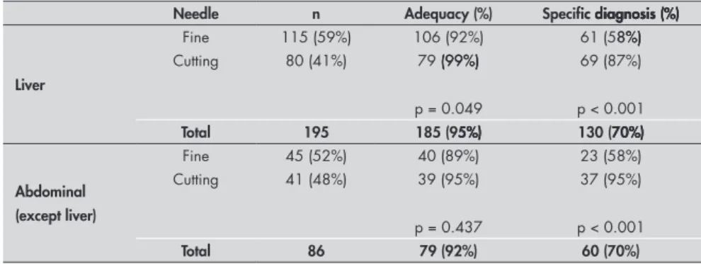
In 195 hepatic lesion biopsies, 185 (95%) obtained diagnostic samples. Fine-needle biopsy was used in 115 (59%) and cutting-needle in 80 (41%). The samples were
consid-ered adequate in 106 (92%) of the fine-needle biopsies and in 79 (99%) of the cutting-needle biopsies (p = 0.049). A specific diagnosis was obtained in 61 (58%) of the fine-needle samples and in 69 (87%) of the cutting-needlecutting-needleutting-needle samples (p < 0.001).
The complications differed according to the location of the lesions and also with the type of needle used. Out of 628 thoracic procedures, pneumothorax was recorded inhorax was recorded inorax was recorded in 76 (16%) of the 486 cases performed using fine needles and in eight (6%) of the 142 cases performed using cutting needles. Three cases of severe hemoptysis were reported, two of them with the use of fine needles. Two cases of hemothorax occurred, both with the use of fine needles. Thoracic drainage was necessary in 24 (4.9%) cases, all of them in procedures performed with fine needles.
There were four major complications (1.31%) reported among the 306 abdominal biopsies (203 intraperitoneal and 103
re-Table 3. Abdominal biopsies guided by computed tomography according to the needle guided by computed tomography according to the needleguided by computed tomography according to the needleaccording to the needlethe needle type, sample adequacy rate and specific diagnosis rateis rate rate
Needle n Adequacy (%) Specific diagnosis (%)ic diagnosis (%) diagnosis (%)
Liver
Fine 115 (59%) 106 (92%) 61 (58%)8%)%)
Cutting 80 (41%) 79 (99%) (99%)(99%)
p = 0.049
69 (87%)
p < 0.001
Total 195 185 (95%)5%)%) 130 (70%)70%)%)
Abdominal (except liver)
Fine 45 (52%) 40 (89%) 23 (58%)
Cutting 41 (48%) 39 (95%)
p = 0.437
37 (95%)
p < 0.001
Total 86 79 (92%) 60 (70%)
Table 4. Biopsies guided by computed tomography according to needle type, sampleaccording to needle type, sampleneedle type, sample adequacy rate and specific diagnosis rateis rate rate
Site Needle n Adequacy (%) Specific diagnosis (%)
Retroperitoneum
Fine 62 (30%) 52 (84%)84%)%) 28 (54%)54%)%)
Cutting 146 (70%) 136 (93%)93%)%)
p = 0.038
117 (86%)86%)%)
p < 0.001
Total 208 188 (90%) 145 (70%)
Musculoskeletal
Fine 20 (15%) 17 (85%)85%)5%) 9 (53%)53%)3%)
Cutting 114 (85%) 103 (90%)90%)%)
p = 0.439
99 (97%)9 (97%)
p < 0.001
Total 134 120 (90%) 108 (81%)
Head and neck
Fine 37 (76%) 26 (70%)70%)%) 17 (65%)65%)5%)
Cutting 12 (24%) 12 (100%)100%)%)
p = 0.045
12 (100%)100%)%)
p = 0.001
Total 49 38 (78%) 29 (60%)
troperitoneal). One case had a large splenicne case had a large splenic case had a large splenicsplenicnic subcapsular hematoma in a pancreatic tail biopsy performed with a cutting needle. Another case consisted of loop perforation in a patient treated for a mixed ovarian cancer with surgery, chemotherapy and radiotherapy. This patient presented a persistently growing retroperitoneal tumor and the procedure was performed with a 22-gauge “Chiba” needle through an anterior approach. The third case had a large peripancreatic hematoma in a cut-ting-needle pancreatic biopsy, and the fourth case was a tumor implant in the abdominal wall after fine-needle aspiration biopsy for an advanced renal tumor.
DISCUSSION
Needle biopsies can shorten the period of hospital stay and decrease the number of operations as well as the treatment costs.1-6
Computed tomography guided biopsy guided biopsyguided biopsy has been widely used as an effective and safewidely used as an effective and safe as an effective and safeas an effective and safean effective and safe method for biopsy guidance, providing excel-lent visualization of the anatomy. It has great versatility and allows safe access to almost any deep-seated lesion in the human body. Through changing the patient’s positionhanging the patient’s position patient’s position and breathing techniques, the versatility and safety of the procedure can be further improved.1,2,14,15
Fine-needle biopsy has long been recog-nized as a useful technique for safely obtain-ing biopsies from deep-seated lesions. This technique has high sensitivity for diagnosing malignant disease.1,3,5,9,11,12 However, the
qual-ity of the tissue provided by cutting needles is superior, thereby allowing diagnoses to be made and further pathological investiga-tions to be performed. Recent reports have described higher success rates from the use of cutting-needle biopsies, which have obtained adequate biopsy samples and have thus en-abled accuracy in the diagnosis and specific histology results.4,5,7-10
The present study was conducted to evalu-ate the general results from CT guided biopsy in a large series, with evaluation of sample adequacy, diagnostic performance and major complications relating to CT guided biopsies routinely done in different body regions with the use of different types of needles in a oncol-ogy center in Brazil.
1. Westcott JL. Direct percutaneous needle aspiration of local-ized pulmonary lesions: result in 422 patients. Radiology. 1980;137(1 Pt 1):31-5.
2. Gazelle GS, Haaga JR. Guided percutaneous biopsy of intraab-dominal lesions. AJR Am J Roentgenol. 1989;153(5):929-35. 3. Ferrucci JT Jr, Wittenberg J, Mueller PR, et al. Diagnosis of
abdominal malignancy by radiologic fine-needle aspiration biopsy. AJR Am J Roentgenol. 1980;134(2):323-30. 4. Ben-Yehuda D, Polliack A, Okon E, et al. Image-guided
core-needle biopsy in malignant lymphoma: experience with 100 patients that suggests the technique is reliable. J Clin Oncol. 1996;14(9):2431-4.
5. Guimarães AC, Chapchap P, de Camargo B, Chojniak R. Computed tomography-guided needle biopsies in pediatric oncology. J Pediatr Surg. 2003;38(7):1066-8.
6. Husband JE, Golding SJ. The role of computed tomography-guided needle biopsy in an oncology service. Clin Radiol. 1983;34(3):255-60.
7. Hooper KD, Abendroth CS, Sturtz KW, Matthews YL, Stevens LA, Shirk SJ. Automated biopsy devices: a blinded evaluation. Radiology. 1993;187(3):653-60.
8. Moulton JS, Moore PT. Coaxial percutaneous biopsy techniqueCoaxial percutaneous biopsy techniqueoaxial percutaneous biopsy technique with automated biopsy devices: value in improving accuracy and negative predictive value. Radiology. 1993;186(2):515-22. Radiology. 1993;186(2):515-22.Radiology. 1993;186(2):515-22.
9. Farias AP, Deheinzelin D, Younes RN, Chojniak R. Computed tomography-guided biopsy of mediastinal lesions: fine versus cutting needles. Rev Hosp Clin Fac Med Univ São Paulo. 2003;58(2):69-74.
10. Hopper KD, Grenko RT, TenHave TR, Hartzel J, Sturtz KW, Savage CA. Percutaneous biopsy of the liver and kidney by using coaxial technique: adequacy of the specimen obtained with three different needles in vitro. AJR Am J Roentgenol. 1995;164(1):221-4.
11. Yu LS, Deheinzelin D, Younes RN, Chojniak R. Computed tomography-guided cutting needle biopsy of pulmonary lesions. Rev Hosp Clin Fac Med Univ São Paulo. 2002;57(1):15-8. 12. Santos GC, Morini SR, Granero LC, Chojniak R, Longatto
Filho A. Fine needle aspiration of thoracic lesions: experience Fine needle aspiration of thoracic lesions: experienceFine needle aspiration of thoracic lesions: experience in a Brazilian cancer center. Pathologica. 1999;91(4):256-9. 13. Cardella JF, Bakal CW, Bertino RE, et al. Quality improvement
guidelines for image-guided percutaneous biopsy in adults. JJ Vasc Interv Radiol. 2003;14(9 Pt 2):S227-30.
14. Tobkes AI, Nord HJ. Liver biopsy: review of methodology andLiver biopsy: review of methodology and complications. Dig Dis. 1995:13(5):267-74.
15. Westcott JL, Rao N, Colley DP. Transthoracic needle biopsy of small pulmonary nodules. Radiology. 1997;202(1):97-103. 16. Smith EH. Complications of percutaneous abdominal
fine-needle biopsy. Review. Radiology. 1991;178(1):253-8.
17. Garcia-Tsao G, Boyer JL. Outpatient liver biopsy: how safe is it? Ann Intern Med. 1993;118(2):150-3.
18. Piccinino F, Sagnelli E, Pasquale G, Giusti G. Complications following percutaneous liver biopsy. A multicentre retrospective study on 68,276 biopsies. J Hepatol. 1986;2(2):165-73. 19. Zech CJ, Helmberger T, Wichmann MW, Holznecht N,
Diebold J, Reiser MF. Large core biopsy of the pancreas under CT fluoroscopy control: results and complications. J Comput Assist Tomogr. 2002;26(5):743-9.
20. Voravud N, Shin DM, Dekmezian RH, Dimery I, Lee JS, Hong WK. Implantation metastasis of carcinoma after percutaneous fine-needle aspiration biopsy. Chest 1992;102(1):313-5. 21. Slywotzky C, Maya M. Needle tract seeding of transitional
cell carcinoma following fine-needle aspiration of a renal mass. Abdom Imaging 1994;19(2):174-6.
Sources of funding: None
Conflict of interest: None
Date of first submission: August 16, 2004
Last received: November 25, 2005
Accepted: November 29, 2005
from 54 to 67% for fine-needle and from 82 to 100% for cutting-needle biopsies, depending on the biopsy site. These better performancessite. These better performances. These better performances for cutting-needle biopsy were statistically significant for most anatomical regions, except for the adequacy of the samples from non-pulmonary thoracic, musculoskeletal and non-hepatic abdominal lesions. In these situations, the numbers of cases and/or differ-ences in the results were smaller. The reported success rate for obtaining adequate samples using needle biopsies ranges in the literature from 70 to 85% for lung lesions and from 80 to 95% for non-lung lesions.1-12 As expected,
for any given site, the sample adequacy and the specific diagnosis rate were always better for cutting-needle biopsies. Fine-needle aspiration biopsy has long been recognized as useful because it is easily performed and provides high rates of diagnoses, especially for docu-mentation of malignancy.1,3,5,9,11,12 More
re-cently, studies have provided reassurances regarding the safety and better diagnostic performance of automated cutting needles, such that their use has increased, especially when a specific benign or malignant diagnosis is desired.4,5,7-10 In our results, the better
per-formance of cutting-needle biopsy could be partially explained by the selection of different types of needle for different lesions based on our initial safety premises: there was a trend towards using fine needles for smaller, deeper
and probably malignant lesions, especially in the lungs, and towards the use of cutting needles in larger, more outlying lesions. On the basis of our acquired experience and other reports, we now favor the use of cutting needles for most lesions, reserving fine needles mainly for patients with coagulation deficits.
Significant complications were reported de-scriptively due to the small number of cases.
The frequency of significant complica-tions was in keeping with the literature. Most of them consisted of pneumothorax relating to lung lesions, with a low rate of tube place-ments.1,9,13,15 The reported incidence of
pneu-mothorax in the literature ranges from 10 to 20%, with tube placement required for 5 to 10% of these cases.1,9,13,15 All tube placements
in our series were required after fine-needle aspiration biopsies but, again, there was a tendency to use fine needles for the difficult smaller and more central lung lesions, which could have increased the complication rate. The safety of the procedure in the abdomen and retroperitoneum was confirmed by the low incidence of major complications that occurred with either type of needle. The numbers of such complications were not enough to correlate with any specific needle type.2,3,14,16-19 A needle tract implant was
observed in a patient with a locally advanced renal carcinoma. This type of complication is rarely reported in the literature and the
risks from it are outweighed by the benefit of avoiding surgery in cases of advanced tumors and benign lesions.20,21 A greater concern is the
potential seeding of initial, surgically curable malignancy: in such cases, despite the rarity of this complication, removal of the needle tract could be performed through surgery.20,21
Minor complications such as local mild pain or subcutaneous hemorrhagic collections may have been underestimated, since this was a retrospective study and these incidents might not have been recorded in post-biopsy subsequent evaluations.
CONCLUSIONS
The present study has reported on the has reported on thehas reported on the experiences of routine use of computed tomo-of routine use of computed tomo- routine use of computed tomo-graphy guided biopsy in a Brazilian oncology center, with demonstration of the effectiveness and safety of the method. Overall, computed tomography guided biopsies have proven to be very useful, through obtaining diagnostic samples in most instances, with low incidence of complications, through providing guid-ance for therapeutic decisions and through avoiding surgery in many instances, especially in cases of diagnosing advanced malignant diseases. Both types of needles used showed satisfactory results but, in our view, cutting-needle biopsy should be used when a specific diagnosis is desired without greater incidence of complications.
RESUMO Biópsia dirigida por tomografia computadorizada: experiência em 1.300 procedimentos
CONTEXTO E OBJETIVO: Biópsia percutânea dirigida por tomografia computadorizada (TC) é amplamente aceita como um procedimento efetivo e seguro para diagnóstico em muitas situações clínicas. Sensibilidade, especificidade e acurácia dependem do sítio de biópsia e do tipo de agulha utilizada. Alguns estudos demonstraram vantagem no uso de agulhas cortantes em relação as agulhas finas tipo “Chiba”. Este estudo apresenta a experiência de um centro de oncologia em biópsias guiada por TC.
TIPO DE ESTUDO E LOCAL: Estudo retrospectivo realizado no Hospital do Câncer, A. C. Camargo, São Paulo, Brasil.
MÉTODOS: 1.300 biópsias consecutivas executadas de julho de 1994 a fevereiro de 2000 foram analisa-das. A indicação foi presença de nódulo ou massa suspeita de neoplasia maligna primária em 845 (65%) casos e de metástase em 455 (35%). 628 biópsias foram realizadas em lesões torácicas, 281 abdominais, 208 retroperitoneais, 134 músculo-esqueléticas, e 49 na região de cabeça e pescoço. 765 (59%) biópsias foram executados com agulha fina tipo “Chiba” de 22 gauge com técnica de punção aspirativa e 535 (41%) com agulha cortante automática tipo “Tru-cut” de 16 gauge ou 18 gauge.
RESULTADOS: Amostra apropriada foi obtida entre 70% e 92% das punções com agulha fina e de 93 a 100% das biópsias com agulha cortante. Diagnósticos específicos variaram entre 54 a 67% para agulha fina e de 82 a 100% para agulha cortante, dependendo do local da biópsia. Os índices de obtenção de amostra adequada e diagnóstico específico sempre foram melhores para a biópsia com agulha cortante. Em 530 biópsias pulmonares ocorreram 84 pneumotóraces (16%), e dois casos de hemotóraces (0,3%). Drenagem foi necessária em 24 casos (4,9%). Em biópsias abdominais ocorreram dois casos de hemor-ragia importante e um caso de peritonite; dois pacientes necessitaram cirurgia. Um implante tumoral no trajeto da biópsia foi observado em portador de carcinoma renal localmente avançado.
CONCLUSÃO: Ambos os tipos de agulha mostraram resultados satisfatórios, mas, em nossa visão, agulha cortante deve ser utilizada quando um diagnóstico específico é desejado, sem levar a maior incidência de complicações.
PALAVRAS-CHAVE: Biópsia por agulha. Tomografia computadorizada por raios X. Neoplasias. Agulhas.Agulhas. Biópsia por agulha fina.
AUTHOR INFORMATION
Rubens Chojniak, MD, PhD. Department of Radiology, Hospital A. C. Camargo, São Paulo, Brazil.
Rony Klaus Isberner, MD. Department of Radiology, Hospital A. C. Camargo, São Paulo, Brazil.
Luciana Marinho Viana, MD. Department of Radiology, Hospital A. C. Camargo, São Paulo, Brazil.
Liao Shin Yu, MD. Department of Radiology, Hospital A. C. Camargo, São Paulo, Brazil.
Alessandro Amorim Aita, MD. Department of Radiology, Hospital A. C. Camargo, São Paulo, Brazil.
Fernando Augusto Soares, MD, PhD. Department of Pathology,Department of Pathology, Hospital A. C. Camargo, São Paulo, Brazil.
Address for correspondence:
Rubens Chojniak
Departamento de Imagem do Hospital do Câncer Rua Professor Antonio Prudente, 211 — Liberdade São Paulo (SP) — Brasil — CEP 01509-900 Tel. (11) 3272-0261
E-mail: [email protected]