Revista da Sociedade Brasileira de Medicina Tropical
http://dx.doi.org/10.1590/0037-8682-0008-2012
Address to: Dra Luisa Caricio Martins. Laboratório de Patologia Clínica das
Doenças Tropicais/NMT/UFPA. Av. Generalíssimo Deodoro 92, Umarizal, 66055-240 Belém, PA, Brasil.
Phone: 55 91 3201-6812 e-mail: [email protected] Received 19 September 2012 Accepted 27 March 2013
Association between histological indings, aminotransferase
levels and viral genotype in chronic hepatitis C infection
Amanda Alves Fecury
[1], Marcella Kelly Costa de Almeida
[1], Kemper Nunes dos Santos
[1],
Andrei da Silva Freitas
[1], Socorro de Fátima Loureiro Dantas
[1], Carlos Araújo da Costa
[1],
Ângelo Barlleta Crescente
[1], Rita Catarina Medeiros de Sousa
[1], Elza Baía de Brito
[1],
Reza Nassiri
[2], Elizabeth Lampe
[3]and Luisa Caricio Martins
[1][1]. Laboratório de Patologia Clínica das Doenças Tropicais, Núcleo de Medicina Tropical, Universidade Federal do Pará, Belém, PA. [2]. Institute of International Health, Michigan State University, Michigan, USA. [3]. Fundação Oswaldo Cruz, Rio de Janeiro, RJ.
ABSTRACT
Introduction: The genomic heterogeneity of hepatitis C virus (HCV) inluences liver disorders. This study aimed to determine
the prevalence of HCV genotypes and to investigate the inluence of these genotypes on disease progression. Methods:
Blood samples and liver biopsies were collected from HCV-seropositive patients for serological analysis, biochemical marker measurements, HCV genotyping and histopathological evaluation. Results: Hepatitis C virus-ribonucleic acid (HCV-RNA) was detected in 107 patients (90.6% with genotype 1 and 9.4% with genotype 3). Patients infected with genotype 1 exhibited higher
mean necroinlammatory activity and ibrosis. Conclusions: HCV genotype 1 was the most prevalent and was associated with greater liver dysfunction.
Keywords: Hepatitis C virus. Genotype. Histopathology.
Hepatitis C virus (HCV) infection is often asymptomatic, but it is chronic in a large (85%) proportion of cases. Approximately 20% of individuals with chronic HCV develop liver damage, cirrhosis or cancer1.
In Brazil, the seroprevalence of HCV is moderate, although rates vary among different regions of the country2. Genotype
1 is predominant, followed by genotypes 3 and 23. In the State
of Pará, the overall prevalence ranges from 0.5 to 2% among blood donor candidates4, and the genotype distribution is similar
to that reported elsewhere in Brazil5.
Hepatitis C virus genotype 1 has been associated with severe liver damage and the response to treatment6. However,
other studies were unable to conirm this association7. Brazilian
studies correlating the different HCV genotypes with histological
and clinical presentation are scarce and have not conirmed an
association between liver disease severity and HCV genotype8,9.
The objectives of this study were to determine the prevalence of HCV genotypes in patients with chronic hepatitis C in the
State of Pará, Brazil and to investigate the inluence of these
genotypes on the biochemical and histopathological parameters of the disease.
A total of 152 adult patients of both genders with positive HCV serology participated between 2008 and 2010. None of the patients was co-infected with hepatitis B virus (HBV) or human
immunodeiciency virus (HIV). The study was approved by the Ethics
Committee on Human Research of the Núcleo de Medicina Tropical. Peripheral blood was collected from each patient for enzyme immunoassays, the measurement of biochemical markers of liver damage and the investigation of viral ribonucleic acid (RNA)
and HCV genotyping by molecular biology techniques. The
ETI-AB-HCVK-4 (DiaSorin, Italy) enzyme-linked immunosorbent assay (ELISA) kit was used to detect speciic HCV antibodies.
RNA was extracted from all samples using the QIAamp Viral RNA kit (Qiagen, Germany). HCV-RNA was detected in the sera of the patients by nested polymerase chain reaction (PCR)10. Viral
genotypes were determined by the restriction fragment length polymorphism (RFLP) technique using the restriction enzymes Ava II and Afa I11.
Alanine transaminase (ALT), aspartate transaminase (AST)
and gamma-glutamyl transpeptidase (γ-GT) were measured
using appropriate kits and an automated analyzer (Katal, Minas Gerais, Brazil).
Liver biopsies were obtained from 107 patients who tested positive for viral RNA. The biopsies were cut and stained with
hematoxylin-eosin and analyzed by a pathologist. The ibrosis stage and inlammatory activity grade were used to diagnose
chronic hepatitis12.
The log-likelihood ratio G-test13 was used to compare
Fecury AA et al - HCV: aminotransferase levels
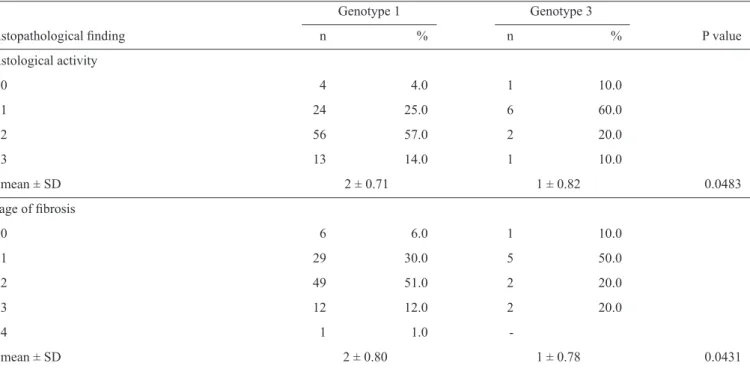
TABLE 1 - Association between hepatitis C virus genotypes 1 and 3 and histopathological indings in the liver.
Genotype 1 Genotype 3
Histopathological inding n % n % P value
Histological activity
0 4 4.0 1 10.0
1 24 25.0 6 60.0
2 56 57.0 2 20.0
3 13 14.0 1 10.0
mean ± SD 2 ± 0.71 1 ± 0.82 0.0483
Stage of ibrosis
0 6 6.0 1 10.0
1 29 30.0 5 50.0
2 49 51.0 2 20.0
3 12 12.0 2 20.0
4 1 1.0 -
mean ± SD 2 ± 0.80 1 ± 0.78 0.0431
SD: Standard deviation.
Differences between histological activity (0 to 3) and stage
of ibrosis (0 or 1) were evaluated by the Mann-Whitney test.
Statistical analysis was performed using the BioEstat 5.0
program (available at http://www.mamiraua.org.br/downloads/
programas), with the level of signiicance set at 5%.
Viral RNA was detected in 107 (70.4 %) of 152 patients. In the remaining 45 (29.6%) patients, the viral load could not be detected by the implemented method, likely because it was very low. In total, 97 (90.6%) of the 107 patients were infected with genotype 1, and 10 (9.4%) were infected with genotype 3. There was no case of mixed infection. No difference in gender distribution or age was observed between the HCV genotypes. The mean age was 45 years.
Liver biopsies were obtained from all 107 patients from whom viral RNA had been isolated, and all samples were submitted to histopathological examination, which revealed chronic hepatitis.
Grade 0 or 1 necroinlammatory activity was observed in 33% (35/107) of these patients, and grade 2 or 3 inlammation was
noted in 67% (72/107). Meanwhile, 40% (43/107) of patients
had stage 0 or 1 ibrosis, and 60% (64/107) had stage 2 to
4 (Table 1). An analysis of the histopathological alterations according to viral genotype using the Mann-Whitney test showed
higher mean necroinlammatory activity and a higher degree of ibrosis among patients infected with genotype 1 (Table 1).
No significant association was observed between the
biochemical markers studied (transaminases, γ-GT) and the
different genotypes. There was also no association between these
markers and histological activity or stage of ibrosis.
The present results showed a high prevalence of HCV genotype 1 (90%), followed by genotype 3 (10%). Genotypes 2,
4, 5 and 6 were not detected. Other studies conducted in northern Brazil reported a similar distribution3. This inding might be
explained by the historic colonization of Pará by Europeans,
Africans and Asians. Genotype 1 is the most prevalent in those regions, followed by genotype 314.
The mean age of the patients in the present cohort was 45
years, a inding consistent with those of other studies conducted
in Brazil8.No difference in age was observed between patients
infected with genotypes 1 and 3. Similar age proiles have been
reported in patients in Piauí, Brazil9.
This study demonstrated an association between histopathological alterations and HCV genotype. The present
indings indicate a signiicant risk of high-grade liver ibrosis and
histological activity in patients infected with genotype 1. Similar results have been reported in studies conducted in other countries6,7.
In general, chronic hepatitis C patients with elevated ALT levels and high serum HCV-RNA titers are considered to have active HCV replication in the liver and a predisposition to liver injury. In addition, serum ALT is currently used as a marker for the degree of histological damage. However, studies have reported equivocal results regarding the association between the degree of histological damage, serum ALT level, HCV-RNA titer and HCV genotype in chronic hepatitis C15. The present study
showed no signiicant correlation between serum ALT, AST or γ-GT levels and histological abnormalities.
In conclusion, the present study conirms previous reports
that showed a high prevalence of genotype 1 in the State of Pará and demonstrates that this genotype is associated with more
severe histological alterations. These indings also suggest that
Rev Soc Bras Med Trop
The authors declare that there is no conlict of interest.
CONFLICT OF INTEREST
FINANCIAL SUPPORT
REFERENCES
Departamento de Ciência e Tecnologia/Ministério da Saúde
(DECIT/MS) andFundação de Amparo à Pesquisa do Estado do Pará (FAPESPA); Grant 035/2007.
1. Aguilera GA, Romero YS, Regueiro BJ. Epidemiology and clinical
manifestations of viral hepatitis. Enferm Infecc Microbiol Clin 2006;
24:264-276.
2. Ministério da Saúde. Secretaria de Vigilância em Saúde, Departamento de
Vigilância Epidemiológica. Hepatites virais: o Brasil está atento. Brasília, DF: Ministério da Saúde; 2008.
3. Campiotto S, Pinho JR, Carrilho FJ, Silva LC, Souto FJ, Spinelli V, et al. Geographic distribution of hepatitis C virus genotypes in Brazil. Braz J
Med Biol Res 2005; 38:41-49.
4. Fonseca JCF, Brasil LM. Hepatitis C virus infection in the Brazilian
Amazon region. Rev Soc Bras Med Trop 2004; 37:S1-S8.
5. Sawada L, Pinheiro AC, Locks D, Pimenta AS, Rezende PR, Crespo DM, et al. Distribution of hepatitis C virus genotypes among different exposure categories
in the State of Pará, Brazilian Amazon. Rev Soc Bras Med Trop 2011; 44:8-12.
6. Singh S, Gupta R, Malhotra V, Sarin SK. Predictors of histological
activity and ibrosis in chronic hepatitis C infection: a study from North India. Ind J Pathol Microbiol 2010; 53:238-243.
7. Lee YS, Yoon SK, Chung ES, Bae SH, Choi JY, Han JY, et al. The
relationship of histologic activity to serum ALT, HCV genotype and HCV
RNA titers in chronic hepatitis C. J Korean Med Sci 2001; 16:585-591.
8. Codes L, Freitas LA, Santos-Jesus R, Vivitski L, Silva LK, Trepo C, et al. Comparative study of hepatitis C virus genotypes 1 and 3 in Salvador, Bahia Brazil. BrazJ Infect Dis 2003; 7:409-417.
9. Veras KN, Jacobina KS, Soares VY, Avelino MA, Vasconcelos CM, Parente JM. Chronic hepatitis C virus in the state of Piauí, northeastern Brazil. Braz J Infect Dis 2009; 13:125-129.
10. Oliveira JM, Rispeter K, Viazov S, Saback FL, Roggendorf M, Yoshida CFT. Differences in HCV antibody patterns in haemodialysis patients infected
with the same virus isolate. J Med Virol 2001; 63:265-270.
11. Hazari S, Acharya SK, Panda SK. Development and evolution of qualitative comparative reverse transcription polymerase chain reaction (RT-PCR) for hepatitis C virus RNA in serum using transcribed thio-RNA
as internal control. J Virol Methods 2004; 116:45-54.
12. Gayotto LCC, Comitê SBP/SBH. Visão histórica e consenso nacional
sobre a classiicação das hepatites crônicas. Gastroenterol Endosc Digest 2000; 19:137-140.
13. Sokal RR, Rohlf FJ. Biometry: the principles and practice of statistics in biological research. 4th edition. New York: Freeman & Company; 2011.
14. Esteban JI, Sauleda S, Quer J. The changing epidemiology of hepatitis C
virus infection in Europe (Review). J Hepatol 2008; 48:148-162.
15. Naito M, Hayashi N, Hagiwara H, Hiramatsu N, Kasahara A, Fusamoto H, et al. Serum hepatitis C virus RNA quantity and histological features of hepatitis C virus carriers with persistently normal ALT levels. Hepatol