Massive Plexiform Neuro
fi
broma of the Neck and
Larynx
Mohammad Kamal Mobashir
1Abd ElRaof Said Mohamed
1Mohammad Waheed El-Anwar
1Ahmad Ebrahim El Sayed
1Mouhamad A. Fouad
21Department of Otorhinolaryngology-Head and Neck Surgery,
Zagazig University, Zagazig, Egypt
2Department of Pathology, Zagazig University, Zagazig, Egypt
Int Arch Otorhinolaryngol 2015;19:349–353.
Address for correspondence Mohammad El-Anwar, MD, Department of Otorhinolaryngology Head and Neck Surgery, Faculty of Medicine, Zagazig University, Zagazig 0020552309843, Egypt
(e-mail: mwenteg@yahoo.com).
Introduction
Laryngeal neurofibromas are extremely rare, accounting for only 0.03 to 0.1% of benign tumors of the larynx.1We report a case of massive neck plexiform neurofibroma (PN) with intralaryngeal extension in a 5-year-old boy with neurofi bro-matosis type 1 (NF-I). The PN was surgically removed.
Review of Literature with Differential
Diagnosis
Histologic subtypes of neurofibromas include the cutaneous, subcutaneous, nodular, and diffuse plexiform variants. PN is a nonmetastatic and locally invasive tumor that can occur on the skin or along the peripheral nerves.2
Sarcomatous degeneration occurs in 5% of neurofibromas,3 and the risk for malignant degeneration of PN into the peripheral nerve sheath tumor is 20%. Furthermore, these spindle cell sarcomas tend to be poorly responsive to therapy,
are frequently metastatic at the time of diagnosis, and are associated with a 28% 5-year survival.
NF-I, also known as von Recklinghausen disease, is an autosomal dominant disease with an incidence of 1 in 2,600 to 3,000 individuals.4
The differential diagnosis of neck mass in children includes cystic hygromas, branchial cleft cysts, thyroglossal duct cysts, dermoid and teratoid cysts, and cystic vascular abnormali-ties.5 Neck PN should be considered in this differential diagnosis and in stridor in children.
Case Report
A 5-year-old boy presented with exertional inspiratory stri-dor and vague throat discomfort on swallowing with no change of voice, hemoptysis, pain, cough, or snoring. The patient had no known systemic or congenital abnormalities, and the developmental milestones were within the normal ranges.
Keywords
►
plexiform
neuro
fi
broma
►
larynx
►
neck
Abstract
Introduction
Laryngeal neuro
fi
bromas are extremely rare, accounting for only 0.03 to
0.1% of benign tumors of the larynx.
Objectives
To report the
fi
rst case of massive neck plexiform neuro
fi
broma with
intralaryngeal (supraglottic) extension in a 5-year-old boy with neuro
fi
bromatosis type 1
and to describe its treatment.
Resumed Report
This massive plexiform neuro
fi
broma was surgically removed,
relieving its signi
fi
cant respiratory obstructive symptoms without recurrence to date.
Conclusion
Massive neck plexiform neuro
fi
broma with supraglottic part was found in
a child with neuro
fi
bromatosis type 1; it should be included in differential diagnosis of
stridor and neck mass in children. It was diagnosed and removed in early in childhood
without recurrence.
received October 22, 2014 accepted
November 10, 2014 published online December 12, 2014
DOI http://dx.doi.org/ 10.1055/s-0034-1396793. ISSN 1809-9777.
Copyright © 2015 by Thieme Publicações Ltda, Rio de Janeiro, Brazil
THIEME
General examination showed a moderately built and nourished child with steady gait and satisfactory vital signs. There were no signs of icterus, clubbing, or anemia. No neurologic or ophthalmologic symptoms were evident. The abdomen had many brown café au lait patches (>6) ranging from 3 to 4 mm in diameter with smooth borders (►Fig. 1). No axillary or inguinal freckling, superficial neurofibromas, or Lisch nodules were evident, as might be expected in NF-I. Audiological evaluation was normal for both ears.
Neck examination revealed large, diffuse, and soft swelling in the right neck with ill-defined edges with no redness, hotness, tenderness, superficial vessels, pigmentation, or ulceration. There were no palpable cervical lymph nodes.
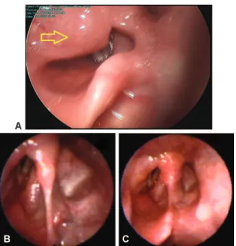
Endoscopy of the larynx revealed a large submucosal pinkish mass covered by apparently normal mucosa located in the right aryepiglottic fold, bulging into the supraglottic area and obstructing the view of the ipsilateral true vocal cord. The right arytenoid appeared to be pushed anteriorly (►Fig. 2). His hypopharynx, left supraglottic area, and vallec-ulae were normal.
Routine laboratory tests, including complete blood count, erythrocyte sedimentation rate, and bleeding profile were normal. There was no clinical evidence (by history, examina-tion, and laboratory tests) of inflammatory and autoimmune disease.
Computed tomography revealed a large, noninfiltrative heterogeneous neck mass. Magnetic resonance examination of the neck revealed a large right mass extending from the
Fig. 1 Postoperative images of the patient. (A) Early while tracheostomy in place. (B) After decannulation. (C) Well-healed neck scar at 2 years postoperatively, with no recurrent neck masses and one café au lait patch (blue arrow). (D) Abdomen showing many brown café au lait patches (>6).
lower neck to the level of the hyoid bone, associated with supraglottic extension involving the right aryepiglottic fold. It encroached on the retrotracheal and prevertebral areas. It was isointense with muscle on T1-weighted imaging, hyper-intense on T2-weighted imaging, and enhanced after gado-linium contrast injection (►Fig. 3).
Radiologic size was massive (652820 mm). It did not affect the cartilaginous framework of the larynx and vocal cords, and no vertebral lytic lesions were detected. The vascular structures of the neck were normal (►Fig. 3).
Based on thesefindings, a tentative diagnosis of benign neck and laryngeal tumor was made. Clinical and imaging features favored a massive neurofibroma. A punch biopsy was not performed, but an excisional biopsy was selected.
The case was then submitted to a physician and pediatri-cian, who ruled out other features of NF-I and who also excluded the possibility of any systemic involvement.
Under general hypotensive anesthesia, the surgical site was prepared with povidone iodine solution and the pa-tient was draped. A collar neck incision of two finger breadths above the suprasternal notch was performed.
The incision was deepened to the subplatysmal plane and the skinflap was raised. Tracheotomy was donefirst. The mass was identified, dissected, and removed with its la-ryngeal part resected after cutting the attachment of supe-rior constrictor muscle to the thyroid cartilage, dissecting the mass from the mucosa without injury of the mucosa and without jeopardizing the great vessels of the neck and laryngeal cartilage framework. We were unable to totally remove the tumor, so near-total removal was done; a residual part at the right lower neck near the apex of the pleura could not be excised and was left. Recovery from general anesthesia was uneventful. Endoscopic examina-tion of the larynx reported immobile right vocal cord (►Fig. 2). The child was extubated (of tracheotomy) after 7 days (►Fig. 1) and was discharged from the hospital with no further complaint. The patient experienced a complete relief of symptoms.
The histopathologic sections showed a lesion composed of bundles of nervefibers arranged in a concentric manner with areas of myxoid changes. Schwann cells andfibroblasts were also seen. A histopathologic diagnosis of PN was established
Fig. 3 Preoperative imaging. (A) Axial MRI shows large diffuse noninfiltrative neck mass. (B) Axial MRI shows the mass encroaching on laryngeal inlet. (C) Axial MRI shows enhancement of the mass. (D) Sagittal MRI shows large mass extending to lower neck. (E) Axial computed tomography (CT) shows large heterogeneous density noninfiltrative neck mass. (F) Axial CT shows encroachment on laryngeal inlet and supraglottic region.
(►Fig. 4). The immunohistochemical staining for S-100 pro-tein was positive and confirmed the diagnosis of a neurofi -broma. Because the child had PN and>6 café au lait patches, he was diagnosed with NF-I.6
The patient was followed for 24 months with no recur-rence or further complaints. Moreover, endoscopic examina-tion of the larynx at 1 and 2 year after excision showed apparently normal laryngeal mucosa without recurrent masses with immobile right vocal cord (►Fig. 1). Apart from the residual piece, postoperative magnetic resonance imaging showed apparently normal larynx and neck (►Fig. 4).
Discussion
To date, fewer than 30 cases of endolaryngeal neurofibromas have been reported in the English literature since its first description by Hollinger in 1950.7Most of these lesions have been reported in the pediatric population and in association with NF-1.1Our patient differs from this conventional profile as he was young (only 5 years old) and had no personal or family history of von Recklinghausen disease. Moreover, the neurofibroma reported here was plexiform type, involving deep neck spaces with intralaryngeal extension. It was
re-moved surgically by an external neck approach without thyrotomy.
To the best of our knowledge, this is thefirst reported case of massive neck PN with intralaryngeal extension as part of NF-1 in a 5-year-old boy with no personal or family history of similar condition. Moussali et al reported a case of PN of the larynx in a 4-year-old child, but it was limited to the larynx with no neck extension.8
PN is rare and seen in only 5 to 15% of cases with NF-I; 50% of the cases of NF-I are inherited as autosomal dominant traits. The area most frequently affected by laryngeal neuro-fibroma is the supraglottic region, with the majority involv-ing the arytenoids and the aryepiglottic fold, followed by the false vocal cords, because these areas are rich in terminal nerve plexuses.9These laryngeal sites were encroached in our case.
Although characteristically benign, PN can cause pain, disfigurement, and functional changes and more importantly may turn malignant. Unlike the other variants, PN carries an increase risk of malignant peripheral sheath tumors.3
As punch biopsy is difficult to perform, a preoperative histologic diagnosis is often been difficult.10We did not do a punch biopsy but decided to perform excisional histopathol-ogy for our patient.
Fig. 4 Postoperative axial magnetic resonance imaging (MRI) showing (A) normal laryngeal inlet and (B) removal of mass from the neck. Postoperative histopathology of the plexiform neurofibroma showing (C) neurofibromas unencapsulated and showing zonation with a more cellular central region containing residual nerve twigs and more myxoid areas at the periphery (100). (B) Typically, the Schwann cells
predominate and are spindled to ovoid and slender with characteristic wavy nuclei (200). (F) Neurofibromas are composed of several elements,
In light of the poor results from medication trials, primary therapy continues to be complete surgical excision of the neurofibroma.11No specific treatment for PN currently exists, aside for surgical resection.12,13
Younger age, the tumor’s location in the head and neck region, and incomplete surgical resection are predictive factors for a higher risk of the tumor progressing and recur-ring.12Our case fell into these categories. Thus, recurrence was expected, particularly as we were unable to totally remove the tumor. In such cases, lifelong follow-up is often warranted.
Neurofibromas have been reported in the literature to be surgically difficult to separate from normal tissue because they lack a well-defined capsule and are instead made up of a mesh of interwoven spindle cells, axons, and collagenfibers.1 Extensive tumors may be associated with massive bleeding.12 Therefore, it is wise not to delay surgery.12,13Surgical excision was decided for our case. Early surgical intervention of PN of the head and neck with a goal of near total resection avoids the loss of function associated with these tumors, such as tracheostomy dependence, swallowing difficulty, and speech problems, and prevents the inexorable progression of sub-stantial cosmetic deformity.13
Removal of PN is a challenging procedure as the lesion may involve multiple nerve fascicles, with serpiginous growth and significant vascularity. Even with surgical excision, PN has a recurrence rate of 20%.12,14
Lateral or median thyrotomy and pharyngotomy have been presented by most authors as the treatments of choice for the surgical excision of these tumors15,16; we preferred cutting the attachment of superior constrictor muscle to the thyroid cartilage, dissecting the mass from the mucosa to preserve the laryngopharyngeal function.
No malignant changes were found in our case. No malignant changes have been noted to occur among isolated neuro-fibromas to date. The progression from a solitary neurofibroma to multiple neurofibromatosis and then transformation into malignancy is theoretically possible but exceedingly rare.10
No recurrence had been detected to date (2 years of follow-up), similar to the studies of Ransom et al,13who did near total removal, and Patil et al,9who did total removal with follow-up of 4 years. However, recurrence was detected after debulking of a massive facial neurofibroma by Asha’ari et al,12but they did not report recurrence after debulking of a parotid neurofibroma.
It is believed that the laryngeal neurofibroma arises from the superior laryngeal branch of the glossopharyngeal nerve.10 But in our reported case, the PN was massive, extending to the lower neck, and its resection was complicat-ed by recurrent laryngeal nerve injury. These suggest the recurrent laryngeal nerve to be the origin of the tumor and ensured that these tumors are unencapsulated and tended to infiltrate and separate the normal nerve fascicles, as was documented before.17
Final Comments
A case of massive neck PN with supraglottic part in a 5-year-old boy with NF-I was reported; it should be included in differential diagnosis of stridor and neck mass in children. Massive neck PN can be surgically removed without recur-rence if diagnosed and removed in early childhood, eliminat-ing the respiratory symptoms. This study highlights the significant of early diagnosis and excision of massive neck PN and its complications.
References
1 Liu J, Wong CF, Lim F, Kanagalingam J. Glottic neurofibroma in an elderly patient: a case report. J Voice 2013;27(5):644–646
2 Hartley N, Rajesh A, Verma R, Sinha R, Sandrasegaran K. Abdominal manifestations of neurofibromatosis. J Comput Assist Tomogr 2008;32(1):4–8
3 Holt GR. E.N.T. manifestations of Von Recklinghausen’s disease. Laryngoscope 1978;88(10):1617–1632
4 Ji Y, Xu B, Wang X, Liu W, Chen S. Surgical treatment of giant plexiform neurofibroma associated with pectus excavatum. J Cardiothorac Surg 2011;6:119
5 Davey S, McNally J. A case of an ectopic cervical thymic cyst. Case reports in clinical medicine 2013;2(2):152–153
6 Ferner RE, Huson SM, Thomas N, et al. Guidelines for the diagnosis and management of individuals with neurofibromatosis 1. J Med Genet 2007;44(2):81–88
7 Hollinger PH, Cohen LL. Neurofibromatosis (von Recklinghausen’s disease) with involvement of the larynx: report of a case. Laryn-goscope 1950;60:193–196
8 Moussali N, Belmoukari S, Elmahfoudi H, Elbenna N, AbdelouafiA, Gharbi A. [An uncommon cause of dyspnea in children. Plexiform neurofibroma of the larynx]. Arch Pediatr 2013;20(6):629–632
9 Patil K, Mahima VG, Shetty SK, Lahari K. Facial plexiform neurofi -broma in a child with neurofibromatosis type I: a case report. J Indian Soc Pedod Prev Dent 2007;25(1):30–35
10 Chen YW, Fang TJ, Li HY. A solitary laryngeal neurofibroma in a pediatric patient. Chang Gung Med J 2004;27(12):930–933
11 Washington EN, Placket TP, Gagliano RA, Kavolius J, Person DA. Diffuse plexiform neurofibroma of the back: report of a case. Hawaii Med J 2010;69(8):191–193
12 Asha’ari ZA, Kahairi A, Shahid H. Surgery for massive paediatric head and neck neurofibroma: two case reports. The International Medical Journal Malaysia 2012;11(2):54–57
13 Ransom ER, Yoon C, Manolidis S. Single stage near total resection of massive pediatric head and neck plexiform neurofibromas. Int J Pediatr Otorhinolaryngol 2006;70(6):1055–1061
14 Gutmann DH. Recent insights into neurofibromatosis type 1: clear genetic progress. Arch Neurol 1998;55(6):778–780
15 Riga M, Katotomichelakis M, Papazi T, Tsirogianni O, Danielides V. Supraglottic laryngeal neurofibroma treated with transoral LASER surgery; a case report and review of the literature. Otorhinolar-yngol Head Neck Surg 2012;48:26–28
16 Rosen FS, Pou AM, Quinn FB Jr. Obstructive supraglottic schwan-noma: a case report and review of the literature. Laryngoscope 2002;112(6):997–1002
17 North KN. Neurofibromatosis 1 in childhood. Semin Pediatr Neurol 1998;5(4):231–242

