Effects of the
fi
bers distribution in the human eardrum: A
biomechanical study
Fernanda Gentil
a,n, Marco Parente
b, Pedro Martins
b, Carolina Garbe
b, Carla Santos
b,
Bruno Areias
b, Carla Branco
c, João Paço
d, Renato Natal Jorge
baLAETA, INEGI
–Faculdade de Engenharia da Universidade do Porto, Clínica ORL–Dr. Eurico Almeida, Widex, ESTSP, Portugal
bLAETA, INEGI
–Faculdade de Engenharia da Universidade do Porto, Portugal
cHospital CUF Infante Santo, Hospital Vila Franca de Xira, Faculdade de Ciências Médicas de Lisboa, Portugal dHospital CUF, Faculdade de Medicina da Universidade de Lisboa, Portugal
a r t i c l e
i n f o
Article history:
Accepted 17 March 2016
Keywords:
Biomechanics Finite element method Eardrum quadrants Eardrumfibers
a b s t r a c t
The eardrum separates the external ear from the middle ear and it is responsible to convert the acoustical energy into mechanical energy. It is divided bypars tensaandparsflaccida. The aim of this work is to analyze the susceptibility of the four quadrants of the pars tensa under negative pressure, to different lamina propriafibers distribution. The development of associated ear pathology, in particular the for-mation of retraction pockets, is also evaluated. To analyze these effects, a computational biomechanical model of the tympano-ossicular chain was constructed using computerized tomography images and based on thefinite element method. Threefibers distributions in the eardrum middle layer were com-pared: case 1 (eardrum with a circular band offibers surrounding all quadrants equally), case 2 (eardrum with a circular band offibers that decreases in thickness in posterior quadrants), case 3 (eardrum without circularfibers in the posterior/superior quadrant).
A static analysis was performed by applying approximately 3000 Pa in the eardrum. Thepars tensaof the eardrum was divided in four quadrants and the displacement of a central point of each quadrant analyzed. The largest displacements of the eardrum were obtained for the eardrum without circular
fibers in the posterior/superior quadrant.
&2016 Elsevier Ltd. All rights reserved.
1. Introduction
The tympanic membrane, is a thin piece of tissue that separates the external ear from the middle ear. The acoustical energy is here transformed in mechanical energy and transmitted to the three ossicles of the middle ear. The tympanic retraction pockets are an ear disorder whose pathophysiology is still not completely under-stood. The retracted segment of eardrum is often known as a retraction pocket. The pathology occurs in 40% of cases in thepars flaccida(theflaccid portion of the tympanic membrane, located in the upper portion of the eardrum, which shows more fragility, due to the lack of thefibrous middle layer, present in thepars tensa) and in 36% of cases in the posterior/superior quadrant of thepars tensa (Paço et al., 2009). Otalgia is occasionally a feature and is due to changes in middle ear pressure or infection. The hearing loss is typically conductive in nature. Several factors are important in the
formation of this pathology, including Eustachian tube dysfunction Danner (2006)and structural changes to the membrane, secondary to repeated bouts of inflammation (Ruah et al., 1992). Three factors must occur for the eardrum to become retracted: (i) negative middle ear pressure; (ii) weakness of the eardrum and (iii) increase in surface area of the eardrum (Ikeda et al., 2011). Thefirst factor can result from an inadequate opening of the Eustachian tube, the pressure within the middle ear can be less than atmospheric pressure and the eardrum can become sucked into the middle ear space. The second one can be related with weakness of the middle layer of the pars tensa in the postero-superior quadrant, or alter-natively, after the perforation by grommet, predisposing these areas to retraction. The third factor, associated with an unusual devel-opment of new cells on the surface of the eardrum, which migrate over the surface and move out along the ear canal. This process of proliferation and migration can result in increase of a retraction pocket. Most common complaints are infected pockets causing otorrhea and conductive hearing loss and can reach up to 45–55 dB in some cases. Progressive retraction of pars tensa may cause the atrophic membrane to drape over the incus and stapes, often resulting in necrosis of these ossicles. It is usually regarded to be a Contents lists available atScienceDirect
journal homepage:www.elsevier.com/locate/jbiomech www.JBiomech.com
Journal of Biomechanics
http://dx.doi.org/10.1016/j.jbiomech.2016.03.030 0021-9290/&2016 Elsevier Ltd. All rights reserved.
n
Correspondence to: Faculty of Engineering, University of Porto–INEGI, Rua Dr. Roberto Frias no 404, 4200-465 Porto, Portugal. Tel.:þ351 914763107; fax:þ351 22 508 1445.
layer remains the same for all quadrants, the circularfiber layer is always distributed in the periphery and differentially in the var-ious quadrants. The same study identified three possible scenar-ios: in 45% of cases the circularfibers involve all quadrants, in 30% of cases the band decreases in thickness in posterior quadrants and in 25% of cases circularfibers are not identified in the pos-terior/superior quadrant.
The aim of this work is to analyze, based on thefinite element method, the effects of the distribution of thefibers of the lamina propria, namely the susceptibility of the four quadrants of thepars tensato negative pressure. It will be investigated the influence of this aspect on the development of ear pathologies, particularly the retraction pockets.
2. Material and methods
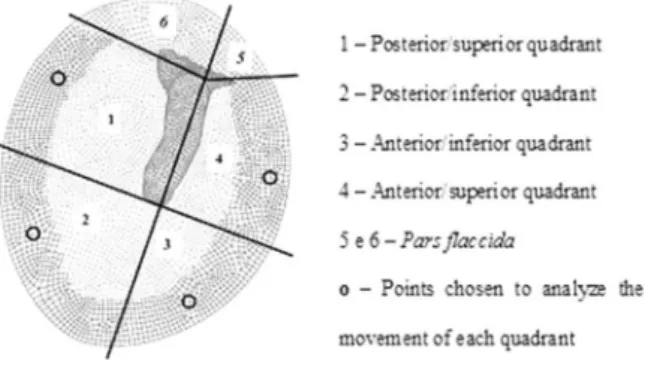
In this study, a geometric model of the tympano-ossicular chain (Gentil et al., 2011) was used to model the middle ear. The eardrum was adapted (Garbe et al., 2009,2010) to the dimensions described in the study ofPaço (2003). It was divided (topographically) into six quadrants. Thepars tensais composed by four quadrants: posterior/superior, posterior/inferior, anterior/superior and anterior/inferior. The
parsflaccidais composed by the remaining two quadrants. After this division, a representative node was chosen from thefinite element mesh (Fig. 1) for each quadrant and the displacements for each node were analyzed.
Fig. 2shows the dimensions of the eardrum model. The height of the eardrum (vertical axis) is 9.7 mm. This is the distance that separates the upper and lower limits along the malleus. The width of the eardrum (horizontal axis), determined as the transversal distance at the umbo, is 8.8 mm. The eardrum has an elliptical shape (Fig. 2a).
Taking into account the dimensions of thepars tensa, the umbo was taken as the reference region. The distance from the umbo to the anterior edge of the ear-drum is 3.9 mm. The distance from the umbo to the posterior edge of the earear-drum is 5.2 mm, and from the umbo to the eardrum lower edge is 4.3 mm (Fig. 2b).
Regarding the dimensions of theparsflaccida, the anterior part has a value of 1.6 mm. The dimension of the posterior part is approximately the double of the previous, with an average of 3.0 mm. The height of theparsflaccidahas a value of 1.7 mm (Fig. 2c).
The discretization of the model was made using ABAQUS software (Hibbit et al., 2004), starting with the discretization of the eardrum and then the ossicles.
The eardrum was discretized using three-dimensional hexahedral elements of eight nodes, of type C3D8, and divided into two parts: theparsflaccida(located at the top and representing 10% of the eardrum total area, approximately) and the
pars tensa(responsible for the vibration of the eardrum and representing near 90% of eardrum total area).
Thepars tensaof the eardrum was considered to be divided into three layers (Garbe et al., 2009) according to its anatomy: layer 1, the external; layer 2, the intermediate; layer 3, internal. From the biomechanical point of view the inter-mediate layer is very important because it is primarily responsible for the stiffness of the eardrum, with radial and circularfibers. The inner layer is continuous with the mucosa of the middle ear, and the external one continues into the external auditory canal.
The intermediate layer of the eardrum has two planes offibers: an external, located in contact with the epidermis which is composed of radialfibers available, and another, arranged in contact with the mucosa that consists of circularfibers. The radialfibers are found throughout the surface of thepars tensa, since the cir-cularfibers are away from the umbo. The plane of radialfibers radiates itsfibers, beginning from the malleus to the periphery of the eardrum. The distribution of radialfibers in every quadrant is identical (Paço et al., 2009).
toPaço (2003), the distribution of circularfibers occurs in three different ways (Fig. 3): Case 1 represents the eardrum with a circular band offibers surrounding all quadrants equally; Case 2 represents the eardrum with a circular band offibers that decreasing in thickness in posterior quadrants; Case 3 represents the eardrum without circularfibers in the posterior/superior quadrant.
The properties of the eardrum were set according to their anatomy. Thepars
flaccidais always considered in the same way, isotropic elastic. For thepars tensa
(Table 1) different properties were used for each layer. The central layer properties were characterized by different types offibers distribution. The material properties were based on previous works, whereE is the Young's modulus, the indexθ
indicates circular direction andrindicates radial direction. The Poisson's ration is assumed equal to 0.3 for all materials and the damping coefficients asα¼0 s 1and β¼0.0001 s (Prendergast et al., 1999;Sun et al., 2002). The density of the eardrum is 1.20Eþ03 Kg/m3.
For the discretization of the ossicles, tetrahedral elements of type C3D4 were chosen, instead of hexahedral as in the eardrum. This takes into account the strongly irregular geometries of the ossicles.
The ossicles were divided into regions according to their properties. The mal-leus was divided into three parts (head, neck and handle); the incus (body, short process and long process); for the stapes, the same properties were applied to its constituent parts (head, neck, anterior and posterior cruras and stapes footplate). The ossicles are assumed as having isotropic behavior, with linear elastic properties obtained from literature (Ferris and Prendergast, 2000; Sun et al., 2002). The Young's modulus of all ossicles of the middle ear was considered 1.41Eþ10 Pa. The density varies according to the constituent parts of the ossicles themselves.
The simulation of the joints between the ossicles, malleus/incus and incus/ stapes were made through mathematical formulations representative of contact (Wriggers, 2002), with a friction coefficient equal to 0.9 (Gentil et al., 2007).
The group formed by the three ossicles (malleus, incus and stapes) is attached on the outside to the eardrum and inside to the oval window by the stapes footplate.
Based on the Yeoh model (Yeoh, 1990), the present work uses hyperelastic non-linear behavior for the ligaments (Gentil et al., 2006,2011;Holzapfel, 2000) being the Hill model (Martins et al., 1998) used for the muscles (Gentil et al., 2013).
Related to the boundary conditions, the eardrum is constrained in order to simulate the tympanic sulcus. Anatomically, theparsflaccidais free. Thepars tensa
is attached at its periphery and in the posterior/superior quadrant the eardrum was constrained in layer 3. In other quadrants the eardrum wasfixed in layer 1 and layer 3, using the nodes shown in the scheme ofFig. 4.
The ossicles are suspended by ligaments and muscles to the tympanic cavity walls. The malleus is attached by the superior, lateral and anterior ligaments and the tensor tympanic muscle; the incus by the superior and posterior ligaments; the stapes by the stapedius muscle and surrounding the periphery of the stapes foot-plate, 78 bar elements werefixed, simulating the annular ligament.
To analyze the retraction pockets formed in the pars tensa a static analysis was performed by applying 3000 Pa in the eardrum. With the sole objective of ana-lyzing the influence of different types offibers distribution in the eardrum central layer on the behavior of the eardrum (umbo), the simulations were carried out applying a uniform sound pressure level (SPL) of 70 dB SPL in the eardrum. The frequency range investigated covers the interval of 100 Hz to 10 kHz (the audibly range).
3. Results
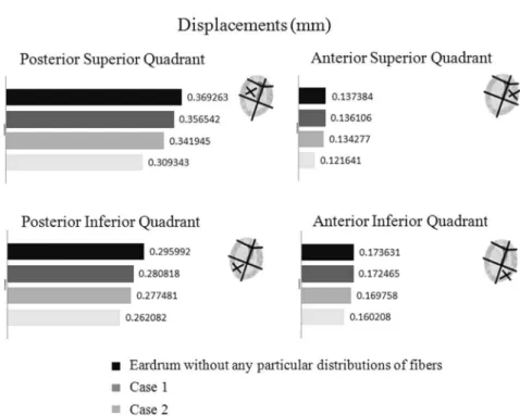
and anterior/superior quadrants of the eardrum (according to the scheme presented inFig. 1), for the threefibers distribution cases considered, and with the eardrum without any particular fibers distributions. The biggest displacements occur in the posterior/superior quadrant and the smallest on the anterior/ superior one.
Thereafter, a dynamical study was made and the frequency range was considered between 100 Hz and 10 kHz. The sound pressure level applied in the eardrum was 70 dB SPL for all results. For the analysis of the results, the displacements obtained in four frequencies of that range (100, 782.8, 1807 and 9659 Hz) were registered and their differences were checked. Fig. 6 shows these displacements of a central point of each quadrant of the eardrum for the threefibers distributions cases considered, and with the eardrum without distribution of fibers. In all results, on average, it was consistently observed
that the displacements obtained with the eardrum without distribution of thefibers have lower amplitude than thefibered cases. The major differences occurred in two posterior quad-rants, not showing significant differences in two anterior quadrants comparing the 3 cases described. In the case where the central layer of the eardrum lacks the range of circular fibers (case 3), the posterior/superior quadrant has greater mobility when compared with the other cases. We can also observed that displacements decrease for higher frequencies.
Fig. 7 shows the displacements of the umbo for all analyzed cases. Despite the displacements in each of the quadrants, in the three considered cases, are different, for the umbo the results obtained are very similar. It may be concluded that the transmis-sion of energy for the considered eardrumfibers distribution does not interfere with the middle ear ossicles movement.
Fig. 2.Dimensions of eardrum in reference to the handle of the malleus, in mm.
Fig. 3.External face–parsflaccidaandpars tensafor Case 1 (eardrum with a circular band offibers surrounding all quadrants equally), Case 2 (eardrum with a circular band offibers that decreasing in thickness in posterior quadrants) and Case 3 (eardrum without circularfibers in the posterior/superior quadrant).
Table 1
Some material properties ofpars tensaof the eardrum.
Material properties
Layers Density Model Poisson's ratio Young's Modulus (N/m2)
Pars tensa E(θ) E(r)
Layer 1 1.20Eþ03 Elastic Isotropic 1.00Eþ07
Layer 2 With circularfibers 1.20Eþ03 Elastic Orthotropic 0.3 2.00Eþ07 3.20Eþ07
Without circularfibers 1.20Eþ03 Elastic Orthotropic 0.3 0.50Eþ07 3.20Eþ07
4. Discussion
In all results, it was consistently observed that the displace-ments obtained with the eardrum without any particular dis-tribution of thefibers have lower amplitude than the cases with fibers distribution was considered (Fig. 6).
The major differences occurred in two posterior quadrants, not showing significant differences in two anterior quadrants com-paring the 3 cases described (Fig. 5).
It can be observed an increased order of magnitude of the eardrum displacements, as (Case 1)o(Case 2)o(Case 3) (Fig. 3). In the case where the intermediate layer of the eardrum lacks the range of circularfibers (Case 3), the posterior/superior quadrant has greater mobility when compared with the other cases. The movement of the eardrum in Case 1 and Case 2 is 92% and 96% of Case 3. The eardrum of Case 2 has higher mobility than on Case 1 (Fig. 6).
Fig. 4.Detail of the boundary conditions in the eardrum.
We have found that regardless of the composition of the lamina propria the posterior/superior quadrant suffered the greatest dis-placement with the application of negative pressure, which can be
attributed to its largest size. Furthermore, it was also observed that the fewer the circular fibers, the greater the eardrum displacement.
Since the posterior/superior quadrant shows great variation in this aspect and may even present a lack of circularfibers in 25% of the population, this quadrant is inevitably more susceptible to the development of pathology related to pressure changes, including retraction pockets, as is observed clinically.
This study allows us to identify a group of patients more prone to developing this pathology, explaining the higher incidence of retraction pockets on the posterior/superior quadrant compared with the other quadrants of thepars tensa.
It also contributes to validate the finite element method to study the pathophysiology of human middle ear, opening a range of possibilities for simulation on future studies.
5. Conclusion
The transmission of energy for the considered eardrumfibers distribution does not interfere with the middle ear ossicles movement, since the displacements of the umbo are similar.
Comparing the 3 cases, the major differences occur in the two posterior quadrants and no significant differences are present in the two anterior quadrants.
Regarding the composition of the lamina propria the posterior/ superior quadrant suffer the greatest movement for a negative pressure, which can be attributed to its largest size. It should also be noted that as a result of this investigation, it was seen that the fibers distribution has a significant importance on the formation of retraction pockets.
Conflict of interest
The authors declare that they do not have conflict of interest.
Acknowledgment
The authors acknowledge the funding of the Project UID/EMS/ 50022/2013 of LAETA, from Fundação para a Ciência e Tecnologia (FCT) of Ministério da Educação e Ciência, Portugal and the sup-port of FCT and Fundo Social Europeu (FSE) under programs POPH-QREN and Grant SFRH/BPD/71080/2010 and SFRH/BD/108292/ 2015 are gratefully acknowledged.
References
Ars, B.M., 1991. Tympanic membrane retraction pockets. Etiology, pathogeny, treatment. Acta Otorhinolaryngol. Belg. 45 (3), 265–277.
Danner, C.J., 2006. Middle ear atelectasis: what causes it and how is it corrected? Otolaryngol. Clin. N. Am. 39 (6), 1211–1219.
Ferris, P., Prendergast, P.J., 2000. Middle-ear dynamics before and after ossicular replacement. J. Biomech. 33, 581–590.
Garbe, C., Gentil, F., Parente, M., Martins, P., Jorge, R.M.N., 2009. Dynamics Analysis of Tympanic Membrane Layers. In: Proceedings of the 7th EUROMECH Solid Mechanics Conference. Ambrósio, J., et al. (Eds.), pp. 227–228.
Garbe, C., Gentil, F., Parente, M., Martins, P., Jorge, R.M.N., Ferreira, A., 2010. Development of Computational Model to Analyze the Influence of Fiber Direction in the Tympanic Membrane. In: Proceedings of the 6th International Conference on Technology and Medical Sciences.
Gentil, F., Jorge, R.M.N., Ferreira, A.J.M., Parente, M.P.L., Martins, P.A.L.S., Almeida, E., 2006. Biomechanical simulation of middle ear using hyperelastic models. J. Biomech. 1, 388–389.
Gentil, F., Jorge, R.M.N., Ferreira, A.J.M., Parente, M.P.L., Moreira, M., Almeida, E., 2007. Study of the effect of contact friction between the ossicles of the middle ear. Revista Internacional de Métodos Numéricos para Cálculo y Diseño en Ingeniería 2, 177–187.
Gentil, F., Parente, M., Martins, P., Garbe, C., Jorge, R.N., Ferreira, A., Tavares, J.M.R.S., 2011. The influence of mechanical behaviour of the middle ear ligaments: a
finite element analysis. Proc. Inst. Mech. Eng. Part H J. Eng. Med. 225, 68–76. Gentil, F., Parente, M., Martins., P., Garbe, C., Paço, J., Ferreira, A., Tavares, J., Natal, R.,
2013. The influence of muscles activation on the dynamical behaviour of the tympano-ossicular system of the middle ear. Comput. Methods Biomech. Biomed. Eng. 16 (4), 392–402.http://dx.doi.org/10.1080/10255842.2011.623674. Hibbit, D., Karlsson, B., Sorenson, P., 2004. ABAQUS Analysis User’s Manual, version
6.5. Hibbit, Karlsson & Sorenson Q7 Inc.
Holzapfel, G.A., 2000. Nonlinear Solid Mechanics. John Wiley & Sons Ltd, United States.
Ikeda, R., Oshima, T., Oshima, H., Miyazaki, M., Kikuchi, T., Kawase, T., Kobayashi, T., 2011. Management of patulous Eustachian tube with habitual sniffing. Otol. Neurotol. 32 (5), 790–793.
Martins, J.A.C., Pires, E.B., Salvador, R., Dinis, P.B., 1998. A Numerical model of passive and active behavior of skeletal muscles. Comput. Methods Appl. Mech. Eng. 151, 419–433.
Paço, J., 2003. Doenças do Tímpano. LIDEL, Lisboa, pp. 57–65.
Paço, J., Branco, C., Estibeiro, H., Oliveira, D., 2009. The posterosuperior quadrant of the tympanic membrane. Otolaryngol.–Head Neck Surg. 140, 884–888. Prendergast, P., Ferris, P., Rice, H., Blayney, A., 1999. Vibro-acoustic modelling of the
outer and middle ear using thefinite-element method. Audiol. Neuro-Otol. 4, 185–191.
Ruah, C.B., Schachern, P.A., Paparella, M.M., Zelterman, D., 1992. Mechanisms of retraction pocket formation in the pediatric tympanic membrane. Arch. Oto-laryngol.–Head Neck Surg. 118 (12), 1298–1305.
Sun, Q., Gan., R., Chang, K., Dormer, K., 2002. Computer-integratedfinite element modeling of human middle ear. Biomech. Model. Mechanobiol. 1, 109–122. Wriggers, P., 2002. Computational Contact Mechanics. John Wiley & Sons Ltd,
United States.
Yeoh, O.H., 1990. Characterization of elastic properties of car-bon-black-filled rub-ber vulcanizates. Rubrub-ber Chem. Technol. 63, 792–805.