Aurelino Fernandes Schmidt Júnior1, Olavo Ribeiro Rodrigues2, Roberto Storte Matheus3,
Jorge Du Ub Kim4, Fábio Biscegli Jatene5
Abstract
Objective: To create a reference map of mediastinal lymph nodes through the analysis of their size, number and distribution in various lymph node stations. Method: A total of 50 cadavers, 38 males and 12 females, were studied. Of those 50, 39 were Caucasian. The mean age was 59.9 ± 14.1 years, the mean height was 173.1 ± 7.6 cm, and the mean weight was 71.0 ± 12.0 kg. A bilateral mediastinal dissection was performed in order to resect and isolate all lymph nodes. The area, as well as the major and minor transverse diameters, of each lymph node was determined by radiographic imaging analysis. Results: In a sample of 485 stations, 1742 lymph nodes were dissected (2.58 ± 1.89 lymph nodes/station), revealing a mean number of 21.2 ± 8.5 lymph nodes on the right and 13.6 ± 6.3 on the left. The lymph node stations 1, 2R, 4R, 5, and 7 were present in more than 90% of the sample. Only the 4R and 7 lymph node stations were always present. The lymph node stations 2L, 3p, and 8 were present in 32, 36, and 54% of the sample, respectively. Mediastinal lymph nodes were present in greater numbers in the 2R, 4R and 7 lymph node stations. In addition, these stations presented the largest mediastinal lymph nodes. Conclusion: Composing a reference map for lymph node sizes was feasible. No alterations were observed in the distribution, number, or size of lymph nodes in the age brackets studied, regardless of gender, race, weight, or height.
Keywords: Lymph nodes; Mediastinum; Cadaver; Anatomy.
* Study carried out at the Universidade de Mogi das Cruzes – UMC, University of Mogi das Cruzes – School of Medicine, Mogi das Cruzes, Brazil, and at the Universidade de São Paulo – USP, University of São Paulo – School of Medicine, São Paulo (SP) Brazil.
1. PhD and Assistant Professor in the Surgery Department of the Universidade de Mogi das Cruzes – UMC, University of Mogi das Cruzes – School of Medicine, Mogi das Cruzes (SP) Brazil.
2. PhD and Adjunct Professor in the Thoracic Surgery Department of the Universidade de Mogi das Cruzes – UMC, University of Mogi das Cruzes – School of Medicine, Mogi das Cruzes (SP) Brazil.
3. Masters and Assistant Professor in the Thoracic Surgery Department of the Universidade de Mogi das Cruzes – UMC, University of Mogi das Cruzes – School of Medicine, Mogi das Cruzes (SP) Brazil.
4. Intern at the Universidade de São Paulo – USP, University of São Paulo – School of Medicine, São Paulo (SP) Brazil.
5. Full Tenured Professor in the Thoracic Surgery Department of the Universidade de São Paulo – USP, University of São Paulo – School of Medicine, São Paulo (SP) Brazil.
Correspondence to: Aurelino Fernandes Schmidt Júnior. Av. Frederico Straube, 512, CEP 08790-310, Mogi das Cruzes, SP, Brasil. Phone/Fax 55 11 4799.8310. E-mail: [email protected]/[email protected]
Introduction
The importance of a study of the medias-tinal lymph node groups derives from their active involvement in various neoplastic and infectious processes. Mediastinal lymph node enlargement is a well known characteristic of such situations.
However, the distribution, size, and number of the mediastinal lymph nodes have yet to be clearly defined. This results from the great variability in the distribution of lymph nodes in certain mediastinal regions. In addition, there is no systematization in the dissection of lymph nodes, previous studies having used different designations for each region. This, together with the fact that different tech-niques, such as autopsy, thoracic surgery, and chest X-ray, have been employed to study the lymph node stations, has made it difficult to draw comparisons among the results.
In addition, the concept of lymph node enlarge-ment is fundaenlarge-mental to the understanding of several diseases. Although there is controversy regarding the definition of an ideal value, various authors have adopted a maximum of 1 cm on the short axis
to define normal lymph node size.(1-6) However, the
patient samples evaluated in Brazilian studies of diseases with mediastinal lymph node involvement are not comparable in size to those evaluated in American, English, and Japanese studies.
One single anatomical study to determine the size and number of mediastinal lymph nodes, conducted
in Japan, involved 40 cadavers.(7) The authors
observed that the larger lymph nodes were located in the subcarinal station (station 7), followed by the tracheobronchial station (station 10R), equivalent to station 4R on the map created by the American Thoracic Society. The mediastinal lymph nodes were larger in the inhabitants of the urban zone than in those of the rural zone, suggesting that local factors can alter these characteristics.(7)
There seems to be some factor that interferes in the size of the lymph nodes of Brazilians. A study carried out with the aim of comparing the role of computed tomography and mediastinos-copy in the mediastinal lymph node staging in lung cancer showed that only 23% of the lymph nodes larger than 20 mm in diameter presented neoplasia, compared with over 80% for lymph nodes of the same size in the Japanese, American and European populations. This could be attributed to the fact
that, due to the high incidence of inflammatory diseases in Brazil, mediastinal lymph node
enlarge-ment is common in this population.(8,9)
The definition of the size and number of medias-tinal lymph nodes found after anatomical dissection has not been established. Studies on lymph node drainage still need to define the number of lymph nodes, which would serve as a reference to charac-terize the radicality of the resection.
Based on the localization of each lymph node in the anatomical examination of the mediastinum, it is possible to define their distribution, size, and number in the various mediastinal stations. In the present study, we will attempt to determine whether any of the data collected correlate with age, gender, weight, height, race, or cause of death. It will soon be possible to establish a reference map to be used as a standard of normality for the population studied.
Methods
A total of 50 adult cadavers, in which death was not the direct result of diseases with medi-astinal lymph node involvement, were dissected during the period from March of 2001 to June of 2003. Of those 50 cadavers, 38 (76%) were males. The age ranged from 36 to 90 years (mean,
59.9 ± 14.1 years; median, 60 years). Thirty-nine
were White, seven were Black, and four were of mixed ethnicity. Heights ranged from 150 cm to
185 cm (mean, 173.1 ± 7.6 cm). Weights ranged
from 31 to 114 kg (mean, 71 ± 12 kg). In 50% of
the cases, the ultimate cause of death, determined in the autopsy, was pulmonary edema.
This study was conducted according to the guidelines for research involving human beings (health research) established in Brazilian National Health Council Resolution nº 196/96,(10) as well as
in Law 8501 (11/30/92), which addresses the use of cadavers. The Ethics Committee for the Analysis of Research Projects of the Clinical Board of the University of São Paulo School of Medicine Hospital das Clínicas approved the present study.
the Union Internationale Contre le Cancer.(11) The
topography of the various mediastinal lymph node stations was preserved by the in situ dissection of the mediastinal structures, according to the
modifi-cations of dissection techniques already described.(12)
The lymph node dissection and resection was radical, that is, all the lymph nodes were removed, together with the circumjacent mediastinal fat, within the defined anatomical limits. The visceral block of the mediastinum was not removed. This made it possible to maintain the syntropy of the lymph node stations, and their limits, with the anatomical structures and divisions of the mediastinum.
The lymph nodes were fixed in an aqueous solu-tion of formalin and separated from the mediastinal fat tissue by a thorough dissection on a plank, after which they were digitally photographed. The lymph nodes were counted on a per-station basis. The possibility of lymph node shrinkage, as a result of the fixation, was studied in 84 lymph nodes, prior to and after seven days of immersion in 10% buff-ered formalin solution. Sizes were compared using a nonparametric test. There were no significant differences between the two time points in terms of the dimensions of the lymph nodes.
All of the lymph nodes were embedded in paraffin and identified according to their station. The paraffin blocks containing the lymph nodes identified in each mediastinal station were stained with hematoxylin and eosin for histological study. The observation of the slides under a magnifying loupe allowed the lymph node counts after fixation, in order to determine the presence or absence of lymph node coalescence.
Histological analysis under optical microscopy was performed by an independent pathologist who was blinded as to the cause of death. Lymphatic tissue without active pathological process was identified. In addition, the analysis allowed the separation of nonlymphatic tissues that had been inadvertently included in the dissection. The nonl-ymphatic tissues were photographed and their images were demarcated. They were then excluded from the measurements.
The images were processed using the Image Toll program for Windows, version 3.0, developed by the Health Science Center of the University of Texas at San Antonio. The measurements were calibrated against a line drawn over a known distance (10 mm) on a millimeter ruler on each image. The perimeter
of each lymph node was demarcated, the area was calculated, and the minimum/maximum diameters were measured by the computer.
Regarding the descriptive analysis, the minimum and maximum values were observed, and the calcu-lation of means and standard deviation was used for quantitative variables. The coefficient of varia-tion (standard deviavaria-tion*100/mean) was calculated in order to determine the parameter that presented the smallest differences among values of compa-rable dimensions. For the qualitative variables, relative and absolute frequencies were calculated. Regarding the statistical analysis, the homoge-neity of the groups in relation to the proportions was tested by using the chi-square test or Fisher’s exact test. This was indicated for the comparison of the proportions when the response fields presented expected frequencies <5.
All of the tests were performed at a significance level of 5%.(13)
Results
A total of 485 stations were dissected. The mean presence of the various stations upon medi-astinal dissection was 74.6%. Stations 2R, 4R and 7 were the most frequently found upon dissec-tion, being resected in 98 and 100% of the cases. Stations 2L, 3p, and 8, in turn, were found only in 32, 36 and 54% of the cases, respectively.
A total of 1742 lymph nodes were found
(mean, 2.58 ± 1.89 lymph nodes per station; mean,
34.8 ± 12.2 lymph nodes per case). The mean
number of lymph nodes found in the right
medi-astinum was 21.2 ± 8.5, compared with 13.6 ± 6.3 in
the left mediastinum. The station containing the most lymph nodes was station 2R, accounting for 15.6% of the lymph nodes dissected (Table 1). Over 158 lymph nodes were added to the number of lymph nodes, when recounted on the slide, totaling 1900 lymph nodes. Consequently, 8.3% coalescent lymph nodes were found.
A total of 73.2% of the lymph nodes were oval- or kidney-shaped. Approximately 9.7% presented an irregular or mixed shape. The lymph nodes were rectangular or triangular in 11% of the cases, and elongated or figure-eight shaped in 6.1%.
pattern at a 95% confidence interval (mean + 2*SD). We found values for the short axis of over 10 mm in most of the stations, except for stations 2L and 9R. Coefficients of variation per area, short axis, and long axis, were determined in order to choose the type of measurement that presented the least vari-ability. The coefficient of variation expresses the variation percentage for each type of measurement. The measurements of the short axis presented fewer variations in most lymph node stations (Table 2).
We attempted to determine whether the number of lymph nodes correlated with the variables age, weight, and height. We found no significant correla-tion with any of these factors. Nor was the presence or absence of stations found to correlate with any of these demographic variables.
We also attempted to determine whether gender, race, and congestion were associated with the number of lymph nodes per station. There were no differences between genders or among races regarding the number of lymph nodes. The cadavers were divided into two groups by cause of death (congestive and noncongestive diseases). No differ-ence was found between these two groups in terms of the number of lymph nodes.
The size of the lymph nodes was analyzed by gender, race, and cause of death. These were not associated with the variations in the lymph node sizes. Lymph node size was not found to be corre-lated with age, height, weight, or coalescence.
The analysis of variance revealed significant difference in the size of the lymph nodes (p < 0.001). In a multiple-comparison test, a significant differ-ence was found between station 7 and the remaining stations (p < 0.05). Station 4R differed significantly from almost of the other stations (p < 0.05), the exceptions being stations 3a, 3p, and 5. The remaining stations did not differ among themselves.
Discussion
This is the first anatomical study conducted in Brazil with the aim of determining the distribu-tion, number, and size of lymph nodes. Although a wider number of studies use tomography, which is a noninvasive procedure with a wide number of patients available, we opted to carry out a dissec-tion study. Since it is a more complicated process, with few cadavers available for study, anatomical studies are fewer in number. Nevertheless, such studies allow the anatomical location to be more clearly defined, and the results are more precise in relation to the definition of size and number. Such studies also allow the histopathological confirma-tion of lymph node tissue free of active pathological processes.
Most authors who have conducted anatomical studies of mediastinal lymph nodes in Brazil had the objective of elucidating aspects of the treatment and prognosis of the bronchogenic carcinoma.
Table 1 - Number of lymph nodes, including means, standard deviations, and percentages of the total, by station.
Station Lymph nodes Mean SD %
1 116 2.58 1.89 6.66
2R 276 5.52 4.48 15.84
2L 55 3.24 1.95 3.16
3a 87 2.29 1.56 4.99
3p 36 1.89 1.33 2.07
4R 229 4.58 3.02 13.15
4L 150 3.49 2.93 8.61
5 188 4.09 2.06 10.79
6 210 5.00 3.37 12.06
7 205 4.10 2.68 11.77
8 57 2.04 1.45 3.27
9R 61 2.03 1.71 3.50
9L 72 2.12 1.74 4.13
Total 1742 2.58 1.89 100.00
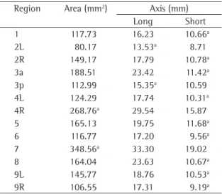
Table 2 - Maximum standard size per area (long axis and short axis).
Region Area (mm2) Axis (mm)
Long Short 1 117.73 16.23 10.66a
2L 80.17 13.53a 8.71
2R 149.17 17.79 10.78a
3a 188.51 23.42 11.42a
3p 112.99 15.35a 10.59
4L 124.29 17.74 10.31a
4R 268.76a 29.54 15.87
5 165.13 19.75 11.68a
6 116.77 17.20 9.56a
7 348.56a 33.30 19.02
8 164.04 23.63 10.67a
9L 145.77 18.76 10.53a
9R 106.55 17.31 9.19a
The number of lymph nodes unilaterally resected during surgical procedures in the treatment of lung neoplasms is smaller when compared to that of specific studies on lymph node dissection. In this study, an average of 21.2 lymph nodes in the right mediastinum, and 13.6 in the left mediastinum, were dissected via thoracotomy. These findings are similar to those of other authors who studied mediastinal lymph nodes (Table 3). Authors who analyzed patients with lung neoplasms found lower numbers of lymph nodes.(7,14,15) This suggests that
the mediastinal lymph node drainage is not radical, that a dissection conducted in a cadaver is an easier approach to the mediastinum, or that the histo-logical analysis did not consider coalescence in the lymph node counts.
The lymph node count was affected by fusion or coalescence; therefore, different numbers were found in the count and recount of dissected lymph nodes on the slides. Lymph node fusions were found frequently (in 32.2% of the stations dissected in the present study).
There were no differences in the number of lymph nodes in relation to age or gender, as was
suggested in one study.(2) The sample presented
a higher mean age. In addition, there were fewer women. This might have resulted in the selection of a population in which the variation in the number of lymph nodes was not significant.
The measurements of the long axis were signifi-cantly higher when granulomatous scar tissue was observed in the lymph nodes. The incidence of granulomatous diseases can affect the reference value adopted for the size of the lymph node.
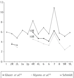
The lymph node sizes were greater than those found by other authors. Figure 1 shows the compar-ison of the mean sizes of the lymph node short axes observed in the present study and reported by other authors, considering the modifications of the descriptors on the maps of the lymph node stations.
The size of the lymph nodes was not uniform among the stations. Therefore, the short axis means for station 7 were significantly higher than the others. Station 4R presented the second greatest mean, which coincides with the observation made by other authors, who reported a significant differ-ence in the dimension of the lymph nodes located in the upper mediastinum when compared to that
of the nodes adjacent to the carina.(20)
Although it was expected that the values obtained for the area would more accurately represent the
lymph node size,(2) the measurements presented
higher coefficients of variation for most stations. Only the lymph nodes of stations 4R and 7 pre-sented lower variation. Therefore, they reprepre-sented a safer parameter for the evaluation of the size alterations in large lymph nodes, which were gener-ally irregular. In most mediastinal stations, the short axis is the value with greater reliability. The data we gathered made it possible to define a reference
0 2 4 6 8 10 12
1 2R 2L 3a 3p 4R 4L 6 6 7 8 9R 9L
10R 10R
Schmidt Kiyono et al.(7)
Glazeret al.(2)
Figure 1 - Mean sizes of the short axis of the mediastinal lymph nodes, by author.
Table 3 - Mean number of mediastinal lymph nodes resected, by author.
Author Numbers of lymph nodes Approach
Kiyono et al.(7) 30.1 (bilateral)
31 (bilateral)
Thoracotomy
Hoksch et al.(16) Video-assisted thoracoscopy
Namori et al.(17) 20 ± 8 (r)a 15 ± 3 (l)a Video-assisted thoracoscopy
Sgawa et al.(18) 40.3 (r)a 37.1 (l)a Video-assisted thoracoscopy
Schmidt(19) 21.2 ± 8.5 (r) 13.6 ± 6.3 (l) Thoracotomy
value for the size of the mediastinal lymph nodes by station. For each station, the lymph nodes sizes were determined as the maximum standard value at a 95% confidence interval (mean + 2*SD). The maximum reference values found for the short axis were greater than 10 mm in most stations, except for stations 2L and 9R (Figure 2).
The population studied, comprising Brazilian
adults, presented 21.2 ± 8.5 mediastinal lymph
nodes on the right and 13.6 ± 6.3 on the left. Stations 1, 2R, 4R, 5, and 7 were present in more than 90% of the cases. Only stations 4R and 7 were always present. Stations 2L, 3p, and 8 were present in 32, 36, and 54% of the cases, respectively. The mediastinal lymph nodes were present in higher numbers in stations 2R, 4R, and 7. These same stations also aggregate the larger mediastinal lymph nodes. The fact that 8.3% of the lymph nodes were coalescent must be considered in the analysis of chest imaging tests and pathological findings.
We were successful in composing a reference map for mediastinal lymph node sizes. No signifi-cant alterations were observed in the distribution, number, or size of lymph nodes among any of the age brackets studied, regardless of gender, race, weight, height, or cause of death.
References
1. Libshitz HI, McKenna RJ Jr. Mediastinal lymph node size in lung cancer. AJR Am J Roentgenol. 1984;143(4):715-8. 2. Glazer GM, Gross BH, Quint LE, Francis IR, Bookstein FL,
Orringer MB. Normal mediastinal lymph nodes: normal and size according to American Thoracic Society mapping. AJR Am J Roentgenol. 1985; 144(2):261-5.
3. McLoud TC, Bourgouin PM, Geenberg RW, Kosiuk JP, Templeton PA, Shepard JA, et al. Bronchogenic carcinoma: analysis of staging in the mediastinum with CT by correlative lymph node mapping and sampling. Radiology, 1992;182(2):319-23.
4. Murray JG, O’Driscoll M, Curtin JJ. Mediastinal lymph node size in an Asian population. Br J Radiol. 1995; 68(808):348-50.
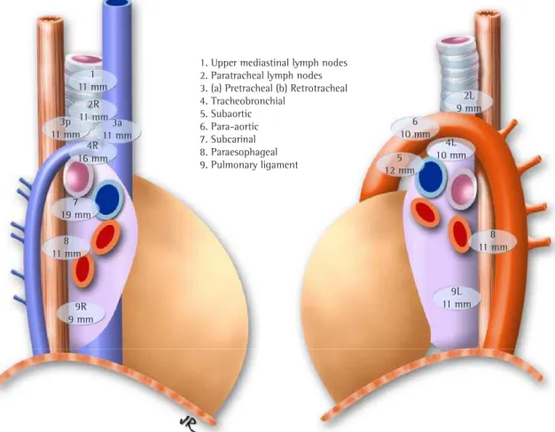
Figure 2 - Map of the mediastinal lymph node stations with the maximum size of the short axis.
1. Upper mediastinal lymph nodes 2. Paratracheal lymph nodes 3. (a) Pretracheal (b) Retrotracheal 4. Tracheobronchial
5. Subaortic 6. Para-aortic 7. Subcarinal 8. Paraesophageal 9. Pulmonary ligament 1
11 mm 2R 11 mm 3p 11 mm
3a 11 mm 4R 16 mm
7 19 mm
8 11 mm
9R 9 mm
2L 9 mm 6
10 mm
5 12 mm
4L 10 mm
8 11 mm
5. Arita T, Matsumoto T, Kuramitsu T, Kawamura M, Matsunaga N, Sugi K, et al. Is it possible to differentiate malignant mediastinal nodes from benign nodes by size? Reevaluation by CT, transesophageal echocardiography, and nodal specimen. Chest. 1996;110(4):1004-8.
6. Erly WK, Borders RJ, Outwater EK, Zaetta JM, Borders GT. Location, size and distribution of mediastinal lymph node enlargement in chronic congestive heart failure. J Comput Assist Tomogr. 2003;27(4):485-9.
7. Kiyono K, Sone S, Sakai F, Imai Y, Watanabe T, Izuno I, et al. The number and size of normal mediastinal lymph nodes: a postmortem study. AJR Am J Roentgenol. 1988;150(4):771-6.
8. Fernandez A. Análise comparativa entre a tomografia axial computadorizada e a mediastinoscopia no estadiamento linfático do câncer do pulmão [Tese]. São Paulo: Universidade de São Paulo; 1992.
9. Fernandez A, Bammann RH, Beyruti R, Junqueira AR, Jatene FB. Avaliação mediastinal no estadiamento do câncer de pulmão. J Pneumol. 1998;24(1):12-22.
10. Conselho Nacional de Saúde. Resolução n° 01/88: normas de pesquisa em saúde. Bioética. 1995;3(2):137-54.
11. Mountain CF, Dresler CM. Regional lymph node classification for lung cancer staging. Chest. 1997;111(6):1718-23. 12. Naruke T. Mediastinal lymph node dissection. In: Pearson FG,
Cooper JD, Deslauriers J, Ginsberg RJ, Hiebert C, Patterson GA, et al, editores. Thoracic Surgery. New York: Churchill Livingstone, 1993. p.909-17.
13. Rosner B. Fundamentals of biostatistics. 2nd ed. Boston: PWS Publishers, 1986. p.584.
14. Rodrigues OR, Antonangelo L, Yagi N, Minamoto H, Schmidt Junior AF, Capelozzi VL, et al. Prognostic significance of argyrophilic nucleolar organizer region (AgNOR) in resected non-small cell lung cancer (NSCLC). Jpn J Clin Oncol. 1997;27(5):298-304.
15. Pereira JCN, da Silva AGP, Soares F, Ab’Saber AM, Schmidt A, Rodrigues OR, et al. Nuclear and environment morphometric profile in tumor size and nodal metastasis of resected typical pulmonary carcinoid. Pathol Res Pract. 2004; 200(6):459-67.
16. Hoksch, B., Ablassmaier, B., Walter, M., Mueller, J.M. The thoracoscopic lymphadenectomy: experiences in a cadaver model. Cardiovasc Eng. 2000;5(2):91-4.
17. Nomori H, Horio H, Naruke T, Suemasu K. What is the advantage of a thoracoscopic lobectomy over a limited thoracotomy procedure for lung cancer surgery? Ann Thorac Surg. 2001;72(3):879-84.
18. Sagawa M, Sato M, Sakurada A, Matsumura Y, Endo C, Handa M, et al. A prospective trial of systematic nodal dissection for lung cancer by video-assisted thoracic surgery: can it be perfect? Ann Thorac Surg. 2002;73(3):900-4.
19. Schmidt Jr AF. Estudo anatômico da distribuição, tamanho e número dos linfonodos mediastinais em brasileiros adultos [tese]. São Paulo: Universidade de São Paulo; 2004. Available from: http://www.teses.usp. br/teses/disponiveis/5/5156/tde-19082005-153022/ 20. Genereux G, Howie JL. Normal mediastinal lymph