Key words:
Prostate; Anabolic Agents; Nandrolone; Image Cytometry
Int Braz J Urol. 2013; 39: 675-82
__________________
Submitted for publication: April 15, 2013
__________________
Accepted after revision: July 25, 2013
Purpose: Many adverse effects have been associated with abuse of anabolic-androgenic steroids (AAS), including disorders of the urogenital tract. The objective of this study is to analyze the morphological modifications in the prostate ventral lobe of pubertal and adult rats chronically treated with AAS, using morphometric methods.
Materials and Methods: We studied 39 male Wistar rats weighing between 400 g and 550 g. The rats were divided into four groups: (a) control rats, with 105 days of age (C105) (n = 7); (b) control rats with 65 days of age (C65) (n = 9), injected only with the vehicle (peanut oil); (c) treated rats, with 105 days of age (T105) (n = 10) and (d) treated rats with 65 days of age (T65) (n = 13). The treated rats were injected with nandrolone decanoate at a dose of 10 mg.Kg-1 body weight. The steroid hormone and the vehicle were administered by intramuscular injection once a week for eight weeks. The rats were killed at 161 days of age (C105 and T105) and 121 days of age (C65 and T65) and the ventral prostate lobe was dissected and processed for histology. The height of the acinar epithelium, the surface densities of the lumen, epithelium and stroma were observed with X400 magnification using an Olympus light microscope coupled to a Sony CCD video camera, and the images transferred to a Sony monitor KX14-CP1. The selected histological areas were then quantified using the M42 test-grid system on the digitized fields. The data were analyzed with the Graphpad software. To compare the quantitative data in both groups (controls and treated) and the outcomes, Student’s t-test was used (p < 0.05 was considered significant).
Results: The weight (p < 0.001) and volume (p = 0.004) of the prostate ventral lobe sho-wed differences between C65 and T65 groups and between C105 and T105 groups. The epithelium height showed no difference between groups C65 and T65 (p = 0.8509), but the T105 group showed an increase of 32% compared to C105 (p = 0.0089). Concerning the lumen, surface density presented no difference between C65 and T65 (p = 0.9031) and a decrease of 19% for T105 compared to C105 (p = 0.0061). There was no difference in epithelium surface density between C65 and T65 (p = 0.7375), but it was 51% higher (p = 0.0065) in T105 compared with C105. Regarding stroma surface density, there were no differences between C65 and T65 or between C105 and T105. Finally, there was no difference in collagen pattern between C105 and T105, but T65 showed a predominance of collagen fibers compared to C65.
Conclusion: The use of anabolic androgenic steroids in rats promotes structural changes in the prostate. We observed structural changes in the weight, volume and epithelium height of the prostate ventral lobe and a predominance of collagen fibers.
The prostate after administration of anabolic androgenic
steroids: a morphometrical study in rats
_______________________________________________
Rafael Arêas Vargas, Leonardo Pires Oliveira, Stephan Frankenfeld, Diogo Benchimol de Souza,
Waldemar Silva Costa, Luciano Alves Favorito, Francisco José Barcellos Sampaio
Urogenital Research Unit of State University from Rio de Janeiro, RJ, Brazil
ABSTRACT
ARTICLE
INFO
INTRODUCTION
Anabolic-androgenic steroids (AAS) are wi-dely used by professional and amateur athletes to improve athletic performance, appearance and mus-cle mass. However, many adverse effects have been associated with AAS abuse, including disorders of the urogenital tract (1). Abuse of anabolic-andro-genic steroids may be an etiological factor in male infertility among recreational power athletes. Ana-bolic-androgenic steroids and endurance exercise can also induce some subclinical alterations in the hypothalamic-pituitary-gonadal (H-P-G) axis (1,2).
Testicular changes resulting from AAS use are well documented in the literature (3). High do-ses of nandrolone decanoate reduce the volume of the testis and length of seminiferous tubules in rats (4). But studies of the structural changes in the prostate after AAS are scarce in the literature (5).
An experimental study with rats demons-trated that changes in the serum levels of testoste-rone can cause morphological alterations in pros-tate tissue (6). The administration of exogenous androgenic-anabolic steroids has been demonstra-ted to have profound effects on the human prostate gland, including an increase in prostatic volume, reduction of urine flow rate and alteration in voi-ding patterns (7).
This study aimed to assess the morphologi-cal modifications in the prostate ventral lobe of pu-bertal and adult rats chronically treated with supra--physiological doses of AAS, using morphometric methods.
MATERIALS AND METHODS
(B) control rats with 65 days of age (C65) (n = 9) injected only with the vehicle (peanut oil); (C) tre-ated rats, with 105 days of age (T105) (n = 10) and (D) treated rats with 65 days of age (T65) (n = 13).
The treated rats (T65 and T105) were in-jected with nandrolone decanoate at a dose of 10 mg.Kg-1 body weight while the control groups
(C65 and C105) received injections of 90% pea-nut oil (diluted in benzoic alcohol) as a vehicle (8). Both the steroid hormone and vehicle were administered by intramuscular injection (at the posterior musculature of the pelvic limb) once a week for eight weeks. This treatment protocol was established to simulate a commonly used protocol in humans during puberty or at early adult age, as previously published elsewhere (8).
The rats were killed by anesthetic over-dose (intraperitoneal thiopental injection) at 161 days of age (C105 and T105) and 121 days of age (C65 and T65), and the ventral lobe of the prostate was dissected and its weight (in grams) and volu-me (in cubic centivolu-meters) were estimated (9).
After the measurements, each prostate was processed for histology. Briefly, the prostate was carefully removed and fixed in 10% buffered for-malin. Then each organ was dehydrated in cres-cent concres-centrations of ethanol baths and routinely processed for paraffin embedding. Non-serial (5 µm thick) sections were obtained at 200 µm intervals. The sections were stained with hematoxylin-eosin (HE) to assess the integrity of the tissue and with Picrosirius Red to identify different collagen types.
after analyzing 250 measurements in at least 25 different fields.
The collagen pattern was qualitatively analyzed by Picro-Sirius Red staining by observing the color of collagen fibers’ birefringence under po-larized light.
Means were statistically compared using the unpaired T-test (p < 0.05) with the Graph Pad Prism software (11).
RESULTS
The weight (p < 0.0001) and volume (p = 0.0004) of the prostate ventral lobe showed differen-ces between groups C65 and T65 and between C105 and T105 (Figure-2).
The epithelium height of the prostate ven-tral lobe showed no difference between groups C65 (20.85 ± 6.241) and T65 (22.16 ± 4.311) (p = 0.8509),
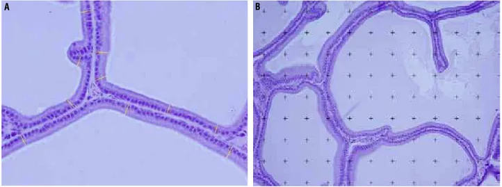
Figure 1 - Photomicrographs of the rat prostate ventral lobe showing the morphometric features. A) The height of the acinar epithelium (yellow) was measured in micrometers in photomicrographs of 400X magnification using the Image J software. HE X400. B) The surface density of ventral lobe prostate lumen, epithelium and stroma were estimated by the point intercepts method, with a grid of 100 points superimposed on magnified images using the Image J software. HE X400.
Figure 2 - Comparative graphs between prostate ventral lobe weight (g) and volume (cm3) of treated and control rats. Data expressed as mean and SD. p < 0.05. The analysis showed difference between C65 and T65 groups, as well as between C105 and T105 groups. Weight: p < 0.001 and volume: p = 0.0004.
but the T105 group (19.19 ± 2.765*) showed a 32% increase compared to C105 (14.11 ± 3.666) (p = 0.0089) (Figure-3 and Table 1).
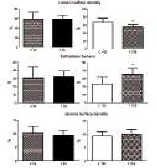
Concerning lumen surface density of the prostate ventral lobe, there was no difference betwe-en C65 and T65 (p = 0.9031) and T105 presbetwe-ented a decrease of 19% compared to C105 (p = 0.0061). The epithelium surface density presented no difference between C65 and T65 (p = 0.7375), but was 51% higher (p = 0.0065) in T105 in comparison to C105. Regarding the stroma surface density, there were no differences between C65 and T65 and between C105 and T105 (Figure-4).
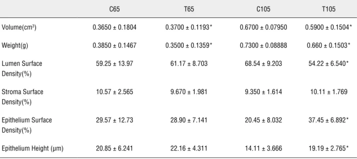
Table-1 reports the numerical values with mean and standard deviations of the structures studied.
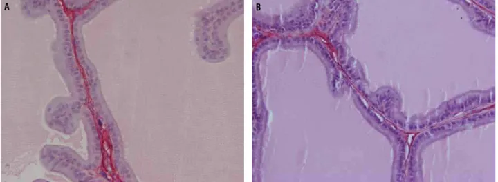
The collagen pattern was qualitatively analyzed by Picro-Sirius Red staining. There was no difference between C105 and T105, but T65 showed a predominance of collagen fibers compa-red to C65 (Figure-5).
DISCUSSION
Anabolic-androgenic steroids are synthe-tic derivatives of testosterone and are important
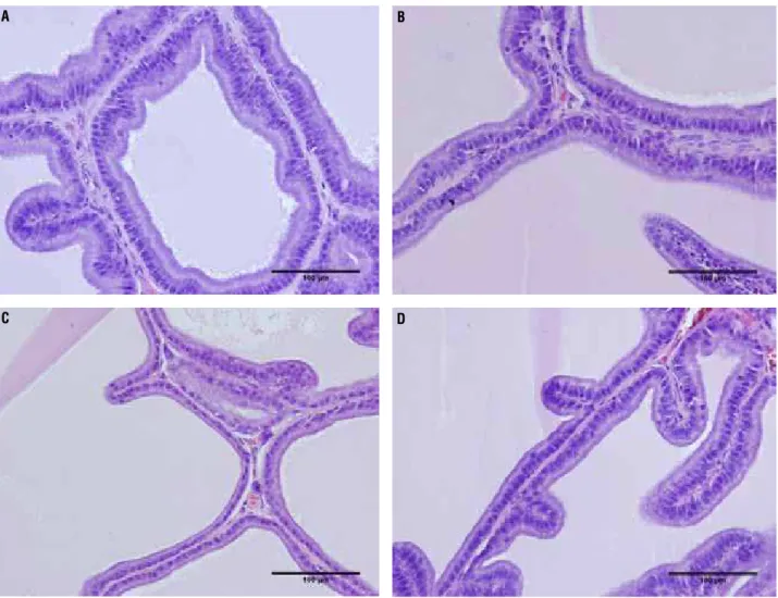
Figure 3 - Photomicrographs of the rat prostate ventral lobe group showing the epithelium height. The epithelium height showed no difference between groups C65 and T65, but the T105 group showed an increased of 32% compared to C105. A) Rat prostate ventral lobe of group C65. HE 400X. B) Rat prostate ventral lobe of group T65 HE 400X. C) Rat prostate ventral lobe of group C105 HE 400X. D) Rat prostate ventral lobe of group T105 HE 400X.
A
C
B
Table 1 - The table shows the numerical values with mean and standard deviation (SD) of the structures studied in the rat prostate ventral lobe treated with anabolic androgenic steroids. C105 - control rats, with 105 days of age; C65 - control rats with 65 days of age, injected only with the vehicle (peanut oil); T105 - treated rats, with 105 days of age and T65 - treated rats with 65 days of age.
C65 T65 C105 T105
Volume(cm3) 0.3650± 0.1804 0.3700 ± 0.1193* 0.6700 ±0.07950 0.5900 ± 0.1504*
Weight(g) 0.3850 ± 0.1467 0.3500 ±0.1359* 0.7300 ± 0.08888 0.660± 0.1503*
Lumen Surface Density(%)
59.25±13.97 61.17± 8.703 68.54±9.203 54.22 ± 6.540*
Stroma Surface Density(%)
10.57±2.565 9.670± 1.981 9.350±1.614 10.11 ± 1.769
Epithelium Surface Density(%)
29.57±12.73 28.90± 7.141 20.45± 8.032 37.45 ± 6.892*
Epithelium Height (µm) 20.85±6.241 22.16± 4.311 14.11±3.666 19.19 ± 2.765*
*p < 0.05 was considered significant.
pharmacologically for treatment of various me-dical conditions such as growth deficiency, some blood disorders and osteoporosis. Androgens play a crucial role in the development of male repro-ductive organs such as the epididymis, vas defe-rens, seminal vesicle, prostate and penis (6). The use of anabolic-androgenic steroids can lead to various body changes, including to the male geni-tal system (12-14).
Many studies have been published repor-ting structural changes of testes after AAS use (3-5). High doses of nandrolone decanoate reduce the testis volume and length of seminiferous tubules in rats (4). It is also clearly established that folli-cle-stimulating hormone (FSH) and luteinizing hormone from the pituitary gland have growth--promoting effects on testis development and that administration of exogenous androgens suppres-ses the serum luteinizing hormone and FSH level in humans and rats (3).
The prostate is influenced by the serum le-vels of testosterone, and the use of AAS can also cause morphological changes in prostate tissue (6). These findings suggest that AAS can provoke
functional changes and diseases of the prostate as well as lowered fertility.
Androgens are needed for puberty, male fertility and male sexual function. High levels of intra-testicular testosterone, secreted by Leydig cells, are necessary for spermatogenesis (15). Tes-tosterone is a critical hormone for prostate deve-lopment, growth and maintenance. The enzyme 5-alpha-reductase seems to play an important role by converting androgens into dihydrotestostero-ne, which acts in the cell nucleus of target organs, such as male accessory glands, skin and prostate. Prostatic morphogenesis is determined not geneti-cally, but by exposure to androgens produced by the testes in the fetus (16).
gets of the action of androgens in the prostate, the reason it is widely used in experiments regarding hormones (16). Besides this, the ventral prostate has only a small quantity of epithelial folds, unlike the other prostate lobes in rats (20). For this reason, we decided to evaluate the morphology of the ventral lobe instead of the others.
A recent study investigated the structural alterations of the rat prostate after treatment with AAS for 14 weeks in animals weighing between 250 and 300 g (5). The authors concluded that nandrolone decanoate causes atrophic changes in the components of the rat prostate. In our study, we observed that the use of AAS causes changes in the prostate morphology after briefer treatment (8 weeks), and that the changes are less severe in pubertal animals and more severe in adults.
Although we used a similar method, our results were quite different from those of Karba-lay-Dust (5). The height of the epithelium increa-sed 32% in the groups of 105-day-old animals, a parameter not altered in the previous study. That study also found that the use of AAS diminished the density of the lumen, epithelium and stroma. In contrast, we analyzed the surface density and found that the lumen diminishes, the epithelium enlarges and the stroma does not change after tre-atment with AAS.
A recent in vitro study investigated the prostate after administration of themethyl-1--testosterone (M1T) (21). The authors showed that M1T had high potency to stimulate the tis-sue weight of the prostate as well as the seminal vesicle (21). That paper shows stimulation of pro-liferation of the prostate epithelium, which is the most androgen-sensitive part of this organ. The prostate epithelium is an extremely sensitive ma-rker for anabolic activity. In our sample, we con-firmed this anabolic activity in the prostate after administration of AAS. The epithelium height of the prostate ventral lobe in the T105 group sho-wed a 32% increase compared to the C105 group. In another study in rats, the authors showed that accessory sex gland weights were increased by androgen treatment in all groups compared with castrates (22).
One of the limitations of this study is the testosterone analysis. We performed serum testos-terone analysis by chemiluminescence but since the kit was not specific for rats, the results were not accurate. For instance, in the control groups some rats had undetectable (lower) levels while others had abnormally higher levels (> 14.0 ng/ mL, when normal levels are around 1.4 ng/mL). The results from treated animals were equally aberrant. Therefore, we disregarded this analysis.
Figure 5 - Photomicrographs showing prostate collagen of rat groups. A) Ventral prostate lobe of group T65. The photomicro-graph shows predominance of collagen (red). Picrosirius Red 400X. B) Ventral lobe of group C65. Decreased presence can be observed of collagen (red). Picrosirius Red 400X.
CONCLUSIONS
In conclusion, the use of anabolic-andro-genic steroids in rats promotes structural changes in the prostate. We observed changes in weight, volume and epithelium height of the ventral lobe and a predominance of collagen fibers. These al-terations seem to be more severe in adult animals.
ACKNOWLEDGMENTS
Supported by grants from the National Council for Scientific and Technological Develop-ment (CNPQ) and the Rio de Janeiro State Resear-ch Foundation (FAPERJ)
CONFLICT OF INTEREST
None declared.
REFERENCES
1. Lucía A, Chicharro JL, Pérez M, Serratosa L, Bandrés F, Legido JC: Reproductive function in male endurance athletes: sperm analysisand hormonal profile. J Appl Physiol. 1996; 81: 2627-36. 2. Feinberg MJ, Lumia AR, McGinnis MY: The effect of anabolic-androgenic steroids on sexual behavior and reproductive tissues in male rats. Physiol Behav. 1997; 62: 23-30.
3. Noorafshan A, Karbalay-Doust S, Ardekani FM: High doses of nandrolone decanoate reduce volume of testis and length of seminiferous tubules in rats. APMIS. 2005; 113: 122-5. 4. Shokri S, Aitken RJ, Abdolvahhabi M, Abolhasani F, Ghasemi
FM, Kashani I, et al.: Exercise and supraphysiological dose of nandrolone decanoate increase apoptosis in spermatogenic cells. Basic Clin Pharmacol Toxicol. 2010; 106: 324-30.
5. Karbalay-Doust S, Noorafshan A: Stereological study of the
ef-10. Gundersen HJ, Bendtsen TF, Korbo L, Marcussen N, Møller A, Nielsen K, et al.: Some new, simple and efficient stereological methods and their use in pathological research and diagnosis. APMIS. 1988; 96: 379-94.
11. Sokol RR, Rohlf FJ: In: Biometry, 3rd (ed.), New York: Freeman WH. 1995.
12. Montes GS: Structural biology of the fibres of the collagenous and elastic systems. Cell Biol Int. 1996; 20: 15-27.
13. Payne JR, Kotwinski PJ, Montgomery HE: Cardiac effects of ana-bolic steroids. Heart. 2004; 90: 473-5.
14. Pereira-Junior PP, Chaves EA, Costa-E-Sousa RH, Masuda MO, de Carvalho AC, Nascimento JH: Cardiac autonomic dysfunc-tion in rats chronically treated with anabolic steroid. Eur J Appl Physiol. 2006; 96: 487-94.
15. Shahidi NT: A review of the chemistry, biological action, and clinical applications of anabolic-androgenic steroids. Clin Ther. 2001; 23: 1355-90.
16. Bruchovsky N, Lesser B, Van Doorn E, Craven S: Hormonal ef-fects on cell proliferation in rat prostate. Vitam Horm. 1975; 33: 61-102.
17. Bronson FH, Matherne CM: Exposure to anabolic-androgenic steroids shortens life span of male mice. Med Sci Sports Exerc. 1997; 29: 615-9.
18. Clark AS, Harrold EV, Fast AS: Anabolic-androgenic steroid ef-fects on the sexual behavior of intact male rats. Horm Behav. 1997; 31: 35-46.
19. Hayashi N, Sugimura Y, Kawamura J, Donjacour AA, Cunha GR: Morphological and functional heterogeneity in the rat prostatic gland. Biol Reprod. 1991; 45: 308-21.
20. Roy-Burman P, Wu H, Powell WC, Hagenkord J, Cohen MB: Genetically defined mouse models that mimic natural aspects of human prostate cancer development. Endocr Relat Cancer. 2004; 11: 225-54.