Key words:
Ischemic Preconditioning; Intercellular Adhesion Molecule-1; P-Selectin; E-Selectin; Ischemia; Reperfusion Injury
Int Braz J Urol. 2012; 38: 842-54
__________________
Submitted for publication: May 10, 2012
__________________
Accepted after revision: October 10, 2012 Objective: To study the effect of ischemia preconditioning (IP) on renal
ischemia/reper-fusion (I/R)-associated functional injury and expression of renal adhesion molecules in rats.
Materials and Methods: The ischemia preconditioning plan adopted in this experiment involved renal warm ischemia for 6 min. and blood fl ow for 4 min., repeated four times. The Wistar rat kidneys used for warm ischemia preconditioning were subjected to 60 min of renal warm ischemia followed by reperfusion. The rat kidneys with ischemia/ reperfusion were compared with the ischemia preconditioning group to observe rat renal function and changes in the expression of renal adhesion molecules ICAM-1, P--Selectin, and E-Selectin.
Results: The expression of rat renal adhesion molecules (ICAM-1, P-Selectin, and E-Se-lectin) with ischemia preconditioning was signifi cantly lower than that of the ischemia/ reperfusion group. Serum creatinine was signifi cantly lower than that in the ischemia/ reperfusion group after 48 hours.
Conclusions: Ischemia preconditioning has a protective effect on renal function. Re-duced expression of renal adhesion molecules is likely a mechanism involved in the observed protection.
INTRODUCTION
Acute kidney injury (AKI) with high mor-tality and morbidity occurs frequently in critical patients. Ischemia is currently the leading cause of AKI in hospitalized patients. Kidney ischemic/ reperfusion (I/R) injury is the major cause of renal injury in ischemic AKI. Avoiding IRI is very diffi -cult in organ transplantation. The occurrence me-chanism of kidney I/R injury is complex and data show that infl ammatory mediators, adhesion mo-lecules, and a variety of cytokines are involved in kidney I/R injury, but the exact pathogenesis has not been elucidated. If the condition in AKI cannot be improved in time, I/R injury may be persistent
for several weeks or even years and evolves always to irreversible chronic kidney disease (CKD).
Ischemia preconditioning (IP) refers to an effective protective measure produced by tissues and organs after a brief ischemia/reperfusion, whi-ch can produce tolerance to iswhi-chemia-reperfusion injury (1-3). Research shows that preconditioning stimulation can protect the target organs or distant parts of organs and tissues (4,5). This phenomenon can be observed in many organs, such as skeletal muscle (6) and the heart (4). In the early stage, kid-ney tolerance can be induced by applying intermit-tent blood fl ow interruptions in the early phase of reperfusion (7). The kidney is a vital organ in which preconditioning can probably produce a
protecti-Effect of ischemia preconditioning on renal ischemia/
reperfusion injury in rats
___
_______________________________________________
____________________________________________
Lian-hui Fan, Long He, Zhi-qiang Cao, Jun Xiang, Long Liu
Department of Urology, The General Hospital of Shenyang Military Region, Shenyang, China
ABSTRACT
ARTICLE
INFO
ve effect. Given the need for high energy and a complex vascular network, kidneys are extremely sensitive to ischemia/reperfusion (8,9). Kidney I/R injury is also an important cause of the delayed recovery of function after renal transplantation (10-12). Animal experiments have confi rmed that local and distant preconditioning are effective in protecting the kidneys (13,14). Meta-analysis indi-cated that IP can reduce serum creatinine, blood urea nitrogen and histological renal damage after kidney I/R injury as compared to controls, sugges-ting that IPC effectively reduces renal damage after kidney I/R injury. Increased secretion of adhesion molecules after renal injury caused adhesion of in-fl ammatory cells to endothelial cells, resulting in release of multiple cytokines. These cytokines can also enhance expression of adhesion molecules which not only may result in aggravation of renal injury but also make the rejection of transplanted organ occurs or aggravates. Nowadays, most stu-dies are limited to the macroscopic analysis of the protective effect of renal function. The mechanism behind the renal ischemia preconditioning pheno-menon is yet to be elucidated.
The infl ammatory cascade caused by ische-mia/reperfusion is an important factor leading to renal I/R injury. Research has shown that leukocyte activation, invasion, adhesion, and impaction evi-dently occur in the tissue of an AKI model. As a result, perfusion is diffi cult to recover, and a variety of hazardous substances (such as free radicals, lyso-somal enzymes, and various cytokines) is released, causing histiocyte damage (15,16). The adhesion of leukocytes to the endothelium is mainly mediated by intercellular adhesion molecule-1. Previous stu-dies have confi rmed that the expression of adhesion molecules increases after kidney I/R injury (17-20). This paper established an ischemia/reperfusion di-sease model in rat and studied the expression le-vel of adhesion molecules in IP. The relationship between adhesion molecule expression and kidney I/R injury after IP was also investigated.
MATERIALS AND METHODS
Animals
One hundred and fi fty 2- to 3-month-old Wistar rats (200 g to 250 g; male or female; Third
Military Medical University Animal Institute) were used in this experiment. Goat anti-rat ICAM-1 an-tibody and goat anti-rat P-Selectin anan-tibody (Bei-jing Zhongshan Biotech Co., Ltd.,. China); Goat anti-rat E-Selectin antibody (Ancell, USA); ASBC immunohistochemistry (IHC) kit (Boster Bio-En-gineering Co., Ltd. Wuhan, China); Biotinylated Goat Anti Rabbit IgG (Boster Bio-Engineering Co., Ltd. Wuhan, China); Rat Creatinine (Cr) ELISA Kit (Shanghai Xitang Biological Technology Co., Ltd., China) were also used. This study was carried out in strict accordance with the recommendations in the Guide for the Care and Use of Laboratory Ani-mals of the National Institutes of Health. The ani-mal use protocol has been reviewed and approved by the Institutional Animal Care and Use Commit-tee (IACUC) of The General Hospital of Shenyang Military Region.
Experimental Methods
The Wistar rats were randomly divided into three groups: sham-operated, ischemia/re-perfusion (I/R), and IP groups. Each group was further sorted by time point at 1, 2, 12, 24, and 48 hours (each subgroup with 10 rats). Sodium pentobarbital (1%) was given to the rats via in-traperitoneal anesthetic injection (30~40 mg/kg). The abdominal cavity was opened under aseptic conditions. The right kidney was excised, and the left renal pedicle was freed. In the sham-operated group, the incisions were closed immediately after 60 min. In the ischemia/reperfusion group, the left renal artery and veins were occluded for 60 min. using microaneurysm clamps, and then the inci-sions were released and closed. In the IP group, the renal artery and veins were occluded for 6 min using microaneurysm clamps and the blood was allowed to fl ow again for 4 min. The whole proce-dure was repeated four times. After these procedu-res were conducted the renal artery and veins were occluded for 60 min. After the warm ischemia, the microaneurysm clamps were released, and the in-cisions were closed.
were taken and fi xed in mercuric chloride (4.5 mL) + formaldehyde (0.5 mL) solution for 5 min., and then stored at 4 ºC in picric acid (5 mL) until pathological examination. Venous blood was col-lected to determine the level of serum creatinine in the vena cava at the time of sacrifi ce.
Immunohistochemical staining
All the renal tissue specimens were dehydrated by automatic tissue dehydration ma-chine, paraffi n embedded, cut into 5 µm sections and mounted onto glass slides. Two percent (v/v) APES in acetone was used as adhesive for fi xed tissue. At least six pieces of sections were cut in each specimen: one of the slides was stained with HE and the rest of specimens were placed at 70 ºC overnight. Immunohistochemical staining was performed according to the product manual. Each kidney specimen was stained with ICAM-1, P-Se-lectin, and E-Selectin antibodies for 10 sections, with three sections extracted randomly thereafter. The concentrations of the primary and secondary antibodies were 1:100 dilution. PBS was used as negative control. The specimens were stained with SABC method. Briefl y, the specifi c steps are as follows (ICAM-1 as an example). (1) slice conven-tional dewaxing to water progressively: Xylene (I), 5 min.; Xylene (II), 5 min.; 100% ethanol, 95% ethanol, 90% ethanol, 85% ethanol, 75% ethanol, 5 min. each; rinse in distilled water, 5 min. (2). 1% hydrogen peroxide was used to treat slices at RT for 15 min. to eliminate endogenous peroxidase activity (3). Wash with distilled water and soak in PBS 3 times for 5 min. each (4). Antigen retrie-val in microwave for 11 min. Gradually cooled to RT and soak in PBS for 5 min. (5). Non-immune
serum of normal animal block at RT for 15 min. to reduce non-specifi c staining (6). Dumping of serum and dropping goat anti-rat ICAM-1 antibo-dy (1:100 dilution). Incubate at 37 ºC for 1 hour and overnight at 4 ºC (7). Wash with 0.01M PBS 3 times for 5 min. each (8). Dropping Biotinylated Goat Anti Rabbit IgG and incubation at 37 ºC for 1 hour (9). Wash with 0.01M PBS 3 times for 5 min. each (10). Add avidin and incubate at 37 ºC for 1 hour (11). Wash with 0.01M PBS 3 times for 5 min. each (12). DAB color developed for 5 min. Termi-nate the chromogenic reation until the yellow par-ticles were observed in the cells under microscope (13). The nuclei were stained with hematoxylin (14). Wash with water for 5 min. Dehydrate with graded alcohol series (70% ethanol, 80% ethanol, 90% ethanol, 95% ethanol, 100% ethanol, 5 min each). Clarify with Xylene for 15 min. Mounting with neutral resin.
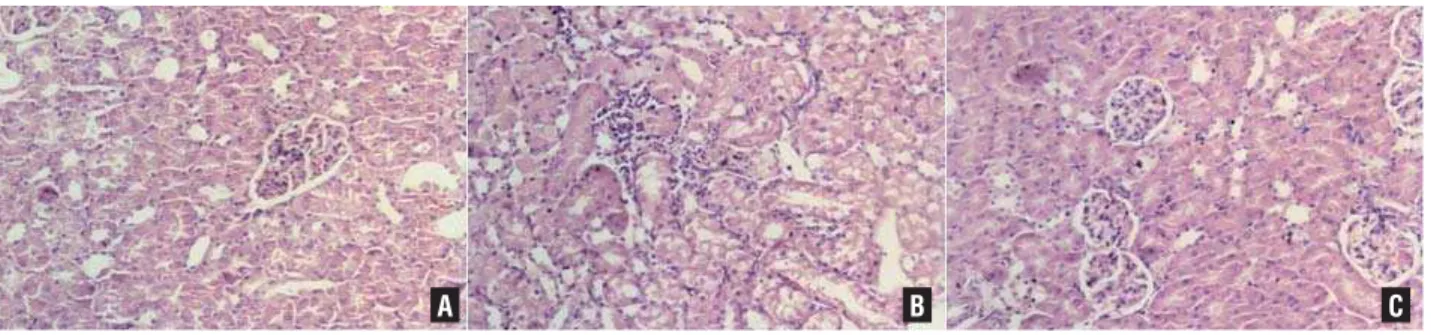
The extent of renal injury was evalua-ted by counting the number of neutrophils in-fi ltration in renal tissue. Three sections were randomly selected for counting the number of neutrophils in fi ve adjacent high-power fi elds. The average was defi ned as the number of positi-ve cells (neutrophils) for each detection indicator (Figure-1).
Statistical analysis
SPSS was used to analyze all data. The results were expressed as mean ± SD. Statistical signifi cance of differences among groups (defi ned as P < 0.05) was evaluated using one-way analy-sis of variance (ANOVA) with Bonferoni correc-tion and Student’s t-test.
Figure 1 - The results for the histological examination. A) Control; B) IR pathology; C) IP.
RESULTS
Changes in ICAM-1 expression after renal IP
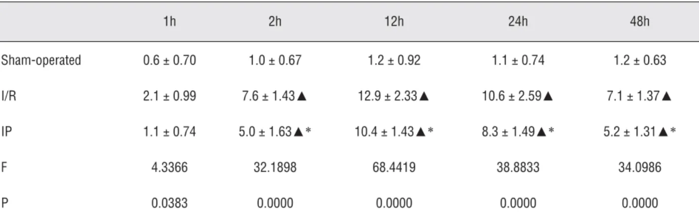
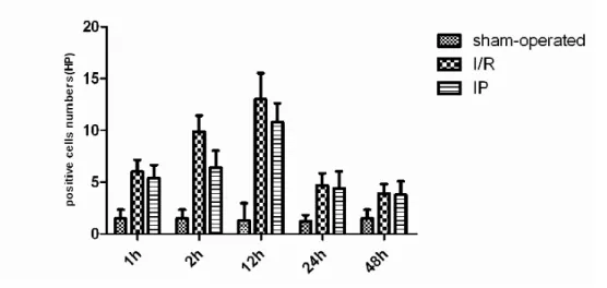
A few positive cells were observed in the sham-operated group. Immunohistochemis-try showed that renal ICAM-1 expression was enhanced after ischemia/reperfusion (Figure-2). The number of ICAM-1-positive cells increased at 2 hours after ischemia/reperfusion and peaked at 12 hours. A downward trend was observed at 24 hours. The expression of ICAM-1 was main-ly observed in the glomerular endothelial cells, peritubular capillaries, and renal proximal tubule epithelial cells. After IP, the number of ICAM-1--positive cells decreased correspondingly. The ex-pression level of ICAM-1 in the IP group was less than that in the I/R group at each time point (p < 0.05; Table-1 and Figure-3).
Changes in P-Selectin expression after renal IP
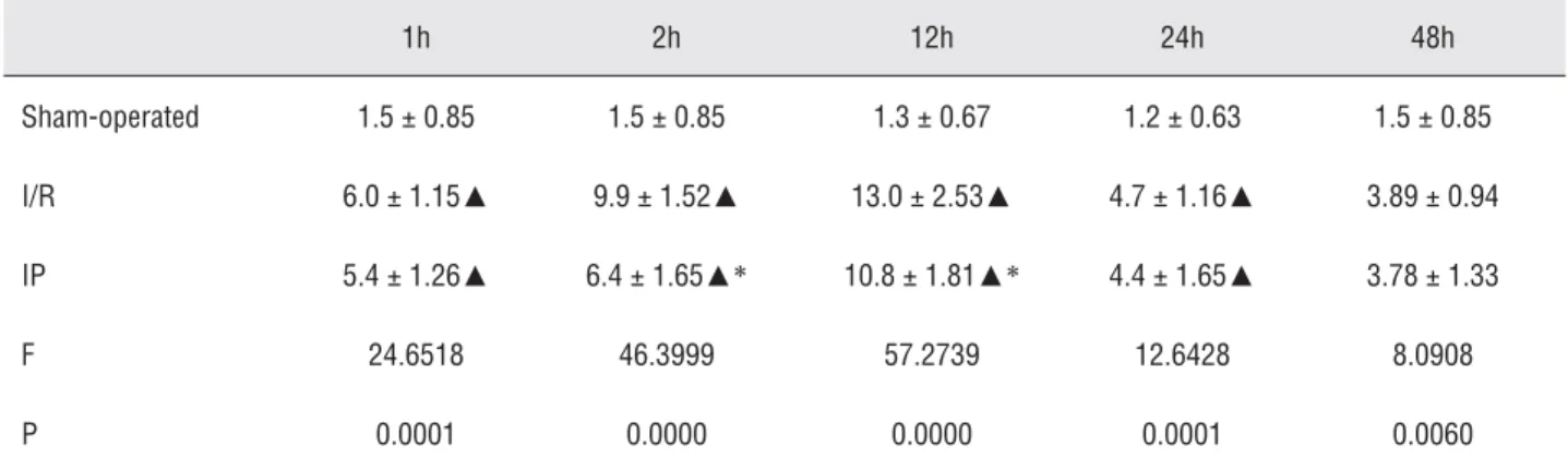
The number of positive cells in the sham--operated group was extremely few (Figure-4). One hour after ischemia/reperfusion, P-Selectin was widely expressed in the kidney, particularly in the glomerular mesangial, capillary loop, tubu-lar, and renal interstitial sections. The expression was most signifi cant in the renal tubular epithelial cells. It peaked 12 hours later and then declined. P-Selectin expression decreased signifi cantly after IP (p < 0.05; Table-2 and Figure-5).
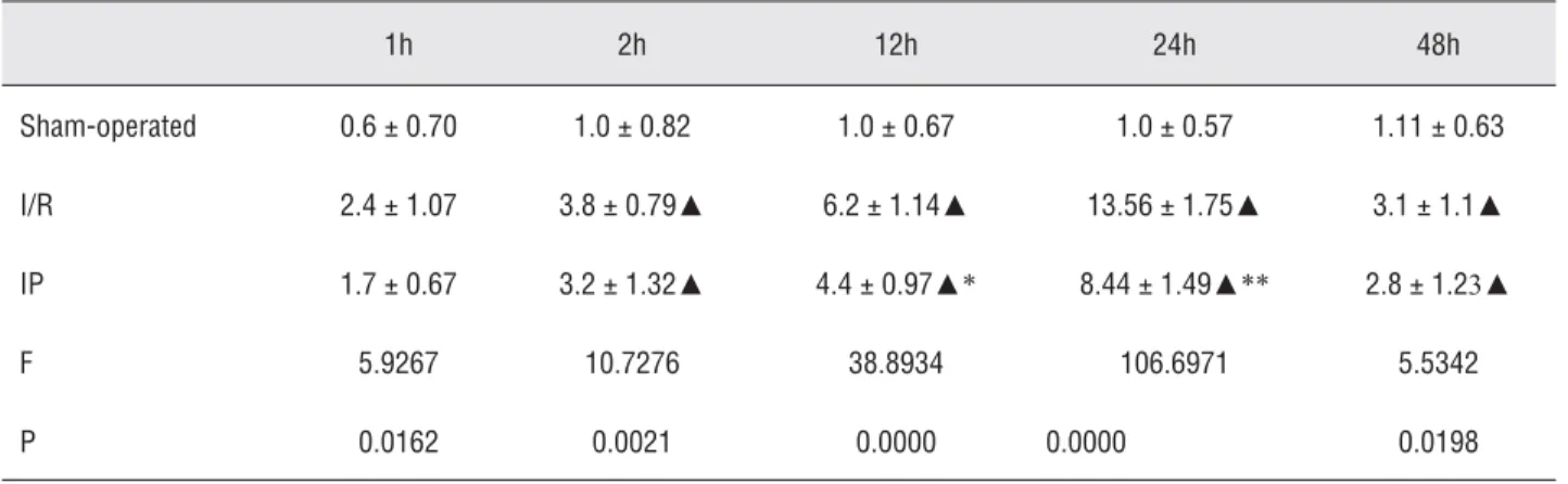
Changes in E-Selectin expression after renal IP
E-Selectin was rarely expressed in the sham-operated group (Figure-6). Two hours after ischemia/reperfusion, the cells expressing E-Selectin increased, specifi cally those in the peritubular capillaries. The expression peaked at
Figure 2 - Histology fi gures of ICAM-1 expression in the three groups. A) Control; B) IR; C) IP.
A B C
Table 1 - Effects of ischemic preconditioning on the expression of ICAM-1 in rat kidney (positive cell number/HP).
1h 2h 12h 24h 48h
Sham-operated 0.6 ± 0.70 1.0 ± 0.67 1.2 ± 0.92 1.1 ± 0.74 1.2 ± 0.63
I/R 2.1 ± 0.99 7.6 ± 1.43▲ 12.9 ± 2.33▲ 10.6 ±2.59▲ 7.1 ± 1.37▲
IP 1.1 ± 0.74 5.0 ± 1.63▲* 10.4 ± 1.43▲* 8.3 ± 1.49▲* 5.2 ± 1.31▲*
F 4.3366 32.1898 68.4419 38.8833 34.0986
P 0.0383 0.0000 0.0000 0.0000 0.0000
Figure 4 - Histology fi gures of P-Selectin expression in the three groups. A) Control; B) IR; C) IP. Figure 3 - Effects of ischemic preconditioning on the expression of ICAM-1 in rat kindney.
A B C
Table 2 - Effects of ischemic preconditioning on the expression of P-Selectin in rat kidney (positive cell number/HP).
1h 2h 12h 24h 48h
Sham-operated 1.5 ± 0.85 1.5 ± 0.85 1.3 ± 0.67 1.2 ± 0.63 1.5 ± 0.85
I/R 6.0 ± 1.15▲ 9.9 ± 1.52▲ 13.0 ± 2.53▲ 4.7 ± 1.16▲ 3.89 ± 0.94
IP 5.4 ± 1.26▲ 6.4 ± 1.65▲* 10.8 ± 1.81▲* 4.4 ± 1.65▲ 3.78 ± 1.33
F 24.6518 46.3999 57.2739 12.6428 8.0908
P 0.0001 0.0000 0.0000 0.0001 0.0060
24 hours and then declined gradually. The number of positive cells in the IP group was signifi cantly reduced (p < 0.05; Table-3 and Figure-7).
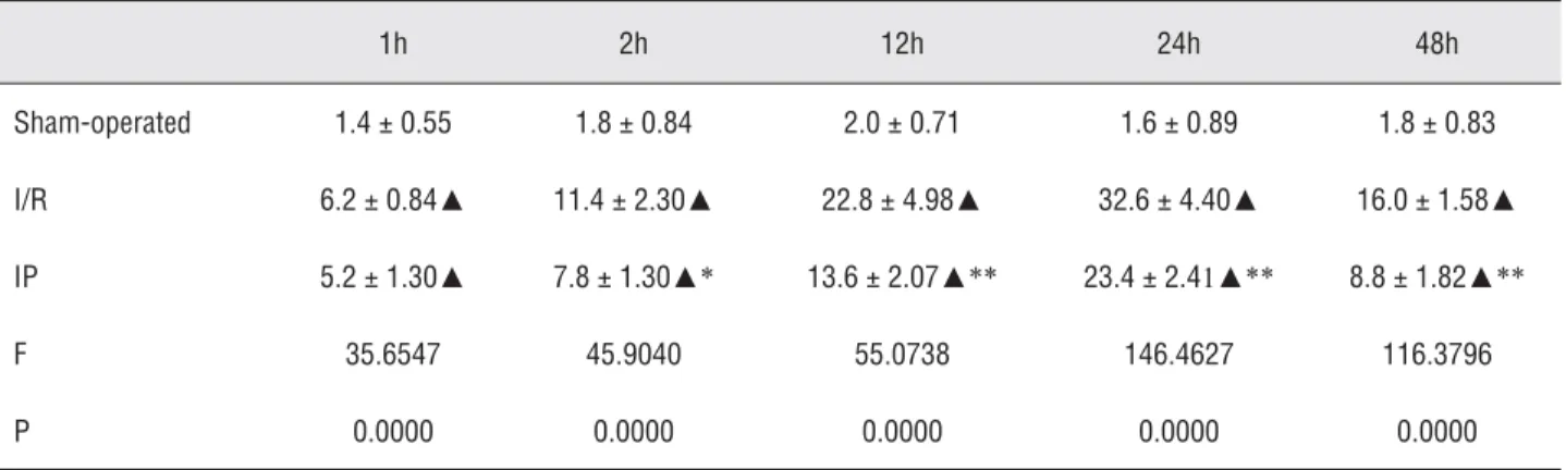
Changes of leukocyte infi ltration in renal tissues
One hour after ischemia/reperfusion, the number of leukocyte infi ltration increased ob-viously. The number peaked at 24 hours and was signifi cantly reduced at 48 hours. The infi ltration occured mainly in vascular, interstitial and tubu-lar areas of the kidney. Compared with those in IR group, the number of leukocyte infi ltration was signifi cantly reduced, especially at 24 hours (p < 0.05; Table-4 and Figure-8).
Renal ischemia/reperfusion and changes in renal function after IP
Serum creatinine levels increased after renal ischemia/reperfusion. In the fi rst 24 hours,
serum creatinine level was signifi cantly higher than at 1, 2, 6, and 12 hours. ARF (acute renal failure) occurred when the creatinine level reached 140 µmoL/L at the fi rst 48 hours. By contrast, the creatinine level in the IP group also increased but at a slower rate. Compared with each correspon-ding time point of IP, no signifi cant difference was observed at the fi rst 1, 2, 12, and 24 hours. Com-pared with the IP group at the fi rst 48 hours, the difference was signifi cant (p < 0.05;Table-5 and Figure-9).
DISCUSSION
Preconditioning is a tolerance or adap-tability to a secondary stimulus exhibited by or-ganisms and organs after primary exposure to a stimulus. This phenomenon known as IP is com-mon in biology. Organs can stand a longer time of
Figure 6 - Histology fi gures of E-Selectin expression in the three groups. A) Control; B) IR; C) IP. Figure 5 - Effects of ischemic preconditioning on the expression of P-Selectin in rat kidney.
warm ischemia after brief ischemia/reperfusion. In 1986, Murry fi rst observed the IP phenome-non in the heart (4). In later studies, many organs such as skeletal muscle have been demonstrated to exhibit IP (6).
In relation to renal preconditioning in a rat model, since the new concept of IP was proposed by Murry and protective effect of IP has been confi rmed in cardiac ischemia-reperfusion injury, many scholars have established the heart IP model and explored its mechanism. If different preconditioning program was used in the renal IP model, the conclusions could not have been the
same. Preconditioning involved the sequential clamping of the left renal artery for 4 min. and its release for 11 min., a total of four times. The results of the above preconditioning program showed that no protection to kidney I/R injury was observed (21). However, Toosy et al. reported that an IP regimen applied only 5 min. before exposure to a 40 min. period of RI in the rat signifi cantly protected it from the functional impairment associated with kidney I/R (22). Furthermore, the results of an IP regimen (three 2 min. periods of ischemia separated by 5 min. of refl ow prior to 45 min. of bilateral renal ischemia followed by
Figure 7 - Effects of ischemic preconditioning on the expression of E-Selectin in rat kidney.
Table 3 - Effects of ischemic preconditioning on the expression of E-Selectin in rat kidney (positive cell number/HP).
1h 2h 12h 24h 48h
Sham-operated 0.6 ± 0.70 1.0 ± 0.82 1.0 ± 0.67 1.0 ± 0.57 1.11 ± 0.63
I/R 2.4 ± 1.07 3.8 ± 0.79▲ 6.2 ± 1.14▲ 13.56 ± 1.75▲ 3.1 ± 1.1▲
IP 1.7 ± 0.67 3.2 ± 1.32▲ 4.4 ± 0.97▲* 8.44 ± 1.49▲** 2.8 ± 1.23▲
F 5.9267 10.7276 38.8934 106.6971 5.5342
P 0.0162 0.0021 0.0000 0.0000 0.0198
Figure 8 - Changes of leucocyte Infi ltrated into renal parenchyma after ischemic preconditioning. Table 4 - Changes of leucocyte Infi ltrated into renal parenchyma after ischemic preconditioning.
1h 2h 12h 24h 48h
Sham-operated 1.4 ± 0.55 1.8 ± 0.84 2.0 ± 0.71 1.6 ± 0.89 1.8 ± 0.83
I/R 6.2 ± 0.84▲ 11.4 ± 2.30▲ 22.8 ± 4.98▲ 32.6 ± 4.40▲ 16.0 ± 1.58▲
IP 5.2 ± 1.30▲ 7.8 ± 1.30▲* 13.6 ± 2.07▲** 23.4 ± 2.41▲** 8.8 ± 1.82▲**
F 35.6547 45.9040 55.0738 146.4627 116.3796
P 0.0000 0.0000 0.0000 0.0000 0.0000
▲P < 0.01 vs. control; * P < 0.05<** P < 0.01 vs. I/R
Table 5 - Changes of serum creatinine after renal ischemic preconditioning (umol/L).
1h 2h 12h 24h 48h
Sham-operated 57.0 ± 3.16 54 ± 4.18 62.4 ± 2.65 60.6 ± 4.22 61.8 ± 5.67
I/R 49.4 ± 2.79 55.4 ± 5.59 58.6 ± 5.98 79.8 ± 7.6 139.0 ± 11.55▲
IP 52.2 ± 5.07 54.6 ± 6.10 59.2 ± 7.85 75.2 ± 7.99 108.0 ± 8.86▲*
F 5.0972 0.0861 0.5996 10.8128 92.7601
P 0.0250 0.9180 0.5647 0.0021 0.0000
24 hours of reperfusion) applied by Cochrane et al. found that IP would ameliorate ischemic renal injury (23). The preliminary experiment of preconditioning method in our study found that the protective effect of prolongation of reperfusion time on renal I/R injury was not that obvious. The results of our preliminary experiment suggested that if the time and frequency of ischemia were fi xed in a repeated cycle of short-time IR the protective effect on kidney would decline with the prolongation of the reperfusion interval. When a certain critical point is exceeded, such as fi xed four 6 min. periods of ischemia separated by more than 10 min. of reperfusion, the role of IP protection would be lost. IP regimen with multiple cycles of short-period ischemia followed by short-period reperfusion had a signifi cant protective effect on kidney I/R injury. At the same time, according to our previous experimental experience with I/R injury, an IP regimen applied 6 min. period of ischemia followed by 48 hours period of reperfusion was the most appropriate method to observe the renal function. Therefore, based on the previous study of the kidney IP model, the following modifi ed IP regimen was used in the present study: the sequential clamping of the renal artery for 6 min. and its release for 4 min., a total of four times; 60 min. of renal ischemia followed by 48 hours of reperfusion. The results of our study showed that this IP regimen
may signifi cantly reduce the incidence of renal I/R injury, suggesting that the experimental animal model was successfully established and may provide a reliable preadaptation program for further studies. According the observations of a preliminary study, changes of experimental data at 1, 2, 12, 24 and 48 hours after IP were obvious. Therefore, these time points were chosen as the observation point. Because one of the aims was to provide experimental data of IP protection for the kidney transplant, no prolonged observation time and the protective effect of later IP were further investigated in the present study.
Most studies have been limited to the ma-croscopic study of the protective effect of renal function in terms of the renal IP phenomenon. Wever conducted a meta-analysis of animal expe-rimental studies on renal IP (24). Creatinine, urea nitrogen, and histological changes were used as indicators of renal IP. Meta-analysis showed that compared with the control group, serum creati-nine and blood urea nitrogen levels decreased signifi cantly and changes in the renal tissue was less important in the IP group. No difference was observed between local and distant preconditio-ning on the protective effect of renal ischemia/ reperfusion. IP signifi cantly improved kidney I/R injury, especially in the advanced window period ( 24 hours). Nevertheless, signifi cant differen-ces were found between studies in terms of the
results of the best protection window. The effect of IP in each animal species varies. Indisputably, renal IP has protective effects to subsequent ische-mic/reperfusion injury. However, the mechanism remains unclear. Mahfoudh-Boussaid (25) used Wistar rats to study IP and found that renal IP re-duces cell lysis and lipid peroxidation and impro-ves renal function. Compared with the ischemia/ reperfusion group, the IP group showed elevated levels of endothelial nitric oxide synthase, nitri-te, and hypoxia inducible transcription factor-1α.
Early preconditioning through reduction of oxi-dative and endoplasmic reticulum stress has been believed to protect against kidney I/R injury. Kim (26) believed that the effect of IP on the renal pro-tective effect is related to the increase in isocitra-te dehydrogenase (IDH1) activity. IDH1 enhanced NADPH levels, elevated the GSH-to-total gluta-thione ratio, and reduced oxidative stress. Chen (3) believed that NF-kappaB is the key medium to reperfusion injury. The activation of NF-kappaB depended on the phosphorylation of its inhibitor. The inhibitor was IkappaB, which had a specifi c NF-kappaB kinase subunit. Animal experiments confi rmed that renal IP reduces renal acute injury by inhibiting IKKbeta activity. The IP protocol adopted in this experiment involved four repeti-tions of warm ischemia for 6 min. and blood fl ow for 4 min. Under the condition of 1 hour warm ischemia and 48 hours perfusion, IP signifi cantly reduced the expressions of renal adhesion mole-cules ICAM-1, P-Selectin, and E-Selectin. At the same time, granulocyte infi ltration in renal tissue after IP was signifi cantly lower than that in the I/R group. Adhesion molecules are the major molecu-les which mediate adhesion of polymorphonuclear leukocytes to endothelial cells. Because of reduced expression of adhesion molecules after IR, adhe-sion of granulocytes to endothelial cells decreased leading to reduction of granulocyte infi ltration in renal tissue. The serum creatinine level of the rats was signifi cantly lower than that of the I/R group. This result proves the explicit protective effect of IP. The mechanism of kidney protection was clo-sely related to the reduction of adhesion molecule expression caused by IP.
We believe that the reduction of adhesion molecule expression is one of the main
when ischemia occurred, expression of adhesion molecules increased with the increasing secre-tion of TNF-α (29) and IL-1 in macrophages and monocytes (30). Increased expression of ICAM-1 induced by IL-1β and TNF-α may lead to an in-crease of leukocyte infi ltration in kidney and sy-nergistically aggravating kidney I/R injury. Zhou (31) used rats to study renal ischemic/reperfusion. He discovered that after ischemia/reperfusion, ICAM-1 expression in renal vascular endothelial cells and P-Selectin expression in renal tubular epithelial cells increase. If anti-P-Selectin antibo-dy was given, kidney I/R injury could be reduced, confi rming the important role of ICAM-1 and P--Selectin in kidney I/R injury. The expression of adhesion molecules could be signifi cantly reduced through IP. The results confi rmed that precondi-tioning had a protective effect on reperfusion kid-neys. The data in another study confi rmed that ischemic preconditioning could prevent postische-mic P-selectin expression in the rat small intes-tine (32). The expression of ICAM-1 of pig lung tissue obviously decreased in group IP than that in control group (33). The study on protective me-chanism of ischemic preconditioning in cultured rat endothelial cells found that changes of post--ischemic expression of adhesion molecules such as ICAM-1 may be related to activation of pro-tein kinase C and production of nitric oxide and free radicals. And this is associated with a lesser adhesion of neutrophils to endothelial cells. Such prevention of neutrophil adhesion may contribute to the protective effect of preconditioning against reperfusion-induced endothelial injury (34). Simi-lar to other methods of blocking the expression of adhesion molecules, we reconfi rmed that the increase in adhesion molecule expression plays an important role in kidney I/R injury. Remote ische-mic preconditioning can signifi cantly reduce the risk of AKI in patients undergoing cardiopulmo-nary bypass-assisted cardiac surgery (35). Howe-ver, the data in some studies indicated that the effect of ischemic preconditioning had limitations and uncertainty. Young et al. reported that 96 adults undergoing high-risk cardiac surgery were randomized to group of remote ischemic precon-ditioning or control. Main endpoints were plasma high-sensitivity troponin T (hsTNT) levels at 6 and
12 hours, worst post-operative acute kidney injury (AKI). The results showed that there were no sig-nifi cant differences between the experimental and control group, suggesting that IP does not reduce hsTNT, AKI in high-risk cardiac surgery (36).
Compared with other methods that block a certain type of adhesion molecule alone, IP is a much more comprehensive method that can reduce a variety of adhesion molecules with higher preci-sion and reliability. The protective effect of whole kidney can be achieved by ischemia/reperfusion, even the protective effect of other organs can be achieved by controlling infl ammatory cytokines. It can also reduce the incidence of multiple organ dysfunction syndrome, which can help meet the needs of clinical and practical use. Unfortunate-ly, some meaningful means of detection can not be performed because of our limited laboratory conditions, making the experimental results in the present study have certain limitations. There also has a certain gap with the complex situation in the actual clinical environment of human body and such fi ndings acquired in the ideal conditions in rats need to further validation at the appropriate internal environment of the human body.
CONFLICT OF INTEREST
None declared.
REFERENCES
1. Yin DP, Sankary HN, Chong AS, Ma LL, Shen J, Foster P, et al.: Protective effect of ischemic preconditioning on liver preservation-reperfusion injury in rats. Transplantation. 1998; 66: 152-7.
2. Hausenloy DJ, Yellon DM: Preconditioning and postcon-ditioning: underlying mechanisms and clinical application. Atherosclerosis. 2009; 204: 334-41.
3. Chen X, Liu X, Wan X, Wu Y, Chen Y, Cao C: Ischemic pre-conditioning attenuates renal ischemia-reperfusion injury by inhibiting activation of IKKbeta and infl ammatory re-sponse. Am J Nephrol. 2009; 30: 287-94.
5. Przyklenk K, Bauer B, Ovize M, Kloner RA, Whittaker P: Regional ischemic ‘preconditioning’ protects remote virgin myocardium from subsequent sustained coronary occlu-sion. Circulation. 1993; 87: 893-9.
6. Hopper RA, Forrest CR, Xu H, Zhong A, He W, Rutka J, et al.: Role and mechanism of PKC in ischemic precondition-ing of pig skeletal muscle against infarction. Am J Physiol Regul Integr Comp Physiol. 2000; 279: R666-76.
7. Liu X, Chen H, Zhan B, Xing B, Zhou J, Zhu H, et al.: Attenu-ation of reperfusion injury by renal ischemic postcondition-ing: the role of NO. Biochem Biophys Res Commun. 2007; 359: 628-34.
8. Safi an RD, Textor SC: Renal-artery stenosis. N Engl J Med. 2001; 344: 431-42.
9. Schrier RW, Wang W: Acute renal failure and sepsis. N Engl J Med. 2004; 351: 159-69.
10. Ojo AO, Wolfe RA, Held PJ, Port FK, Schmouder RL: De-layed graft function: risk factors and implications for renal allograft survival. Transplantation. 1997; 63: 968-74. 11. Perico N, Cattaneo D, Sayegh MH, Remuzzi G: Delayed
graft function in kidney transplantation. Lancet. 2004; 364: 1814-27.
12. Ojo AO, Held PJ, Port FK, Wolfe RA, Leichtman AB, Young EW, et al.: Chronic renal failure after transplantation of a nonrenal organ. N Engl J Med. 2003; 349: 931-40. 13. Cochrane J, Williams BT, Banerjee A, Harken AH, Burke TJ,
Cairns CB, et al.: Ischemic preconditioning attenuates func-tional, metabolic, and morphologic injury from ischemic acute renal failure in the rat. Ren Fail. 1999; 21: 135-45. 14. Wever KE, Warlé MC, Wagener FA, van der Hoorn JW,
Masereeuw R, van der Vliet JA, et al.: Remote ischaemic preconditioning by brief hind limb ischaemia protects against renal ischaemia-reperfusion injury: the role of ad-enosine. Nephrol Dial Transplant. 2011; 26: 3108-17. 15. Wever KE, Menting TP, Rovers M, van der Vliet JA,
Ron-gen GA, Masereeuw R, et al.: Ischemic preconditioning in the animal kidney, a systematic review and meta-analysis. PLoS One. 2012; 7: e32296.
16. Zhang XL, Selbi W, de la Motte C, Hascall V, Phillips A: Re-nal proximal tubular epithelial cell transforming growth fac-tor-beta1 generation and monocyte binding. Am J Pathol. 2004; 165: 763-73.
17. Feng L, Ke N, Cheng F, Guo Y, Li S, Li Q, et al.: The pro-tective mechanism of ligustrazine against renal ischemia/ reperfusion injury. J Surg Res. 2011; 166: 298-305. 18. Satoh S, Suzuki A, Asari Y, Sato M, Kojima N, Sato T, et
al.: Glomerular endothelium exhibits enhanced expression of costimulatory adhesion molecules, CD80 and CD86, by warm ischemia/reperfusion injury in rats. Lab Invest. 2002; 82: 1209-17.
19. Zhou T, Li X, Wu P, Zhang D, Zhang M, Chen N, et al.: Effect of anti-P-selectin monoclonal antibody on renal ischemia/ reperfusion injury in rats. Chin Med J (Engl). 2000; 113: 790-3.
20. Ogawa T, Nussler AK, Tuzuner E, Neuhaus P, Kaminishi M, Mimura Y, et al.: Contribution of nitric oxide to the protec-tive effects of ischemic preconditioning in ischemia-reper-fused rat kidneys. J Lab Clin Med. 2001; 138: 50-8. 21. Islam CF, Mathie RT, Dinneen MD, Kiely EA, Peters AM,
Grace PA: Ischaemia-reperfusion injury in the rat kidney: the effect of preconditioning. Br J Urol. 1997; 79: 842-7. 22. Toosy N, McMorris EL, Grace PA, Mathie RT: Ischaemic
preconditioning protects the rat kidney from reperfusion injury. BJU Int. 1999; 84: 489-94.
23. Cochrane J, Williams BT, Banerjee A, Harken AH, Burke TJ, Cairns CB, et al.: Ischemic preconditioning attenuates func-tional, metabolic, and morphologic injury from ischemic acute renal failure in the rat. Ren Fail. 1999; 21: 135-45. 24. Wever KE, Menting TP, Rovers M, van der Vliet JA,
Ron-gen GA, Masereeuw R, et al.: Ischemic preconditioning in the animal kidney, a systematic review and meta-analysis. PLoS One. 2012;7: e32296.
25. Mahfoudh-Boussaid A, Zaouali MA, Hadj-Ayed K, Miled AH, Saidane-Mosbahi D, Rosello-Catafau J, et al.: Ischemic preconditioning reduces endoplasmic reticulum stress and upregulates hypoxia inducible factor-1α in ischemic kid-ney: the role of nitric oxide. J Biomed Sci. 2012; 19: 7. 26. Kim J, Kim JI, Jang HS, Park JW, Park KM: Protective role
of cytosolic NADP(+)-dependent isocitrate dehydrogenase, IDH1, in ischemic pre-conditioned kidney in mice. Free Radic Res. 2011; 45: 759-66.
27. Koo DD, Welsh KI, West NE, Channon KM, Penington AJ, Roake JA, et al.: Endothelial cell protection against isch-emia/reperfusion injury by lecithinized superoxide dis-mutase. Kidney Int. 2001; 60: 786-96.
28. Singbartl K, Ley K: Protection from ischemia-reperfusion induced severe acute renal failure by blocking E-selectin. Crit Care Med. 2000; 28: 2507-14.
29. Donnahoo KK, Meng X, Ayala A, Cain MP, Harken AH, Mel-drum DR: Early kidney TNF-alpha expression mediates neutrophil infi ltration and injury after renal ischemia-reper-fusion. Am J Physiol. 1999; 277: R922-9.
30. Savage CO, Brooks CJ, Adu D, Richards G, Howie AJ: Cell adhesion molecule expression within human glomerular and kidney organ culture. J Pathol. 1997; 181: 111-5. 31. Zhou T, Sun GZ, Zhang MJ, Chen JL, Zhang DQ, Hu QS, et
32. Davis JM, Gute DC, Jones S, Krsmanovic A, Korthuis RJ: Ischemic preconditioning prevents postischemic P-selectin expression in the rat small intestine. Am J Physiol. 1999; 277: H2476-81.
33. Zhang CF, Chen SX, Guo HZ, Luo WJ: Protective mecha-nism of ischemic preconditioning to the lung ischemia-reperfusion injury. Zhong Nan Da Xue Xue Bao Yi Xue Ban. 2005; 30: 64-7.
34. Beauchamp P, Richard V, Tamion F, Lallemand F, Lebreton JP, Vaudry H, et al.: Protective effects of preconditioning in cultured rat endothelial cells: effects on neutrophil adhe-sion and expresadhe-sion of ICAM-1 after anoxia and reoxygen-ation. Circulreoxygen-ation. 1999; 100: 541-6.
35. Ferraro PM, Gambaro G: Ischemic preconditioning and the risk of acute kidney injury. Kidney Int. 2012; 82: 243; au-thor reply 243.
36. Young PJ, Dalley P, Garden A, Horrocks C, La Flamme A, Mahon B, et al.: A pilot study investigating the effects of re-mote ischemic preconditioning in high-risk cardiac surgery using a randomised controlled double-blind protocol. Basic Res Cardiol. 2012; 107: 256.
_____________________
Correspondence address: