Epilepsy-induced electrocardiographic
alterations following cardiac ischemia and
reperfusion in rats
J.G.P. Tavares
1,2,3, E.R. Vasques
4, R.M. Arida
5, E.A. Cavalheiro
6, F.R. Cabral
7,
L.B. Torres
7, F.S. Menezes-Rodrigues
1, A. Jurkiewicz
1, A. Caricati-Neto
1,
C.M.G. Godoy
8and S. Gomes da Silva
7,9 1Departamento de Farmacologia, Universidade Federal de Sa˜o Paulo, Sa˜o Paulo, SP, Brasil 2Universidade Iguac¸u, Campos V, Itaperuna, RJ, Brasil 3Faculdade de Minas, Muriae´, MG, Brasil 4Departamento de Gastroenterologia, LIM 37, Faculdade de Medicina, Universidade de Sa˜o Paulo, Sa˜o Paulo, SP, Brasil 5Departamento de Fisiologia, Universidade Federal de Sa˜o Paulo, Sa˜o Paulo, SP, Brasil 6Departamento de Neurologia e Neurocirurgia, Universidade Federal de Sa˜o Paulo, Sa˜o Paulo, SP, Brasil 7Hospital Israelita Albert Einstein, Instituto do Ce´rebro, Sa˜o Paulo, SP, Brasil 8Departamento de Cieˆncia e Tecnologia, Universidade Federal de Sa˜o Paulo, Sa˜o Jose´ dos Campos, SP, Brasil 9Nu´cleo de Pesquisas Tecnolo´gicas, Programa Integrado em Engenharia Biome´dica,Universidade de Mogi das Cruzes, Mogi das Cruzes, SP, Brasil
Abstract
The present study evaluated electrocardiographic alterations in rats with epilepsy submitted to an acute myocardial infarction (AMI) model induced by cardiac ischemia and reperfusion. Rats were randomly divided into two groups: control (n=12) and epilepsy (n=14). It was found that rats with epilepsy presented a significant reduction in atrioventricular block incidence following the ischemia and reperfusion procedure. In addition, significant alterations were observed in electrocardiogram intervals during the stabilization, ischemia, and reperfusion periods of rats with epilepsy compared to control rats. It was noted that rats with epilepsy presented a significant increase in the QRS interval during the stabilization period in relation to control rats (P,0.01). During the ischemia period, there was an increase in the QRS interval (P,0.05) and a reduction in the P wave and QT intervals (P,0.05 for both) in rats with epilepsy compared to control rats. During the reperfusion period, a significant reduction in the QT interval (P,0.01) was verified in the epilepsy group in relation to the control group. Our results indicate that rats submitted to an epilepsy model induced by pilocarpine presented electrical conductivity alterations of cardiac tissue, mainly during an AMI episode.
Key words: Epilepsy; Heart; Acute myocardial infarction; Ischemia; Reperfusion
Introduction
Epilepsy is the most common chronic neurological disorder, and people with epilepsy are two to three times more likely to die prematurely when compared with people without epilepsy (1-3). The most common epilepsy-related category of death is sudden unexpected death in epilepsy (SUDEP) (3,4), a leading cause of mortality in patients with epilepsy. Although the cause of SUDEP is still unknown, one of the main mechanisms of SUDEP is autonomic dysregulation (i.e., cardiac abnormalities during and between seizures) (5-7). Suspected mechanisms include cardiac arrhythmia, central and/or obstructive apnea, neurogenic pulmonary edema, and primary cessation of
brain activity (3,8-11). In fact, clinical seizures are often accompanied by intense autonomic changes, and there is evidence that sympathetic activity is increased even interictally in patients with temporal lobe epilepsy (10,12,13). Ictal tachycardia is the more frequent arrhyth-mia (14,15), but ictal bradycardias, high-grade atrioven-tricular block, and asystole are also observed (10,16,17). Curiously, ictal bradyarrhythmia also appears to be associated with a higher risk of SUDEP (10,18). Several case reports have described asystole or bradycardia during epileptic seizures and have linked these conditions to a higher risk of SUDEP (19,20). Trying to explain these
Correspondence: S. Gomes da Silva:,sgomesilva@hotmail.com.; J.G.P. Tavares:,padrao.tavares@hotmail.com..
cardiac conduction abnormalities, P-Codrea Tigaran et al. (21) performed a prospective setting case-control study of all SUDEP cases in Denmark and verified that the SUDEP cases displayed significant fibrosis of the myocardium. The authors raised a hypothesis that this fibrosis could be the consequence of myocardial ischemia as a direct result of repetitive epileptic seizures, which, associated with the ictal sympathetic storm, may lead to lethal arrhythmias. Other studies also identified that epilepsy might be a risk and an adverse prognostic factor for acute myocardial infarction (AMI) (22,23).
Considering that the mechanisms underlying SUDEP remain uncertain and that AMI risk and prognosis in epilepsy have been poorly explored, it would be useful to obtain, in an animal model, additional information about cardiac alterations associated with epilepsy under AMI conditions. Thus, we evaluatedin vivoventricular arrhyth-mias (VA), atrioventricular block (AVB), and mortality in rats with epilepsy submitted to an AMI model induced by cardiac ischemia and reperfusion.
Material and Methods
Induction of epilepsy
Twenty-six male Wistar rats (280±30 g) were used in this study. All experimental protocols described below were approved by the Ethics Committee of the Universidade Federal de Sa˜o Paulo (#1130/11). The colony room was maintained at 21±26C with a 12:12-h light/dark schedule, and the animals were given food and waterad libitumthroughout the experiments. Rats were randomly divided into 2 groups: control (n=12) and epilepsy (n=14). Rats from the epilepsy group were submitted to a temporal lobe epilepsy model induced by a single administration of pilocarpine hydrochloride (350 mg/kg,ip; Sigma, USA) (24). Scopolamine methylni-trate (1 mg/kg, sc; Sigma) was administered 30 min before pilocarpine injection to limit peripheral cholinergic effects (24). The systemic administration of potent muscarinic agonist pilocarpine in rats promotes sequential behavioral and electrographic changes that build up progressively into a limbicstatus epilepticus (SE), which lasts 24 h. During the SE, two rats died. Following this acute period of the epilepsy model, the surviving animals (n=12) were continuously monitored over 24 h for detection of spontaneous recurrent seizures, using a video system. After the first spontaneous recurrent seizure had been detected, animals were monitored over the following 120 days by two researchers in the experimental room for observation periods ranging from 2 to 4 h. During this period, the only observation was whether seizures were present in the rats (to ensure that they were epileptic rats).
Ischemia and reperfusion protocol
After 120 days, animals from the control and epilepsy
groups were submitted to an AMI model induced by cardiac ischemia and reperfusion similar to that previously described (25). All rats were anesthetized with urethane (1.2 g/kg,ip) and fixed in the supine position. After intubation with a catheter (Jelco 14G, USA), rats were mechanically venti-lated with room air with a stroke volume of approximately 10 ml/kg (,82 breaths min) using a ventilator (Insight EFF
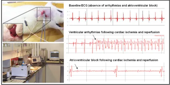
312, Brazil). Under clean dissection, indwelling polyethylene catheters were inserted into a common carotid artery. The body temperature was maintained at 37.56C with a heated operating platform and appropriate heating lamps, and was monitored routinely via a rectal thermometer. The electro-cardiogram (ECG) was recorded using a biopotential amplifier by means of needle electrodes placed subcuta-neously on the limbs. The chest was opened with a left thoracotomy and the heart was gently exteriorized using pressure on the abdomen. A ligature (4/0 braided silk suture attached to a 10-mm micropoint reverse-cutting needle, Ethicon K-890H, USA) was placed around the left anterior descending coronary artery, approximately 2 mm from its origin. A polyethylene tube was threaded over the suture and the heart was replaced in the chest cavity with the ligature ends exteriorized. After a stabilization period of 15 min, the ligature was tied, characterizing the ischemic period, and, following a period of coronary occlusion (10 min), the reperfusion was obtained by cutting the suture (Figure 1). Successful ligation of the coronary artery was validated by ECG alterations (increase in R wave and ST segment) and reduction in arterial pressure (26,27).
Monitoring, recording, and ECG waveform analysis
The ECG of the rats was monitored and recorded from the beginning of the stabilization period using a commercial acquisition system (AqDados 7.02; Lynx Tecnologia Ltda., Brazil). The recorded raw data were evaluated using the commercial software included in the acquisition system (AqDAnalysis 7, Lynx Tecnologia Ltda.; Figure 1). Heart rate as well as incidence of reperfusion-induced VA, AVB, and lethality were measured. To simplify the presentation of the results, ventricular fibrillation, torsades de pointes, and ventricular tachycardia parameters were considered only as VA. ECG measurements were evaluated according to Lambeth conventions (28,29). P duration was manually measured as the time from the beginning of the upstroke of the P wave until its return to the isoelectric baseline. QRS duration was measured from the beginning of the Q wave to the peak amplitude of the downward deflection of the S wave. PR interval was measured from the beginning of the upstroke of the P wave until the maximal amplitude of the R wave. RR interval was measured as the time between consecutive R wave peaks. QT interval was measured from the beginning of the Q wave until the T wave returned to the isoelectric baseline (30).
Data analysis
AVB, and lethality were compared with Fisher’s exact test. Statistical analyses of ECG data were carried out by analysis of variance followed by Bonferroni’s post hoc
test. Values were considered significant when P,0.05. Statistical analyses were performed with the Prism 5.0 software (GraphPad, USA), and data are reported as means±SE.
Results
Both control and epilepsy groups showed no arrhyth-mias during stabilization. The arrhytharrhyth-mias were observed and measured during ischemia and reperfusion (Table 1). No significant difference was detected in the incidence of VA and lethality between studied groups [VA inci-dence=83.3% in the control group (10/12) and 75% in the epilepsy group (9/12) P=1.00; lethality=66.7% in the control group (8/12) and 58.3% in the epilepsy group (7/12) P=1.00]. However, the incidence of AVB was significantly lower in the epilepsy group [25% (3/12)] than in the control group [83.3% (10/12) P=0.012; Table 1].
In the ECG waveform analysis, it was noted that rats with epilepsy presented a significant increase in the QRS interval during the stabilization period in relation to control rats (P,0.01; Figure 2). During the ischemia period, a significant increase in the QRS interval (P,0.05) and
reduction in P wave and QT intervals (P,0.05 in both) were found in rats from the epilepsy group compared to rats from the control group. During the reperfusion period, a significant reduction in the QT interval (P,0.01) was verified in the epilepsy group in relation to the control group.
Discussion
The present study evaluated electrocardiographic alterations in rats with epilepsy submitted to an AMI model induced by cardiac ischemia and reperfusion. Rats with epilepsy presented a significant reduction in AVB incidence following the ischemia and reperfusion proce-dure. In addition, significant alterations were observed in ECG intervals during the stabilization and ischemia and reperfusion periods of rats with epilepsy compared to control rats. Our results indicated that the rats submitted to an epilepsy model induced by pilocarpine presented electrical conductivity alterations of the cardiac tissue, mainly during an AMI episode.
During the stabilization period, no abnormality in P wave or PR, RR, and QT intervals was observed in rats with epilepsy. However, a significant increase in the interval of the QRS complex was noted in these animals, which is indicative of conduction abnormalities. This result showed that the period of ventricular depolarization is
Figure 1.Images of the experiment with ischemia and reperfusion and ECG measurements.
Table 1. Incidence of ventricular arrythmias (VA), atrioventricular block (AVB) and lethality during reperfusion in 12 rats in each group.
Groups VA AVB Lethality
Control 10/12 10/12 8/12
Epilepsy 9/12 3/12* 7/12
already altered in the epilepsy group following stabilization. This alteration could be related to different factors, such as pathological fibrosis of the myocardium, morphological abnormalities of the cardiac conduction system, or an inappropriate autonomous nervous system response (3). Indeed, some studies have suggested that SUDEP cases in humans displayed significant fibrosis of the myocardium, which is known to be the substrate leading to discontinuous propagation and spatial dispersion of cardiac conduction (31,32). Although no pathological analyses have been performed in the present study, our experimental finding is in agreement with reports showing a widening QRS complex as a common cardiac abnormality in patients with epilepsy (33).
During the ischemia and reperfusion periods, signifi-cant alterations in PR and RR intervals between studied groups were not observed. However, changes in the incidence of AVB and in the P, QRS, and QT intervals were found during the periods of ischemia and/or reperfusion in animals with epilepsy. These results show that the period of depolarization is different between animals with epilepsy and controls in conditions of tissue injury. We cannot explain the exact mechanism of the decrease in AVB incidence in epileptic rats. However, it might be related, at least in part, to the KV 1.1 potassium channel, the deficiency of which leads to aberrant parasympathetic neurotransmission and consists of a candidate mechanism for AVB in epileptic animals (12). In the control group, increases in the duration of cardiac atrial or ventricular electrical activities (P wave and QT interval) occur during periods of ischemia and reperfusion compared to the stabilization period. In contrast, these enlargements, which could lead to atrial fibrillation or
ventricular arrhythmias, such as torsades de pointes, were not observed in the epilepsy group during ischemia and reperfusion, leading to a significant difference between groups. This result is interesting and intriguing. It is possible that this particular ECG alteration in rats with epilepsy is related to a compensatory cardiac mechanism. Another possible explanation could be attributed to the seizure effects on gene expression. The molecular mechanism for the malignant brain-heart relationship has remained elusive until the recent discovery of arrhythmogenic mutations. Mutations in potassium (KCNH2, KCNQ1, KCNJ2) and calcium channel subunit genes have been identified to cause short QT syndrome (SQTS) (34), which is a rare, sporadic, or autosomal dominant disorder characterized by markedly accelerated cardiac repolarization and is manifested by a dramatically shortened QT interval, atrial and ventricular arrhythmias, and sudden cardiac death. Although it has been recog-nized for some time that prolongation of cardiac repolar-ization is a substrate for arrhythmias, the link between accelerated repolarization and arrhythmia has only recently been established (34).
Considering that variations in ventricular action poten-tial can lead to increased or decreased QT interval and arrhythmias, it has been established that QT dispersion is a significant predictor of cardiovascular mortality (5,35). In our study in an animal model, a clear correlation between QT enlargement and high incidence of VA and deaths was found in the control group during ischemia and reperfusion. Although the high incidence of mortality in both epilepsy and control groups was noted, the lower QT interval did not modify the mortality incidence in the epilepsy group during ischemia and reperfusion, demonstrating that epilepsy did
Figure 2.Duration of P wave, and PR, QRS, QT and RR intervals in rats from the control and epilepsy groups during stabilization (Stab), ische-mia (Isch) and reperfusion (Rep). *P,0.05, compared to the control group;#P
not represent a high risk of lethal arrhythmias in this model. Nevertheless, it is important to point out that the period of injury has been greatly extended (10 min of ischemia) in our experiments in order to observe significant differences in lethality between the groups. It is probable that a less aggressive stimulus could reveal more subtle differences. Despite this, conductivity alterations of the cardiac tissue were detected in rats with epilepsy following an ischemia and reperfusion protocol. Our results suggest that epilepsy
can result in electrical conductivity alterations of the cardiac tissue, mainly during an AMI episode.
Acknowledgments
Research supported by CNPq, FAPESP (# 09/06953-4) and Instituto Nacional de Neurocieˆncia Translacional (INNT).
References
1. Duncan JS, Sander JW, Sisodiya SM, Walker MC. Adult epilepsy. Lancet 2006; 367: 1087-1100, doi: 10.1016/ S0140-6736(06)68477-8.
2. Pezzella M, Striano P, Ciampa C, Errichiello L, Penza P, Striano S. Severe pulmonary congestion in a near miss at the first seizure: further evidence for respiratory dysfunction in sudden unexpected death in epilepsy. Epilepsy Behav 2009; 14: 701-702, doi: 10.1016/j.yebeh.2009.02.012. 3. Scorza FA, Cysneiros RM, de Albuquerque M, Scattolini M,
Arida RM. Sudden unexpected death in epilepsy: an important concern.Clinics2011; 66 (Suppl 1): 65-69, doi: 10.1590/S1807-59322011001300008.
4. Bell GS, Sander JW. Sudden unexpected death in epilepsy. Risk factors, possible mechanisms and prevention: a reappraisal.Acta Neurol Taiwan2006; 15: 72-83. 5. Damasceno DD, Savergnini SQ, Gomes ER, Guatimosim S,
Ferreira AJ, Doretto MC, et al. Cardiac dysfunction in rats prone to audiogenic epileptic seizures. Seizure 2013; 22: 259-266, doi: 10.1016/j.seizure.2013.01.006.
6. Stollberger C, Finsterer J. Cardiorespiratory findings in sudden unexplained/unexpected death in epilepsy (SUDEP). Epilepsy Res 2004; 59: 51-60, doi: 10.1016/ j.eplepsyres.2004.03.008.
7. Surges R, Thijs RD, Tan HL, Sander JW. Sudden unexpected death in epilepsy: risk factors and potential pathomechanisms.Nat Rev Neurol2009; 5: 492-504, doi: 10.1038/nrneurol.2009.118.
8. Bealer SL, Little JG. Seizures following hippocampal kindling induce QT interval prolongation and increased susceptibility to arrhythmias in rats.Epilepsy Res2013; 105: 216-219, doi: 10.1016/j.eplepsyres.2013.01.002.
9. Nashef L, Walker F, Allen P, Sander JW, Shorvon SD, Fish DR. Apnoea and bradycardia during epileptic seizures: relation to sudden death in epilepsy.J Neurol Neurosurg Psychiatry1996; 60: 297-300, doi: 10.1136/jnnp.60.3.297. 10. Kerling F, Dutsch M, Linke R, Kuwert T, Stefan H, Hilz MJ.
Relation between ictal asystole and cardiac sympathetic dysfunction shown by MIBG-SPECT. Acta Neurol Scand 2009; 120: 123-129, doi: 10.1111/j.1600-0404.2008.01135.x. 11. Scorza FA, Arida RM, Cysneiros RM, Terra VC, Sonoda EY, de Albuquerque M, et al. The brain-heart connection: implications for understanding sudden unexpected death in epilepsy.Cardiol J2009; 16: 394-399.
12. Glasscock E, Yoo JW, Chen TT, Klassen TL, Noebels JL. Kv1.1 potassium channel deficiency reveals brain-driven cardiac dysfunction as a candidate mechanism for sudden unexplained death in epilepsy.J Neurosci2010; 30: 5167-5175, doi: 10.1523/JNEUROSCI.5591-09.2010.
13. Hilz MJ, Platsch G, Druschky K, Pauli E, Kuwert T, Stefan H, et al. Outcome of epilepsy surgery correlates with sympathetic modulation and neuroimaging of the heart. J Neurol Sci2003; 216: 153-162, doi: 10.1016/j.jns.2003. 07.004.
14. Leutmezer F, Schernthaner C, Lurger S, Potzelberger K, Baumgartner C. Electrocardiographic changes at the onset of epileptic seizures. Epilepsia 2003; 44: 348-354, doi: 10.1046/j.1528-1157.2003.34702.x.
15. Mayer H, Benninger F, Urak L, Plattner B, Geldner J, Feucht M. EKG abnormalities in children and adolescents with symptomatic temporal lobe epilepsy.Neurology2004; 63: 324-328, doi: 10.1212/01.WNL.0000129830.72973.56. 16. Almansori M, Ijaz M, Ahmed SN. Cerebral arrhythmia
influencing cardiac rhythm: a case of ictal bradycardia. Seizure 2006; 15: 459-461, doi: 10.1016/j.seizure.2006. 05.008.
17. Altenmuller DM, Zehender M, Schulze-Bonhage A. High-grade atrioventricular block triggered by spontaneous and stimulation-induced epileptic activity in the left temporal lobe.Epilepsia2004; 45: 1640-1644, doi: 10.1111/j.0013-9580.2004.34403.x.
18. Kalume F, Westenbroek RE, Cheah CS, Yu FH, Oakley JC, Scheuer T, et al. Sudden unexpected death in a mouse model of Dravet syndrome.J Clin Invest2013; 123: 1798-1808, doi: 10.1172/JCI66220.
19. Rossetti AO, Dworetzky BA, Madsen JR, Golub O, Beckman JA, Bromfield EB. Ictal asystole with convulsive syncope mimicking secondary generalisation: a depth electrode study.J Neurol Neurosurg Psychiatry2005; 76: 885-887, doi: 10.1136/jnnp.2004.051839.
20. Serafini A, Gelisse P, Reana V, Crespel A. Cardiac asystole during a cluster of right temporo-parietal seizures.Seizure 2011; 20: 181-183, doi: 10.1016/j.seizure.2010.10.031. 21. P-Codrea Tigaran S, Dalager-Pedersen S, Baandrup U,
Dam M, Vesterby-Charles A. Sudden unexpected death in epilepsy: is death by seizures a cardiac disease? Am J Forensic Med Pathol2005; 26: 99-105.
22. Reuterwall C, Hallqvist J, Ahlbom A, De Faire U, Diderichsen F, Hogstedt C, et al. Higher relative, but lower absolute risks of myocardial infarction in women than in men: analysis of some major risk factors in the SHEEP study. The SHEEP Study Group.J Intern Med1999; 246: 161-174, doi: 10.1046/j.1365-2796.1999.00554.x.
132: 2798-2804, doi: 10.1093/brain/awp216.
24. Turski WA, Cavalheiro EA, Schwarz M, Czuczwar SJ, Kleinrok Z, Turski L. Limbic seizures produced by pilocar-pine in rats: behavioural, electroencephalographic and neuropathological study. Behav Brain Res 1983; 9: 315-335, doi: 10.1016/0166-4328(83)90136-5.
25. Guarini S, Martini MC, Bertolini A. Reperfusion-induced arrhythmias and lethality are reduced by a 2 KDa heparin fragment. Life Sci 1995; 57: 967-972, doi: 10.1016/0024-3205(95)02031-D.
26. Di Diego JM, Antzelevitch C. Cellular basis for ST-segment changes observed during ischemia.J Electrocardiol2003; 36 (Suppl): 1-5, doi: 10.1016/j.jelectrocard.2003.09.001. 27. Yan GX, Lankipalli RS, Burke JF, Musco S, Kowey PR.
Ventricular repolarization components on the electrocardio-gram: cellular basis and clinical significance. J Am Coll Cardiol 2003; 42: 401-409, doi: 10.1016/S0735-1097(03)00713-7.
28. Walker MJ, Curtis MJ, Hearse DJ, Campbell RW, Janse MJ, Yellon DM, et al. The Lambeth Conventions: guidelines for the study of arrhythmias in ischaemia infarction, and reperfusion. Cardiovasc Res 1988; 22: 447-455, doi: 10.1093/cvr/22.7.447.
29. Huggins CE, Bell JR, Pepe S, Delbridge LM. Benchmarking ventricular arrhythmias in the mouse - revisiting the
‘Lambeth Conventions’ 20 years on. Heart Lung Circ 2008; 17: 445-450, doi: 10.1016/j.hlc.2008.08.006. 30. Mitchell GF, Jeron A, Koren G. Measurement of heart rate
and Q-T interval in the conscious mouse. Am J Physiol 1998; 274: H747-H751.
31. Tigaran S, Molgaard H, McClelland R, Dam M, Jaffe AS. Evidence of cardiac ischemia during seizures in drug refractory epilepsy patients.Neurology2003; 60: 492-495, doi: 10.1212/01.WNL.0000042090.13247.48.
32. Opeskin K, Burke MP, Cordner SM, Berkovic SF. Comparison of antiepileptic drug levels in sudden unex-pected deaths in epilepsy with deaths from other causes. Epilepsia1999; 40: 1795-1798, doi: 10.1111/j.1528-1157. 1999.tb01600.x.
33. Zijlmans M, Flanagan D, Gotman J. Heart rate changes and ECG abnormalities during epileptic seizures: prevalence and definition of an objective clinical sign.Epilepsia2002; 43: 847-854, doi: 10.1046/j.1528-1157.2002.37801.x. 34. Tristani-Firouzi M. The long and short of it: insights into the
short QT syndrome.J Am Coll Cardiol2014; 63: 1309-1310, doi: 10.1016/j.jacc.2013.11.021.