CASE REPORT
einstein. 2011; 9(4 Pt 1):527-9
Does negative retroperitoneal CT in adolescents with
paratesticular rhabdomyosarcoma preclude the need of
retroperitoneal lymph node dissection?
A tomografia de retroperitôneo normal em adolescentes com rabdomiossarcoma
paratesticular afasta necessidade de linfadenectomia?
Eulalio Damazio1, Eliana Caran2, Valdemar Ortiz3, Antonio Macedo Junior3
ABSTRACT
We report on a 16-year-old male with paratesticular rhabdomyosarcoma who underwent retroperitoneal lymph node dissection due to a stage I tumor (normal retroperitoneal computed tomoghaphy). The surgical finding was three enlarged nodes, positive for metastatic disease. Patient was referred to adjuvant chemotherapy. This case suggests that the Intergroup Rhabdomyosarcoma Study Group IV protocol is subject to questions regarding adolescents with paratesticular rhabdomyosarcoma, and that negative retroperitoneal CT does not preclude the need of lymph node dissection.
Keywords: Rhabdomyosarcoma; Testicular neoplasms;
Lymphadenectomy/methods; Tomography, x-ray computed; Case reports
RESUMO
Apresentamos o caso de um adolescente de 16 anos com rabdomiossarcoma paratesticular, submetido à linfadenectomia retroperitonial por tumor clínico estágio I (tomografia computadorizada retroperitonial normal), cujo resultado cirúrgico demonstrou três linfonodos aumentados e positivos para doença metastática; o paciente foi encaminhado para tratamento quimioterápico adjuvante. Este caso sugere que o protocolo IntergroupRhabdomyosarcoma Study Group IV é questionável para adolescentes com rabdomiossarcoma paratesticular, e que a tomografia computadorizada de abdome negativa para linfonodos não deve afastar a necessidade de linfadenectomia retroperitoneal.
Descritores: Rabdomiossarcoma; Neoplasias testiculares;
Linfadenectomia/métodos; Tomografia computadorizada por raios X; Relatos de casos
INTRODUCTION
Rhabdomyosarcoma (RMS) is the most common soft tissue sarcoma in childhood, with peaks of
incidence between the ages of 2 to 4 years and 15 to 19 years. It accounts for about 6.5% of all pediatric malignancies with an annual incidence of 4 to 7 cases per million(1). Paratesticular rhabdomyosarcoma (PT-RMS) arises from the mesenchymal tissues of the spermatic cord, epididymis, testis and testicular tunics, and represents 7% of all RMS(2). The current multimodal treatment has allowed significant improvement in disease control, with a survival rate up to 80% in 2 years(3).
The suggested use of local treatments, such as retroperitoneal lymph node dissection (RPLND) is controversial because PT-RMS is considered a systemic disease(2). Data from the Intergroup Rhabdomyosarcoma Study Group (IRS-I to IRS-III) suggest that negative retroperitoneal computed tomography (CT) could downstage pathological stage II patients as clinical stage I and interfere in patient’s survival rate.
We report on a 16-year-old patient with PT-RMS,
who presented negative retroperitoneal CT-scan and the RPLND performed 15 days later confirmed positive nodes not detected in the 7-mm CT slices.
CASE REPORT
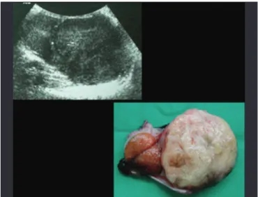
A 16-year-old male presented with a 7-month history of slow growing mass in the right scrotum. The ultrasonography (US) suggested a paratesticular tumor (Figure 1). Laboratory tests showed ß-HCG < 1.2 IU/L (< 5) and alpha-fetoprotein of 0.92 ng/mL (0.5 to 5.5). A radical inguinal orchiectomy was performed and pathological examination revealed a 6-cm paratesticular embryonal RMS. Chest x-ray was normal; the retroperitoneal 7-mm slice CT reviewed
1 Hospital Lucano – Teresina (PI), Brazil.
2 Instituto de Oncologia Pediátrica, Universidade Federal de São Paulo – UNIFESP, São Paulo (SP), Brazil. 3 Department of Urology, Universidade Federal de São Paulo – UNIFESP, São Paulo (SP), Brazil.
einstein. 2011; 9(4 Pt 1):527-9
528 Damazio E, Caran E, Ortiz V, Macedo Junior A
by a senior radiologist at our institution was negative for lymph nodes (Figure 2). We decided to perform a modified RPLND because of the post-pubertal presentation. During dissection three enlarged nodes were identified (Figures 3 and 4) and confirmed to be metastatic by pathological evaluation. Patient was referred to adjuvant chemotherapy.
Figure 1. Sonographic and macroscopic aspect of right testis and paratesticular tumor
Figura 2. Retroperitoneal 7 mm sections CT-scan
Figure 3. Intraoperative retroperitoneal aspect before and after lymph node dissection
Figure 4. Enlarged metastatic lymph nodes removed
DISCUSSION
The Intergroup Rhabdomyosarcoma Study Group (IRSG) established the guidelines for management
of rhabdomyosarcoma (RMS)(3). Multimodal
treatment has considerably improved the outcome of patients with PT-RMS. The superficial location enables detecting early signs and symptoms. Furthermore, the paratesticular site often allows curative surgery with complete excision of the tumor. The high proportion of non-metastatic embryonal tumors that represent the favorable histological subtype may be responsible for high treatment responsiveness.
einstein. 2011; 9(4 Pt 1):527-9
Does negative retroperitoneal CT in adolescents 529
retroperitoneal lymph node (RPLN) status in adults with PT-RMS staged with CT scanning was incorrect for 58% of their patients(4). Yet, there are controversies in the management of PT-RMS, specifically if CT scanning can exclude retroperitoneal involvement. Wiener et al.(5) compared patients treated on IRSG III (n = 100) or IRSG IV (n = 134) and found that there was a significant change in the distribution of patients with group I versus II tumors from III to IRSG-IV (group I, 68% in IRSG-III versus 82% in IRSG-IV). This was the result of decreased node recognition when CT was used to stage in IRSG-IV and was most notable for adolescents (> 10 years).
Some authors confirmed that the majority of patients classified as groups I and II are prepubertal, while most patients in groups III and IV are postpubertal(6). Consequently, the 5-year event-free survival rates in the prepubertal and postpubertal series are also different and in a series with 44 patients, reviewed by Ferrari et al., the survival rate was 91 and 60%, respectively(6). These data suggest the important prognostic role of age and imply biological differences between PT-RMS of childhood and of adolescence, which may be used as an argument for more aggressive approach when treating adolescents. In the same study, abdominal CT correctly staged only 8 of 19 cases (42%). If the therapeutic regimen and subsequent outcome are determined based on sites and volume of disease, then staging is important. This fact is true for PT-RMS, as Wiener et al. demonstrated that node involvement is associated with decreased patient survival, and increased risk of relapse and death(7).
Therefore, if clinical staging is poor and the therapeutic regimen is altered based on pathological staging, RPLND would appear to confer a benefit based on improved staging alone. In our case, a negative CT-scan reviewed by an experienced radiologist would have taken us to propose a wrong chemotherapy regimen if surgery was not performed. Nevertheless, arguments against RPLND for PT-RMS do exist. For clinical group I patients some argue that chemotherapy is able to eradicate micrometastases and omitting RPLND will reduce short and long-term morbidity without affecting survival. We believe that morbidity of modern nerve sparing RPLND is minimal and acceptable and most importantly, stage II patients should receive additional therapy (RPLND, radiation or more intensive chemotherapy) beyond the standard administration of cyclophosphamide, doxorubicin and vincristine. Another argument in favor of RPLND was made by Raney et al. in a review of paratesticular cases from the Intergroup
Rhabdomyosarcoma Studies groups I and II(8).
The authors were uncertain about how effective chemotherapy was in eliminating undetected, non-radiated micrometastases in the regional lymph nodes. They reported on 6 patients who died of the disease, usually due to spread to the lung, after complete remission obtained following the Intergroup Rhabdomyosarcoma Studies guidelines, and despite further individualized treatment combining surgery, chemotherapy and radiation therapy(8).
Retroperitoneal lymphadenectomy can provide a more accurate staging of the disease and define the ideal regimen of chemotherapy. This fact has been observed by other authors and therapeutic results with RPLND and postoperative chemotherapy were excellent with 8 of 9 pathological group I and 9 of 10 pathological group II patients for disease-free survival in the long-term follow-up(9-11).
REFERENCES
1. Wexler LH, Helman LJ. Rhabdomyosarcoma and the undifferentiated sarcomas. In: Pizzo PA, Poplack DC. Principles and practice of pediatric oncology. 3rd ed. Philadelphia: Lippincott-Raven; 1997. p.799-829.
2. Raney RB Jr, Tefft M, Lawrence W Jr, Ragab AH, Soule EH, Beltangady M, et al. Testicular sarcoma in childhood and adolescence: a report from the Intergroup Rhabdomyosarcoma studies I and II, 1973-1983. Cancer. 1987;60(9):2337-43.
3. Raney RB, Chintagumpala M, Anderson J, Pappo A, Qualman S, Wharam M, et al. Results of treatment of patients with superficial facial rhabdomyosarcomas on protocols of the Rhabdomyosarcoma Study Group (IRSG), 1984-1997. Pediatr Blood Cancer. 2008;50(5):958-64.
4. Hermans BP, Foster RS, Bihrle R, Little S, Sandler A, Einhorn LH, et al. Is retroperitoneal lymph node dissection necessary for adult paratesticular rhabdomyosarcoma? J Urol. 1998;160(6 Pt1):2074-7.
5. Wiener ES, Anderson JR, Ojimba JI, Lobe TE, Paidas C, Andrassy RJ, et al. Controversies in the management of paratesticular rhabdomyosarcoma: is staging retroperitoneal lymph node dissection necessary for adolescents with resected paratesticular rhabdomyosarcoma? Semin Pediatr Surg. 2001;10(3):146-52.
6. Ferrari A, Casanova M, Massimino M, Luksch R, Piva L, Fossati-Bellani F. The management of paratesticular rhabdomyosarcoma: a single institutional experience with 44 consecutive children. J Urol. 1998;159(3):1031-4. 7. Wiener ES, Lawrence W, Hays D, Lobe TE, Andrassy R, Donaldson S, et al.
Retroperitoneal node biopsy in paratesticular rhabdomyosarcoma. J Ped Surg. 1994;29(2):171-7.
8. Raney RB, Crist WM, Maurer HM, Foulkes MA. Prognosis of children with soft tissue sarcoma who relapse after achieving a complete response. A report from the Intergroup Rhabdomyosarcoma Study I. Cancer. 1983;52(1):44-50.
9. Tomaszewski JJ, Sweeney DD, Kavoussi LR, Ost MC. Laparoscopic retroperitoneal lymph node dissection for high-risk pediatric patients with paratesticular rhabdomyosarcoma. J Endourol.2010;24(1):31-4.
10. Skolarus TA, Bhayani SB, Chiang HC, Brandes SB, Kibel AS, Landman J, et al. Laparoscopic retroperitoneal lymph node dissection for low-stage testicular cancer. J Endourol. 2008;22(7):1485-9.