Primeira submissão em 16/07/07 Última submissão em 23/10/07 Aceito para publicação em 26/11/07 Publicado em 20/02/08
Nonalcoholic fatty liver disease: histopathological
evaluation and interobserver agreement
Doença hepática gordurosa não-alcoólica: avaliação histopatológica e concordância interobservador
Cynthia Koeppel Berenstein1;Caroline Guimarães Cardoso2; Nivaldo Hartung Toppa3; Virginia Hora Rios Leite4
Background: Nonalcoholic fatty liver disease comprises a spectrum of lesions ranging from steatosis to cirrhosis, with nonalcoholic steatohepatitis being the progressive form of the disease. Alcohol intake, viral hepatitis and other liver diseases must be excluded. Liver biopsy is the gold standard for diagnosis of the disease and is the only method able to differentiate nonalcoholic steatohepatitis from simple steatosis, to grade inflammation and to stage fibrosis. Aims: To analyze the histopathological findings and evaluate interobserver agreement in biopsies previously diagnosed as steatosis or steatohepatitis. Methods: Seventy needle biopsies were analyzed according to Brunt et al.(4), with modifications in the grading and staging components. Clinical data of patients
were collected. Interobserver agreement was calculated based on histopathological findings. Results: Mild nonalcoholic steatohepatitis (grade 1) was the most common form. If fibrosis was detected, stage 1 was the most frequent. Interobserver agreement was very good for macrovesicular steatosis (KW = 0,82) and good for lobular inflammation (KW = 0,68) and fibrosis (KW = 0,73). Conclusions: The classification of Brunt et al., with modifications, can be applied to diagnosis not only of nonalcoholic steatohepatitis but also of nonalcoholic fatty liver disease, representing a reliable method for use in the daily practice of pathologists.
resumo
abstract
key words
unitermos Nonalcoholic
steatohepatitis
Nonalcoholic fatty liver
disease
Steatosis
Liver
1. Master degree; Departamento de Anatomia Patológica e Medicina Legal, Faculdade de Medicina da Universidade Federal de Minas Gerais (UFMG). 2. Physician; UFMG.
3. Doctorate; Laboratório Analys de Anatomia Patológica.
4. Doctorate; Departamento de Anatomia Patológica e Medicina Legal, Faculdade de Medicina da UFMG.
Work conducted at the Department of Pathological Anatomy of UFMG, supported by Conselho Nacional de Desenvolvimento Cientíico e Tecnológico (CNPq), and based on the master’s thesis Doença hepática gordurosa não-alcoólica: avaliação histopatológica em biópsias por agulha e concordância interobservador, presented in 2007 to UFMG.
Introdução: A doença hepática gordurosa não-alcoólica compreende um espectro de lesões que variam da esteatose à cirrose, sendo a esteatoepatite não-alcoólica a forma progressiva da doença. Uso de álcool, hepatites virais e outras doenças hepáticas devem ser excluídos. A biópsia hepática é o padrão-ouro para o diagnóstico da doença, sendo o único método capaz de diferenciar a esteatoepatite da esteatose, graduar a inlamação e estadiar a ibrose. Objetivos: Analisar os achados histopatológicos e avaliar a concordância interobservador em biópsias previamente diagnosticadas como esteatose ou esteatoepatite. Materiais e métodos: Setenta biópsias por agulha foram analisadas segundo Brunt et al.(4), com modiicações nos componentes determinantes da atividade e no estadiamento. Os informes clínicos dos pacientes foram coletados. A concordância interobservador foi calculada com base nos achados histopatológicos. Resultados: Esteatoepatite não-alcoólica discreta (grau 1) foi a forma mais comum. Se havia ibrose, o estágio 1 foi o mais freqüente. Concordância interobservador foi muito boa para esteatose macrovacuolar (KW = 0,82) e boa para inlamação lobular (KW = 0,68) e ibrose (KW = 0,73). Conclusões: A classiicação de Brunt et al., com modiicações, pode ser usada para o diagnóstico não somente da esteatoepatite não-alcoólica, mas também da doença hepática gordurosa não-alcoólica, representando método coniável para uso na rotina diária dos patologistas.
Esteatoepatite não-alcoólica
Doença hepática gordurosa não-alcoólica
Materials and methods
In a retrospective study, needle liver biopsies sent to Analys Laboratory between 1999 and 2006 with histological diagnosis of steatosis or steatohepatitis were selected. Only cases whose exam request did not report abusive alcohol use (more than 20 g/day) or positive serology for virus B and/or C were selected. Clinically, these patients presented elevated aminotransferase levels and/or alterations upon abdominal ultrasound that justiied a liver biopsy. A total of 195 cases were identiied.
After detailed analysis of the records, 89 cases were
excluded: sample belonging to a transplanted liver (n = 10),
heterozygous hemochromatosis (n = 5), alcoholism (n =
35), presence of autoantibodies (n = 4), alcoholism and
C virus infection (n = 2), drug-associated hepatitis (n =
1), no blocks available (n = 3), record not found (n = 7),
and impossibility to contact the responsible physician
(n = 12). Biopsies with fewer than ive portal tracts and
ive centrolobular veins (n =28) were excluded because
they were considered to be non-representative(13). Steatosis
was a basic condition for inclusion, and cases with less than 5% of steatotic hepatocytes were deined as normal and
excluded(5) (n = 8).
The remaining 70 samples were submitted to routine processing: fixation in 10% formalin, dehydration in ethanol, xylene clearing, and embedding in paraffin. The specimens were cut with a microtome and the slides were stained with hematoxylin-eosin, Masson/Gomori’s trichrome, Perls’ stain and picrosirius.
The slides were examined by two experienced pathologists who were unaware of the clinical/laboratory data. A specially prepared protocol was illed out based
on the parameters of Brunt et al.(4, 6, 7), with modiications.
Steatosis was deined as mild (5%-33% of hepatocytes affected), moderate (33%-66%), and marked (> 66%). Microvesicular steatosis was classiied as present or absent, and its location in the acinus was recorded. Hepatocyte ballooning and glycogenated nuclei were scored as absent, occasional or frequent, and their topography was also recorded. Lobular inlammatory activity was classiied as absent, mild (1-2 foci at 20x magniication), moderate (3-4 foci at 20x magniication), and marked (> 4 foci at 20x magnification). Portal inflammatory infiltrate was classiied as absent, mild, moderate or marked, and its cellular phenotype was recorded. If no lobular activity was found, the case was classiied as having only steatosis. For grading the histopathologic lesions of NASH, differing
Introduction
Nonalcoholic fatty liver disease (NAFLD) is considered to be the most common liver disease in the United States and has been related to insulin resistance and metabolic
syndrome(27). NAFLD comprises a spectrum of lesions ranging
from steatosis, steatohepatitis and cirrhosis to hepatocellular
carcinoma(4, 6, 18, 33). Ludwig et al.(23) were the irst to describe
nonalcoholic steatohepatitis (NASH), which is currently considered to be the progressive form of NAFLD.
NAFLD is suspected when clinical examination demonstrates hepatomegaly, and/or abdominal ultrasound suggests fat accumulation in the liver, and/or mild (two to three times the reference value) and persistently elevated aminotransferase levels in individuals presenting no other
cause of these alterations(24). However, NAFLD may also
occur in patients with normal aminotransferase levels(32).
Liver biopsy is considered to be the gold standard for the diagnosis of NAFLD and is the only method able to differentiate NASH from simple steatosis, to grade
inlammation and to stage ibrosis(34).
Histopathological classification systems have been
proposed by Matteoni et al.(25) for NAFLD and by Brunt
et al.(4, 6) for NASH. Few studies evaluating inter- and intraobserver variation of NAFLD are available. Younossi et al.(34) used the system of Matteoni et al.(25) and found moderate interobserver agreement for steatosis grade, location of steatosis, ibrosis stage, hepatocyte ballooning and glycogenated nuclei. Intraobserver agreement was better than interobserver agreement. Inflammation presented poor agreement between pathologists. Mendler et al.(26), proposing a scoring and grading system for NAFLD, also found that intraobserver was better than interobserver
agreement. In 2005, Kleiner et al.(21) published a report on
a histological scoring system for NAFLD, which was the irst and only study evaluating interobserver variation using the
parameters of Brunt et al.(4). Again, inlammation showed
poor interobserver agreement. It is important to remember that NAFLD-related pathologic features display considerable
sample variability(29).
Herein we analyze the histopathological findings of liver needle biopsies and determine interobserver agreement in the histological evaluation of cases with histological diagnosis of steatosis or steatohepatitis, with or without ibrosis and with no history of alcohol abuse, with emphasis on the minimal criteria for the diagnosis of
NASH using the classiication proposed by Brunt et al.(4, 6),
from Brunt(6), the intensity of lobular inlammation was the most important feature. Ballooning and steatosis were also necessary for the diagnosis of NASH, but their intensity was not taken into account. The presence of lipogranulomas, Mallory hyalines, apoptosis and hepatic iron was recorded. If present, iron accumulation was graded. The intensity of ibrosis was divided into four categories: absent, stage 1 (ibrosis limited to perivenular area), stage 2 (perivenular ibrosis and few scattered septa), stage 3 (altered lobular architecture with septal bridging and incipient nodule formation), and stage 4 (cirrhosis). Periportal ibrosis was not considered an element in stage 2 as proposed by
Brunt(6), and scattered septa were introduced to differentiate
cases with little ibrosis from cases with abundant ibrosis (which are considered stage 3).
The following data were obtained from the records: weight, height, body mass index (BMI), presence of obesity, alcohol abuse, associated diseases (arterial hypertension, diabetes mellitus and/or insulin resistance [DM/IR], and hypercholesterolemia), serum alanine aminotransferase (ALT), aspartate aminotransferase (AST) and gamma-glutamyltranspeptidase (GGT), serology for B and C virus, presence of autoantibodies, use of hepatotoxic drugs, glycemia, total cholesterol, serum high-density lipoprotein (HDL), serum low-density lipoprotein (LDL), serum very low-density lipoprotein (VLDL), triglycerides, and abdominal ultrasound result.
With respect to metabolic syndrome components, patients with BMI ≥ 30 or those reported by the physician to be obese but for whom no data regarding weight and/or height were available were classiied as obese. Arterial hypertension was deined as blood pressure higher than 130x85 mmHg on two
or more occasions or when patients used antihypertensive drugs. IR was diagnosed when impaired fasting glucose was ≥ 110 mg/dl, and type 2 diabetes mellitus (DM2) was deined as fasting glycemia > 126 mg/dl. Patients using oral hypoglycemic drugs were also considered to be diabetic. Elevated cholesterol levels were deined as total cholesterol > 200 mg/dl and HDL < 40 mg/dl for men and < 50 mg/dl for women. Hypertriglyceridemia was diagnosed when triglycerides levels > 150 mg/dl. Patients presenting at least three of these features were classiied
as having metabolic syndrome(16).
The results were submitted to descriptive analysis using the SPSS 9.0 statistical program. Interobserver agreement was evaluated using the weighted kappa test (Kw)(20, 30) of the Stata 7.0 program. The K
w coeficient was
interpreted as follows: < 0.2 = low agreement, 0.21 to 0.4 = weak agreement, 0.41 to 0.6 = moderate agreement, 0.61 to 0.8 = good agreement, and > 0.8 = very good
agreement(30).
The institution’s ethics committee approved the study (ETIC 534/04).
Results
Twenty-ive (35.7%) of the 70 patients were women and 45 (64,3%) were men. The mean age was 47,2 years for women and 40,4 years for men. Clinical and laboratory
characteristics of the patients are shown in Table 1.
Information regarding the presence of obesity was available for 47 patients. Twenty-ive (53.2%) of these patients were obese. Seventeen of the 25 obese patients presented BMI > 30 and in the remaining eight cases only the clinical record
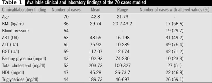
Table 1
Available clinical and laboratory findings of the 70 cases studied
Clinical/laboratory finding
Number of cases
Mean
Range
Number of cases with altered values (%)
Age 70 42.8 21-73
-BMI (kg/m2) 36 29.74 20.2-43.2 17 (56.6)
Blood pressure 64 - - 19 (29.7)
AST (U/l) 63 48.55 16-198 31 (49.2)
ALT (U/l) 65 75.92 10-289 49 (75.4)
GGT (U/l) 59 117.07 12-574 42 (71.2)
Fasting glycemia (mg/dl) 43 102.93 74-230 10 (23.3) Total cholesterol (mg/dl) 53 203.73 100-327 27 (51)
HDL (mg/dl) 47 45.28 26-73.7 22 (46.8)
of obesity was available (BMI not calculated). Nineteen of the 22 non-obese subjects had BMI < 30 and three were clinically described as non-obese.
Information regarding the glycemic proile was obtained for 45 patients. Twelve (26.6%) subjects were classiied as type 2 diabetic or resistant to insulin. Glycemia values were available for 10 of these patients and in the remaining two cases the physician informed about the presence of DM/IR. Seventeen of the 70 patients (24.3%) presented criteria fulilling the classiication of metabolic syndrome. The AST/ALT ratio was less than one in 59 (84.2%) cases.
Biopsies presented a mean of 10.57 portal tracts and of 8.87 centrolobular veins. These structures were not counted in two cases classiied as cirrhosis.
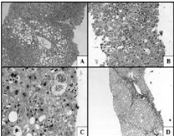
Table 2 summarizes the histological indings and Figure shows some histopathological aspects. In 46 (65.7%) cases,
Figure – Histopathological aspects of NAFLD. A: macrovesicular steatosis around the centrolobular vein. There is a small focus of lobular activity under the vein (HE, 100x); B: moderate NASH with scattered ballooned and steatotic cells (HE, 20“0x); C: Mallory bodies in an extremely ballooned cell (HE, 400x); D: fibrosis stage 3 (Gomori’s thricrome, 50x)
macrovesicular steatosis was diffusely distributed. When diffuse, moderate and marked steatosis predominated (36/46), but cases of mild steatosis (10/46) showing a diffuse distribution in the acinus were also observed. Steatosis was restricted to zones 2 and 3 in 22 (31.4%) cases. The topography of steatosis was not determined in two (2.9%) cases classiied as cirrhosis.
Microvesicular steatosis was associated with mild steatosis in 13/29 (44.8%) cases, with moderate steatosis in 14/23 (60.9%), and with marked steatosis in 15/18 (83.3%). In addition, microvesicular steatosis was observed in 11/20 (55%) patients who presented no associated lobular inlammatory activity.
Hepatocyte ballooning was found in the three zones in 43 (61.4%) cases and in zones 2 and 3 in 25 (35.7%). The topography of ballooning was not evaluated in two cases classiied as cirrhosis.
The portal inflammatory infiltrate consisted of mononuclear cells in 58.8% of biopsies and of mononuclears and neutrophils in 41.2%. Lobular inlammation, deined as present when the inflammatory focus contained neutrophils, was detected in 50 cases (71.4%). These patients were classiied as having NASH. Two patients presented marked activity and corresponded to the cases of cirrhosis. No inlammatory foci were identiied in 20 samples (28.6%). Lipogranulomas were not considered as indicators of inlammatory activity.
Thirty-one (62%) of the 50 patients classiied as having NASH were males. NASH was observed in 68.9% (31/45) of men and 76% (19/25) of women. The following patients presented lobular inflammatory activity: 18
Table 2
Results of the histological analysis of
the 70 liver biopsies
Variable
n
%
Macrovesicular steatosis
Discreet 29 41.4 Moderate 24 34.3 Marked 17 24.3 Microvesicular
steatosis
Present 42 60
Hepatocyte ballooning
Occasional 39 55.7 Frequent 31 44.3 Portal inflammation Absent 34 48.6 Discreet 31 44.3 Moderate 3 4.3 Lobular
inflammation
Discreet 43 61.4 Moderate 5 7.1
Marked 2 2.9 Lipogranuloma Present 25 35.7 Mallory hyaline Present 10 14.3 Apoptosis Present 17 24.3 Glycogenated nuclei Occasional 18 25.7 Frequent 15 21.4 Absent 47 67.1 Hepatic iron* Present 22 33,3 Fibrosis Discreet 12 17.1 Moderate 7 10
Marked 2 2.9 Cirrhosis 2 2.9
(72%) obese and eight (81.8%) non-obese, 11 (57.9%) hypertensive and 34 (75.5%) non-hypertensive, six (50%) DM/IR and 27 (81.8%) non-DM/IR, 20 (74%) with hypercholesterolemia and 17 (65.4%) with normal total cholesterol levels, 16 (72.7%) with low HDL levels and 17 (68%) with HDL levels within normal limits, and 22 (84.6%) with hypertriglyceridemia and 20 (55.5%) with normal serum triglyceride levels. Only six (35.3%) of the 17 patients with metabolic syndrome presented some degree of ibrosis.
Mallory hyaline was not detected in cases presenting only steatosis. In all cases they were present there was some degree of ibrosis. Mallory hyalines were frequently detected in extremely ballooning cells.
Glycogenated nuclei were located in zone 1 in 23 (69.7%) samples and in all zones in nine (27.3%). No topographic assessment was performed in one case (3%) of cirrhosis. No signiicant association was observed between
the occurrence of DM/IR and glycogenated nuclei (X2 =
0.89; p = 0.34).
Iron accumulation determined with Perls’ stain was absent in 44 (62.9%) biopsies. Iron accumulation was detected in the cytoplasm of hepatocytes in four (5.7%) cases, in Kupffer cells in six (8.6%) and in the two cell types in 12 (17.1%). In four cases the material was not suficient for iron investigation. Hepatic siderosis was classiied as grade 1 in 14 (87.5%) biopsies and as grade 2 in two (12.5%).
The weighted kappa test showed very good agreement
for the diagnosis of macrovesicular steatosis (KW = 0.82) and
good agreement for the detection of lobular inlammation
(KW = 0.68) and ibrosis (KW = 0.73).
Discussion
Predominance of NASH among women was reported
up to the 1990’s. More recent reports(1, 3) have shown a
similar frequency of NAFLD and NASH in both genders. Herein 45 (64.3%) of the patients with NAFLD were men, conirming the lack of predominance among women as initially believed. The prevalence of NASH was 76% (19/25) among women and 68.9% (31/45) among men. Women were on average almost a decade older (47.2 years) than men (40.4 years) and presented a greater tendency toward inlammation, which might be explained by the lack of the
protective role of hormones(10).
The rates observed for diabetes, obesity and systemic
arterial hypertension agree with literature data(1, 3, 22, 23, 25, 28).
The same was not observed for hypertriglyceridemia whose
prevalence was below reported values(1, 23). Adams et al.(1)
observed low serum HDL levels in 33% (117/359) of cases, a percentage lower than that found in the present study, which was 46.8% (22/47). However, only 84 of their 435
patients were biopsied(1), and no histological conirmation
of NAFDL was therefore obtained. Regarding serum total
cholesterol, Ludwig et al.(23) observed hypercholesterolemia
in 36% of patients with NASH, in contrast to the 51% found in the present study. The metabolic syndrome component most frequently associated with NAFLD was hypertriglyceridemia, followed by obesity. Only 17 (24.3%) of the 70 patients met criteria for the classiication of metabolic syndrome. This inding shows that NAFLD should be included in the diagnostic hypothesis even in the absence of criteria for metabolic syndrome, since euglycemic and lean patients without dyslipidemias may present some form
of NAFLD. Researchers have been warning since 1994(3)
that the group affected by this liver disease is much larger than the group of middle-aged obese women originally
described in 1989(23).
Most patients (59-84.2%) showed an AST/ALT ratio of less than one. Three of the four cases with a ratio higher than 1.4 had at least moderate ibrosis, a inding indicating
more severe disease(17).
The classiication for NASH proposed by Brunt et al.(4) was
chosen since it is the irst and most widely used international classiication system. The classiication system described
by the Cleveland group(25, 34) does not clarify whether
inlammation is present in category 3 (steatosis + ballooning). In addition, different interpretations as to which categories correspond to steatohepatitis have been reported. Some
authors(12, 17, 31, 35) only considered categories 3 and 4 as NASH,
whereas others(11) regarded categories 2 to 4 as representative
of steatohepatitis.
was highly frequent, being observed in 60% of cases.
In the literature, this association ranges from 34.1%(19)
to 59%(14). Mendler et al.(26) also observed macro- and
microvesicular steatosis in most cases. Brunt(7) reported that
microvesicular steatosis generally occurs in more severe cases. Microvesicular steatosis was observed in cases with no lobular inlammation.
About the use of steatosis grades in the grading of NASH,
we agree with Mendler et al.(26), who omitted steatosis
from their score stating that it is only used to establish the diagnosis of NAFLD but is not correlated with the degree
of steatohepatitis. Brunt et al.(4) deined that in mild NASH
(grade 1) steatosis might be moderate at most, and that in marked NASH (grade 3) steatosis predominantly affects
more than 66% of hepatocytes. Kleiner et al.(21) also uses
the intensity of steatosis in his NAFLD Activity Score (NAS). We observed 12 cases of mild NASH (grade 1) associated with marked steatosis. In addition, one case of marked NASH (grade 3) presented mild steatosis. Thus, 13 (18.5%)
cases did not meet the classiication criteria of Brunt et al.(4).
Furthermore, several reports have shown that steatosis tends
to decrease with the progression of the disease(8) and might
be absent in advanced cases of cirrhosis. It would thus be a contradiction to say that marked NASH can only occur with severe steatosis.
Hepatocyte ballooning was observed in 100% of the samples. However, the inding of ballooning alone does not indicate a diagnosis of NASH. This alteration is a manifestation of cell damage and occurs in response to the most variable injuries, thus being a nonspeciic lesion. In the present study, ballooning was also identiied in cases presenting only steatosis, a inding supporting its importance as a basic lesion of NAFLD and not only related to NASH. Ballooning was classiied as occasional
and frequent. Kleiner et al.(21) divides ballooning in two
categories: those with few balloon cells and those with prominent ballooning. Great dificulties in grading it were encountered during the study since this subclassiication is extremely subjective. Since the minimal criteria of NASH
include ballooning(6, 7, 14) the determination of its presence
would be suficient.
Studies have reported prevalence of inlammation in
NAFLD ranging from 25%(15) to 97%(14), with the rate of
71.4% (50/70) being within the reported range. Mild lobular inlammation in 61.4% (43/70) of the samples supports the concept that mild inlammation is present in
most cases of NASH(7). Lobular inlammation as described
by Brunt et al.(4) was the only parameter used to grade
NASH, with mild inlammation corresponding to mild NASH (grade 1) and so on. Steatosis and hepatocyte ballooning are fundamental for the diagnosis of NASH, but are present at variable degrees. Thus, the presence of these two parameters is obligatory, but their quantiication did not inluence NASH grading.
Lipogranulomas, Mallory hyalines, apoptosis or glycogenated nuclei are not considered fundamental for the classiication of NASH.
Some modiications were introduced in the classiication
of Brunt et al.(4) regarding the staging of ibrosis since this
system includes focal and extensive alterations in the same category, with a biopsy showing focal septal ibrosis being classiied as the same stage as a biopsy presenting extensive septal ibrosis. One of the characteristics of NASH is its
pattern of ibrosis in zone 3(4, 7). Brunt’s system(4) classiies
cases presenting focal or extensive periportal ibrosis as stage 2, with no reference to ibrosis at zone 3. We therefore modiied her original staging and adopted the system
described in the Methods section.
When present, ibrosis was mild in most cases (17.1%).
Prevalence ranging from 18%(30) to 38.7%(22) has been
reported in the literature. Cirrhosis was rare (2.9%), with
studies reporting prevalence of 0%(6) to 26%(14). Most cases
were classiied as stage 1. This inding suggests that ibrosis arises as a consequence of injuries that occur in this area.
The weighted kappa test showed excellent results, with very good agreement for the diagnosis of macrovesicular
steatosis (KW = 0.82) and good agreement for the diagnosis
of lobular inlammation (KW = 0.68) and ibrosis (KW =
0.73). Similarly to the literature, inlammation was the
variable that showed the largest disagreement(21, 26, 34).
Most of the present results were better than those reported
in the literature(21, 26, 34). However, one criticism might be
made: if inlammation is the variable showing the lowest agreement, can it be used as the main parameter for the deinition of NASH grade? We observed that well-trained pathologists obtained agreement in 68% of cases. The Gleason system, which is the most widely used for grading prostate cancer, shows a kappa index ranging from 0.47
to 0.64(2). Nevertheless, this system is widely used. We
therefore believe that, as done in the prostate, NASH can be graded with relative safety by trained pathologists.
1. ADAMS, L. A. et al. The natural history of nonalcoholic fatty liver disease: a population-based cohort study.
Gastroenterology, v. 129, n. 1, p. 113-21, 2005. 2. ALLSBROOK, W. C. et al. Interobserver reproducibility
of Gleason grading of prostatic carcinoma: urologic pathologists. Hum Pathol, v. 32, n. 1, p. 74-8, 2001. 3. BACON, B. R. et al. Nonalcoholic steatohepatitis: an
expanded clinical entity. Gastroenterology, v. 107, n. 4, p. 1103-9, 1994.
4. BRUNT, E. M. et al. Nonalcoholic steatohepatitis: a proposal for grading and staging the histological lesions. Am J Gastroenterol, v. 94, n. 9, p. 2467-74, 1999.
5. BRUNT, E. M. et al. Nonalcoholic steatohepatitis: histologic features and clinical correlations with 30 blinded biopsy specimens. Hum Pathol, v. 35, n. 9, p. 1070-82, 2004.
6. BRUNT, E. M. Nonalcoholic steatohepatitis. Semin Liver Dis, v. 24, n. 1, p. 3-20, 2004.
7. BRUNT, E. M. Nonalcoholic steatohepatitis: definition and pathology. Semin Liver Dis, v. 21, n. 1, p. 3-16, 2001. 8. BUGIANESI, E. et al. Expanding the natural history of
nonalcoholic steatohepatitis: from cryptogenic cirrhosis to hepatocellular carcinoma. Gastroenterology, v. 123, n. 1, p. 134-40, 2002.
9. BURT, A. D.; MUTTON, A.; DAY, C. P. Diagnosis and interpretation of steatosis and steatohepatitis. Semin Diagn Pathol, v. 15, p. 246-58.1998
10. CLARK, J. M.; BRANCATI; F. L., DIEHL, A. M. Nonalcoholic fatty liver disease. Gastroenterology, v. 122, n. 6, p. 1649-57, 2002.
11. CONTOS, M. J.; SANYAL, A. J. The clinicopathologic spectrum and management of nonalcoholic fatty liver disease. Adv Anat Pathol, v. 9, n. 1, p. 37-51, 2002.
12. COTRIM, H. P. et al. Clinical and histopathological features of NASH in workers exposed to chemicals with or without associated metabolic conditions. Liver Int, v. 24, n. 2, p. 131-5, 2004.
13. CRAWFORD, A. R.; LIN, X.; CRAWFORD, J. M. The normal adult human liver biopsy: a quantitative reference standard. Hepatology, v. 28, n. 2, p. 323-31, 1998.
References
14. DIEHL, A. M.; GOODMAN, Z.; ISHAK, K. G. A clinical and histologic comparison with alcohol-induced liver injury.
Gastroenterology, v. 95, n. 4, p. 1056-62, 1988. 15. DIXON, J. B.; BHATHAL, P. S.; O’BRIEN, P. E.
Nonalcoholic fatty liver disease: predictors of nonalcoholic steatohepatitis and liver fibrosis in the severely obese. Gastroenterology, v. 121, n. 1, p. 91-100, 2001.
16. NCEP. Executive summary of the third report of the National Cholesterol Education Program expert panel on detection, evaluation, and treatment of high blood cholesterol in adults (Adult Treatment Panel III). JAMA, v. 285, n. 19, p. 2486-97, 2001.
17. FALCK-YTTER, Y. et al. Clinical features and natural history of nonalcoholic steatosis syndromes. Semin Liver Dis, v. 21. n. 1, p.17-26, 2001.
18. FASIO, E. et al. Natural history of nonalcoholic steatohepatitis: a longitudinal study of repeat liver biopsies. Hepatology, v. 40, n. 4, p. 820-6, 2004.
19. GRAMLICH, T. et al. Pathologic features associated with fibrosis in nonalcoholic fatty liver disease. Hum Pathol, v. 35, n. 2, p. 196-9, 2004.
20. JAKOBSSON, U.; WESTERGREN, A. Statistical methods for assessing agreement for ordinal data. Scand J Caring Sci, v. 19, n. 4, p. 427-31, 2005.
21. KLEINER, D. E. et al. Design and validation of a histological scoring system for nonalcoholic fatty liver disease.
Hepatology, v. 41, n. 6, p. 1313-21, 2005.
22. LEE, R. G. Nonalcoholic steatohepatitis: a study of 49 patients. Hum Pathol, v. 20, n. 6, p. 594-8, 1989. 23. LUDWIG, J. et al. Nonalcoholic steatohepatitis: Mayo Clinic
experiences with a hitherto unnamed disease. Mayo Clin Proc, v. 55, n.7, p. 434-8, 1980.
24. MARCHESINI, G. et al. Nonalcoholic fatty liver disease: a feature of the metabolic syndrome. Diabetes, v. 50, n. 8, p. 1844-50, 2001.
25. MATTEONI, C. A. et al. Nonalcoholic fatty liver disease: a spectrum of clinical and pathological severity.
Gastroenterology, v. 116, n. 6, p. 1413-9, 1999. 26. MENDLER, M. H.; KANEL, G.; GOVINDARAJAN, S.
Proposal for a histological grading system for non-alcoholic fatty liver disease. Liver Int, v. 25, n. 2, p. 294-304, 2005.
mainly neutrophils, which was quantiied according to the
classiication of Brunt et al.(4). It is of the utmost importance
that a grading system for NASH includes minimal criteria for establishment of the diagnosis, such as the presence of macrovesicular steatosis, hepatocyte ballooning and mixed
lobular inlammatory foci. The classiication of Brunt et al.(4)
meets these criteria. When ibrosis is present in NASH, it is generally mild and the growing deposition of collagen in zone 3 accompanied by septum formation determines its stage.
The classiication of Brunt et al.(4), initially developed for
27. NEUSCHWANDER-TETRI, B. A.; CALDWELL, S. H. Nonalcoholic steatohepatitis: summary of an AASLD Single Topic Conference. Hepatology, v. 37, n. 5, p. 1202-19, 2003.
28. POWELL, E. E. et al. The natural history of nonalcoholic steatohepatitis: a follow-up study of 42 patients for up to 21 years. Hepatology, v. 11, n. 1, p. 74-80, 1990. 29. RATZIU, V. et al. Sampling variability of liver biopsy in
nonalcoholic fatty liver disease. Gastroenterology, v. 128, n.7, p. 1898-906, 2005.
30. SIM, J.; WRIGHT, C. C. The kappa statistic in reliability studies: use, interpretation, and sample size requirements. Phys Ther, v. 85, n. 3, p. 257-68, 2005.
31. SIQUEIRA, A. C. G. et al. Non-alcoholic fatty liver disease and insulin resistance: importance of risk factors and
histological spectrum. Eur J Gastroenterol Hepatol, v. 17, n. 8, p. 837-41, 2005.
32. SORRENTINO, P. et al. Silent non-alcoholic fatty liver disease – a clinical histological study. J Hepatol, v. 41, n. 5, p. 751-7, 2004.
33. YOSHIOKA, Y. et al. Nonalcoholic steatohepatitis: cirrhosis, hepatocellular carcinoma, and burnt-out NASH.
Gastroenterology, v. 39, n. 12, p. 1215-8, 2004. 34. YOUNOSSI, Z. M.; DIEHL, A. M.; ONG, J. P. Nonalcoholic
fatty liver disease: an agenda for clinical research.
Hepatology, v. 35, n. 4, p. 746-52, 2002.
35. YOUSSEF, W. I.; McCULLOUGH, A. J. Steatohepatitis in obese individuals. Best Prac Research Clin Gastroenterol, v. 16, n. 5, p. 733-47, 2002.
Mailing address