305
Optimization of methods to assess mitochondrial DNA
in archival paraffin-embedded tissues from mammary
canine tumors
Otimização dos métodos para avaliar o DNA mitocondrial obtido a partir de tumores
mamários caninos incluídos em parafina
Angélica C. Bertagnolli1; Valdemar Máximo2, 3; Bárbara van Asch2, 4; António Amorim2, 4; Luis Cirnes2; Paula Soares2, 3; Geovanni D. Cassali1
A presente comunicação descreve as modificações usadas para extrair e amplificar o DNA mitocondrial obtido de amostras de tumores mamários caninos fixados em formol tamponado a 10% e incluídos em parafina. Os componentes epiteliais e mesenquimais (condromixóide e condróide), bem como a mama normal adjacente, foram microdissectados manualmente de 19 tumores mamários (10 tumores mistos benignos e nove carcinomas em tumores mistos). O DNA foi extraído utilizando-se o Invisorb® Spin Tissue Mini Kit com modificações do protocolo proposto pelo fabricante. Um fragmento de 273-pb foi amplificado por reação em cadeia da polimerase (PCR) e seqüenciado em seqüenciador automático. O fragmento foi analisado em 100% das amostras, entretanto modificações como lise adicional, redução do volume das soluções de extração e PCR, aumento da temperatura de anelamento e do número de ciclos de amplificação foram necessárias. Em seis amostras os produtos iniciais de PCR foram diluídos e reamplificados para obtenção de sucesso.
resumo
abstract
In this study we describe the alterations used to extract and amplify mitochondrial desoxyribonucleic acid (DNA) from formalin-ixed parafin-embedded samples of canine mammary tumors. The epithelial and mesenchymal components (chondromyxoid and chondroid) of each tumor, as well as the normal mammary gland tissues, were manually microdissected from 19 mixed canine mammary tumors (10 benign mixed tumors and nine carcinomas arising in mixed tumors). DNA was extracted by Invisorb®
Spin Tissue Mini Kit, with protocol changes proposed by the manufacturer. A 273-bp fragment was ampliied by polymerase chain reaction (PCR) and submitted to automatic sequence analysis. The fragment was successfully analyzed in 100% of the samples. However, an additional lysis step, the reduction of volume in buffer solutions and PCR, a higher annealing temperature and an increase in the number of PCR cycles were required. The initial PCR products were diluted and re-ampliied in six
unitermos key words
Neoplasia
Cão
Mesenquimal Neoplasia
Dog
Mesenchymal
Primeira submissão em 06/05/08
Última submissão em 10/10/08
Aceito para publicação em 09/11/08
Publicado em 20/08/08 J Bras Patol Med Lab • v. 44 • n. 4 • p. 305-308 • agosto 2008
1. Laboratory of Comparative Pathology, Department of General Pathology, Instituto de Ciências Biológicas of Universidade Federal de Minas Gerais (UFMG). 2. Instituto de Patologia e Imunologia Molecular da Universidade do Porto (IPATIMUP), Portugal
3. Department of Pathology, Faculdade de Medicina da Universidade do Porto (FMUP). 4. Faculdade de Ciências da Universidade do Porto (FCUP).
306
In general, desoxyribonucleic acid (DNA) recovered from
formalin-ixed parafin-embedded tissues is highly degraded(10)
and may be unsuitable for most molecular techniques such as polymerase chain reaction (PCR). Although some studies have shown that PCR can be successfully performed with DNA
recovered from formalin-ixed parafin-embedded-tissues(2, 3, 5, 9),
results have still been limited in some laboratories.
In this study, we describe our experience concerning standardization of mitochondrial DNA (mtDNA) extraction techniques, ampliication and sequencing of 10% formalin-ixed parafin-embedded tissues, microdissected from mformalin-ixed canine mammary tumors.
Nineteen canine mammary tumors (10 benign mixed
tumors and nine carcinomas arising in mixed tumors)(8) were
retrieved from the iles of the Laboratory of Comparative Pathology, Department of General Pathology, Instituto de Ciências Biológicas of Universidade Federal de Minas Gerais (UFMG). The tissues used in this study had been ixed in 10% neutral buffered formalin (without knowledge of total time ixation) and parafin-embedded.
Seven 10 µm-thick sections were cut from formalin-ixed parafin-embedded tumors and mounted on polylysine-coated slides. The microtome used to cut the sections was kept clean and excess parafin and tissue fragments were wiped from the blade holder with xylene between blocks. The sections were dewaxed twice, with two changes in xylene (15 min each) and washed with 100% and 70% etanol (twice, for 5 min).
Microdissection target areas were previously marked on hematoxilin-eosin stained slides. The epithelial (neoplastic epithelial proliferations) and the mesenchymal (chondro-myxoid and chondroid) components of each tumor and the normal residual glandular parenchyma were manually removed from each of the sections using a scalpel, under stereomicroscopic observation. We veriied that the addition of deionized water to the the area to be microdissected facilitates tissue recovery of the material. The tissues were scraped into 1.5-ml tubes and submitted to digestion
and exctraction using an Invisorb® Spin Tissue Mini Kit
(Invitek). The procedures were performed according to the manufacturer’s protocol with the following modiications: the suggested volumes of reagents (lysis, binding and wash buffer, proteinase K) were reduced to half, the elution buffer volume was reduced to 33 µl, an additional lysis step was performed, and mixtures were homogenized by gentle pipetting instead of vortexing.
The digestion included the incubation of samples in a lysis buffer (200 µl) and 20 µl proteinase K (10 µl/ml) at
56°C, with gentle agitation in a thermomixer. An overnight incubation was usually enough to obtain total digestion of the tissue, indicated by a clear solution. In the cases where the samples were still turbid and tissue fragments were visible, an additional lysis step was performed by a new addition of 20 µl proteinase K for another eight hours at 56°C.
After tissue digestion, the tubes were centrifuged for 2 min at maximum speed in order to precipitate all non-lysed material, and 100 µl binding buffer T were added. The solutions were homogenized with gentle pipetting to minimize DNA breakage. The suspensions were transferred onto spin columns placed in 2-ml receiver tubes and incubated for 1 min. The spin columns were centrifuged at 12,000 rpm for 2 min and the iltrate was discarded. The column was placed back in the receiver tube, 200 µl wash buffer were added and centrifuged as in the previous step. An additional centrifugation was performed to remove any residual ethanol. For DNA elution the spin columns were placed in new 1.5-ml receiver tubes and 33 µl of pre-heated elution buffer D were added and incubated for 5 min. A inal centrifugation was performed for 2 min at 10,000 rpm. A total volume of 33 µl of eluted DNA was recovered from each sample.
The DNA extract was stored at 4°C for 24 hours and used for PCR ampliication.
For successful ampliication of degraded DNA samples it is recommendable that the amplicon size be relatively
small(1). However, numerous known polimorphisms strongly
constrain primer design in this region. The primers designed in this work for the ampliication of a 273-bp fragment of the hypervariable mitochondrial D-loop represent a compromise for obtaining the smallest and most potentially informative amplicons.
PCR amplifications were optimized by varying the number of cycles and the annealing temperature. The annealing temperatures tested were 56°C, 57°C, 59°C and 60°C. Reactions were performed with 30, 35 and 40 cycles. PCR optimization reactions were tested for each possible combination of annealing temperature and number of cycles. The best results were obtained with an annealing temperature at 60°C with 35 cycles.
The optimized PCR was performed in a 25-µl total
volume, using 11 µl of Quiagen® Multiplex PCR Kit;
11 µl of H20; 1 µl of each primer (2.5 mM); 1 µl of DNA template; and 1 µl of sterile deionized water. The PCR program consisted of an initial denaturation step at 95°C for 15 min followed by 35 cycles at 95°C for 5 min; 60°C
307
for 1.3 min and 72°C for 1 min; and a inal extension step at 72°C for 10 min.
DNA isolated from the peripheral blood of a dog was used as positive control, and sterile deionized water was used as negative control. The PCR products were run in 6% polyacrilamide electrophoresis gel and visualized after silver staining. A DNA ladder was run as a molecular marker in each gel.
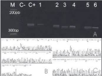
Electrophoresis showed that the 273-bp fragment of the mitochondrial canine control region was ampliied in 52 out of 57 samples (91%). Five samples (three chondroid, two epithelial and one normal mammary tissue) yielded low PCR product, and a second round of PCR ampliication was performed. One microliter from a 100-fold diluted aliquot of the initial PCR product was used as template. This reaction was performed following the same procedure as in the irst round. The use of products from the irst PCR round as starting template produced bands that could be
visualized in the electrophoresis gel (Figure 1A) and were
suficient for subsequent sequencing.
The PCR products were analyzed by automated
sequencing (Figure 1B). In order to clean the PCR product
from primers and nucleotides, PCR products were puriied using a Sephacryl HR300 column (Pharmacia, Biotech, Saclay, France), and sequenced using BigDye Terminator v 3.0 sequencing kit. The sequencing reactions were performed in both directions using 2.6 µl of the puriied PCR products, 0.25 mM of primer, 0.6 µl of sequencing kit and deionized water to complete 5 µl of total reaction volume. The thermocycler program comprised an initial denaturation step at 96°C for 2 min, followed by 35 cycles at 96°C for 15 s, 50°C for 9 s, 60°C for 2 min and a inal extension step at 60°C for 10 min. Sequencing reaction products were puriied using Sephadex G-50 ine columns (Pharmacia) and eluted in deionized formamide. DNA sequencing was carried out in an ABI Prism 3130 Genetic Analyzer (Applied Biosystems Inc., Foster City, CA, USA) using POP-6, and was used for the capillary eletrophoresis separation matrix.
The sequences obtained were compared to the reference sequence, indicated as haplotype A19 (Gen Bank accession entry: NC_002008) and conirmed the ampliication of the predicted 273-bp fragment of the
hypervariable D-loop control region in 48 cases (Figures
1B and 1C). Dificulties in deducing the sequences were
observed in nine samples, which were re-submitted to sequencing reactions with higher and lower amounts of puriied PCR product, according to the intensity of
the band previously detected in polyacrilamide gel electrophoresis. A total of 2.4 µl and 2.6 µl were used for sequencing in samples with high and low intensity bands, respectively. In all the samples it was possible to successfully conirm the DNA sequence.
The analysis of mtDNA offers advantages over nuclear DNA due to a larger number of copies per cell, what potentiates the recovery of DNA from dificult or
degraded materials(4, 7). The literature describes several
successful protocols for analysis of shorter and longer mtDNA fragments recovered from archival ixed
parafin-embedded human tissues(1, 6, 11). In our series of cases, the
success of mtDNA analysis was achieved by the use of a DNA extraction kit with modiications in several steps of the process: lysis (additional lysis step), extraction (reduction of buffer volumes and homogenization by gentle handling), ampliication (high-annealing temperature and increase in the number of PCR cycles) and sequencing (adjustment of the volume of the PCR product).
The samples had been ixed in 10% buffered formalin, but no information on the handling of specimens before tissue fixation (tissue amount, degree of autolysis), fixation-related factors (temperature, and duration of fixation,) and post-fixation procedures storage
(temperature and duration of storage)(10) was available.
Several of the samples may well have been subjected to conditions that contributed to the partial degradation of the nucleic acids.
Figure 1 – A: Evaluation of DNA amplification by polyacrylamide gel electrophoresis. C+= positive control (peripheral blood of a dog); C-= negative control; 1, 2, 3, 4, 5 and 6 = PCR products (273bp) from second round PCR, M, molecular weight marker. 1.2 microliters of PCR products were loaded in each lane; B and C: Confirmation of amplification by direct sequencing of PCR product. B = Sample 1; C = Positive control. Aliquots of the amplicons were purified and sequenced as describe under Material and Methods. Picture in the ‘5 → 3’ orientation is the sense strand of the amplicon from mesenchymal component (case 5)
308
Mailing address
Geovanni Dantas Cassali Av. Antônio Carlos, 6.627 CEP 31270-901 – Belo Horizonte-MG Tel.: (31) 3499-2891
Fax: (31) 3499-2879 e-mail: [email protected]
References
1. ALONSO, A. et al. Usefulness of microchip electrophoresis for the analysis of mitochondrial DNA in forensic and ancient DNA studies. Electrophoresis, v. 27, n. 24, p. 5101-9, 2006.
2. DUDDY, S. K; GOROSPE, S; BLEAVINS, M. R. Genetic analysis of multiple loci in micro samples of fixed paraffin-embedded tissue. Toxicol Sci, v. 46, p. 317-23, 1998.
3. IHLASEH, S. M. et al. Microdissecção e captura a laser na investigação do gene TP53 em tecidos incluídos em parafina. J Bras Patol Med Lab, v.43, n.1, p.61-67, 2007.
4. ISENBERG, A. R. Forensic mitochondrial DNA analysis: a different crime-solving tool. FBI L. Enforcement Bull,
v. 71, n. 8, at 16, 2002.
5. LIBÓRIO, T. N. et al. Evaluation of the genomic DNA extracted from formalin-fixed, paraffin-embedded oral samples archived for the past 40 years. J Bras Patol Med Lab, v. 41, n. 6, p. 405-10, 2005.
6 LU, C. et al. Multiplex STR and mitochondrial DNA testing
for paraffin-embedded specimen of healthy and malignant tissue: interpretation issues. Int Congress Series, v. 1288, p. 648-50, 2006.
7. MIETHING, F. Effect of fixation to the degradation of nuclear and mitochondrial DNA in different tissues. J Histochem Cytochem, v. 39, p. 351-4, 2006. 8. MISDORP, W. et al. Histological classification of mammary
tumors of the dog and the cat. Second Series, Vol. VII. Washington, DC: Armed Forces Institute of Pathology,1999.
9. SIMONATTO, L. E. Avaliação de dois métodos de extração de DNA de material parafinado para amplificação em PCR. J Bras.Patol Med Lab, v. 43, n. 2, p. 121-7, 2007.
10. TOKUDA, Y. et al. Fundamental study on the mechanisms of DNA degradation in tissues fixed in formaldehyde.
J Clin Pathol, v. 43, p. 748-51, 1990.
11. ZHU, W. et al. Mitochondrial DNA mutations in breast cancer tissue and in matched nipple aspirate fluid.
Carcinogenesis, v. 26, n. 1, p. 145-52, 2005. The major limitation in this analysis was the dificulty
in obtaining high quality DNA in some samples (mainly from cartilaginous component), probably due to lower cell content present. In addition to the improvements in the extraction procedure, the re-ampliication step using the initial PCR product as template was crucial to achieving success.
Improvement and routine control of pre-, during and post-ixation procedures should be considered in medicine
veterinary laboratories so that, in the future, tissue banks may consistently offer high-quality DNA.
Acknowledgments
This research was supported by Coordenação de Aperfeiçoamento de Pessoal de Nível Superior (CAPES), Conselho Nacional de Desenvolvimento e Cientíico e Tecnológico (CNPq) and Fundação de Amparo à Pesquisa do Estado de Minas Gerais (FAPEMIG).