Peripherally inserted central venous catheters: alternative or
first choice vascular access?
Cateteres venosos centrais de inserção periférica: alternativa ou primeira
escolha em acesso vascular?
Marcelo Kalil Di Santo1
*
, Diogo Takemoto1, Robert Guimarães Nascimento1, Ariele Milano Nascimento2,
Érika Siqueira3, Caio Túlio Duarte4, Marco Antônio Caldas Jovino1, Jorge Agle Kalil1
Abstract
Background: Peripherally inserted central catheters (PICC) are intravenous devices inserted through a supericial or deep vein of an upper or lower extremity and advanced to the distal third of the superior vena cava or proximal third of the inferior vena cava. hey ofer the advantages of greater safety for infusion of vesicant/irritant and hyperosmolar solutions and enable administration of antibiotics, prolonged parenteral nutrition (PPN), and chemotherapy agents. hey also involve reduced risk of infection compared to other vascular catheters and are more cost-efective than centrally inserted venous catheters (CICVC). Objectives: To present the results of our team’s experience with US-guided and luoroscopy-positioned PICC placement at the Hospital and Maternidade São Luiz (HMSL) Itaim, Rede D’or, Brazil. Methods: his was a prospective, non-randomized study, conducted from February 2015 to November 2016. he institution’s preestablished protocol was followed when vascular access was requested. Indications, prevalent diseases, type of catheter implanted, technical success, and complications related to the catheters were analyzed and inclusion and exclusion criteria are described. Results: A total of 256 vascular accesses were requested, and 236 PICCs (92.1%) and 20 CICVCs (7.9%) were implanted. he main indications were as follows: prolonged antibiotic therapy (52%), PPN (19.3%), and diicult venous access (16%). Technical successes was achieved in 246 catheter placements (96.1%). he right basilic vein was the most common vein punctured for access, in 192 patients (75%), followed by the right brachial vein, in 28 patients (10.9%). Conclusions: Ultrasound-guided and luoroscopy-positioned PICC placement had a low incidence of complications, reduced infection rates, and proved safe and efective in cases of diicult vascular access. PICCs can be considered the devices of choice for central vascular access.
Keywords: central catheter; central venous access; ultrasound-guided puncture; vascular access.
Resumo
Contexto: Os cateteres venosos centrais de inserção periférica (PICC) são dispositivos intravenosos, introduzidos através de uma veia supericial ou profunda da extremidade superior ou inferior até o terço distal da veia cava superior ou proximal da veia cava inferior. Apresentam maior segurança para infusão de soluções vesicantes/irritantes e hiperosmolares, antibioticoterapia, nutrição parenteral prolongada (NPT) e uso de quimioterápicos; demonstram reduzido risco de infecção em comparação a outros cateteres vasculares e maior relação custo/benefício se comparados ao cateter venoso de inserção central (CVCIC). Objetivos: Apresentar os resultados de implantes de PICCs ecoguiados e posicionados por luoroscopia realizados no Hospital e Maternidade São Luiz (HMSL) Itaim, Rede D’or, Brasil. Métodos: Estudo prospectivo, não randomizado, realizado entre fevereiro de 2015 e novembro de 2016. Utilizou-se protocolo pré-estabelecido pela instituição em casos de solicitação de acesso vascular. Foram analisadas indicações, doenças prevalentes, tipo do cateter implantado, sucesso técnico, complicações relacionadas ao cateter, e estabelecidos critérios de inclusão e exclusão. Resultados: Solicitados 256 acessos vasculares, sendo implantados 236 PICCs (92,1%) e 20 CVCICs (7,9%). Principais indicações: antibioticoterapia prolongada (52,0%), NPT (19,3%) e acesso venoso difícil (16,0%). Houve sucesso técnico em 246 cateteres implantados (96,1%). A veia basílica direita foi a principal veia puncionada em 192 pacientes (75,0%), seguida da braquial direita em 28 pacientes (10,9%).
Conclusões: O implante dos PICCs ecoguiados e posicionados por luoroscopia demonstrou baixa incidência de complicações, reduzidos índices de infecção e é seguro e eicaz em casos de acessos vasculares difíceis, sendo esses cateteres considerados dispositivos de escolha em acesso vascular central.
Palavras-chave: cateter central; acesso venoso central; punção ecoguiada; acesso vascular.
1Rede D’or Hospital e Maternidade São Luiz – HMSL Itaim, Serviço de Cirurgia Vascular e Endovascular, São Paulo, SP, Brazil. 2Hospital da Beneicência Portuguesa de São Paulo, Cirurgia Geral, São Paulo, SP, Brazil.
3Rede D’or Hospital e Maternidade São Luiz – HMSL Itaim, Terapia Infusional, São Paulo, SP, Brazil. 4Centro Universitário São Camilo – CUSC, São Paulo, SP, Brazil.
Financial support: None.
Conlicts of interest: No conlicts of interest declared concerning the publication of this article. Submitted: December 28, 2016. Accepted: April 12, 2017.
INTRODUCTION
Peripherally inserted central catheters (PICC) are intravenous devices that are inserted via a supericial or deep vein in an extremity and advanced as far as the distal third of the superior vena cava or proximal third of the inferior vena cava. They can measure from 20 to 65 cm in length and have calibers varying from 1 to 6 French (Fr). They can have from one to three lumens and may be valved (proximal or distal) or nonvalved. They are lexible and radiopaque, have smooth, uniform walls, and can be made from silicone, polyethylene, polyurethane, or carbothane. They are inserted by percutaneous puncture using split sheaths, made from metal or plastic, and are discarded after use.
A PICC was described in the literature for the irst time in 1929 by the German doctor Werner Theodor Otto Forssmann who inserted a cannula into his own antecubital vein and used it to introduce a 65 cm catheter up to the right atrium, conirming the anatomic location by X-ray. This procedure earned him the 1956 Nobel prize for medicine and introduced an alternative option for central venous access via a peripheral access.1 The technique began to be used
in Brazil in the 1990s, initially for applications in neonatology, because of the small diameter of the catheter and the lexibility of the material (silicone), and was later widely adopted in intensive care, oncology, and home care.2
Indications and contraindications for the device have been established; it is recommended that insertion be guided using ultrasonography and positioning of the tip guided with luoroscopy, thereby ensuring greater safety during puncture and positioning and increasing patient comfort during the procedure.
The principal advantages of PICCs are as follows: the beneits of inserting the catheter under local anesthesia, combined or not with sedation; reduction of patient discomfort, by avoiding multiple vein punctures; the possibility of bedside insertion; provision of a safe access for administration of antibiotics; prolonged parenteral nutrition (PPN); an excellent access for administering chemotherapy; increased maximum indwell time and reduced risk of contamination compared with other devices; preservation of the peripheral venous system; and possibility of use in home treatment applications.
One feature of fundamental importance to prevention of complications and iatrogenic events is the fact that the catheter is inserted peripherally, which can potentially prevent occurrence of pneumothorax or hemothorax. Additionally, they are less expensive
than surgically inserted central venous catheters (SICVCs).3,4
The main dificulties and disadvantages with using PICCs are related to the need for an intact vascular network with suficient caliber for implantation; the need for special training for insertion and maintenance of the catheter; the need for rigorous monitoring of the device; and the need for radiography to locate the tip of the catheter.3,4 Evidence has shown that these devices are not free from complications, such as deep venous thrombosis (DVT), thrombophlebitis, occlusion of the catheter, arterial pseudoaneurysms, and infections.5-8 On the other hand, using this type
of catheter avoids venous dissection and exposes the patient to less pain and fewer complications inherent to the procedure.
To present the results of our team’s experience with US-guided and luoroscopy-positioned PICC placement at the Hospital and Maternidade São Luiz (HMSL) Itaim, Rede D’or, São Paulo, SP, Brazil.
METHODS
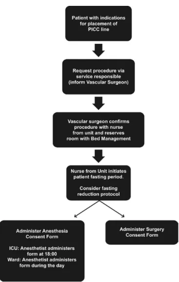
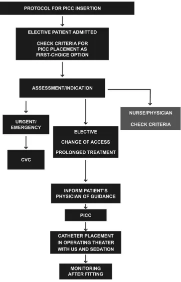
We conducted a prospective, non-randomized study from February 2015 to November 2016, with Research Ethics Committee approval. Our institution’s preestablished protocol was followed whenever vascular access was requested (Figures 1 and 2). The inclusion criteria adopted were: patients admitted to wards or the intensive care unit (ICU) with indications for PPN, infusion of vesicant and/or irritant drugs, dificult access with loss of daily access, chemotherapy, prolonged antibiotic therapy for periods greater than 4 days, and patients on heparin and/or with thrombocytopenia. Contraindications for catheter insertion and/or for the study included pediatric patients, bilateral thrombophlebitis or DVT of upper extremities, cephalic vein as only access option bilaterally, women with mastectomies, presence of arteriovenous istulas in the extremity to be punctured/catheterized and emergency situations. The following variables were analyzed: indications, prevalent diseases, type of catheter implanted, technical success, and complications related to the catheter.
An appropriate peripheral vein in the upper extremity was selected and punctured with the aid of an ultrasound unit in B mode (Mindray – Hemocat).
We used out-of-plane puncture; the appropriate puncture site on the upper limb was chosen as proposed by Dawson,9 delimiting ideal zones for insertion with
ultrasound guidance (the Zone Insertion Method, ZIM). After placement of a metallic guidewire graduated in centimeters, the (Peel-Away) dilation sheath was inserted and then the selected catheter was inserted after sectioning to length, with the appropriate preparatory measures. The inal length of the catheter was calculated using the length of the graduated guidewire. The next stage of the procedure is to evaluate low and backlow through the catheter; followed by transoperative angiography to test the positioning and check that the tip is correctly placed
before ixing the catheter with a Statlock device (Figures 3 and 4).
RESULTS
During the study, 256 vascular accesses were requested and 236 PICCs (92.1%) and 20 SICVCs (7.9%) were implanted. There were 155 female patients (60.5%) and 101 male patients (39.5%), with a mean age of 70.2 years. Within the hospital, 176 patients were in the ICU (68.7%) and 80 were in wards (31.3%). The most common indications for catheter placement were: prolonged antibiotic therapy (52.0%), PPN (19.3%), and dificult venous access (16.0%). Other indications seen with lower frequency were administration of vesicant/irritant medications (8.0%), risk of bleeding (3.3%), and administration of chemotherapy (1.4%).
Figure 2. Protocol for PICC line insertion after request for catheter placement. CVC = central venous catheter; US = ultrasonography.
The catheters used were silicone with valves (PICC Groshong BARD), polyurethane without valves
(Power PICC BARD), or carbothane with valves
(Biolo Hemocat), from 5 to 6 Fr.
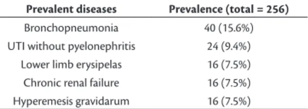
The clinical diseases most often seen in the patients who underwent vascular access are shown in Table 1 in order of prevalence.
The placement procedure for 246 catheters (96.1%) was technically successful, deined as achieving a catheter position in the interior of the superior vena cava.
In 10 catheters (3.9%) it was not possible to achieve an adequate position within the interior of this vein due to technical failures during the initial learning curve: incorrect catheter length (PPN patients in whom a PICC position in the superior or inferior vena cava is obligatory) and failure to advance the catheter despite adequate vein patency (such as, for example, dificulties caused by valve friction).
In 192 patients (75.0%), the right basilic vein was chosen for insertion, followed, in descending order of frequency, by the right brachial vein in 28 patients (10.9%), the left brachial vein in 19 patients (7.4%) and, as the inal option, the left basilic vein in 17 (6.7%).
There were 14 complications related to the procedure in our sample of patients, including two fractures of catheters with distal valves (0.8%), seven catheter obstructions (2.7%), six of polyurethane catheters without valves and one of a valved carbothane catheter, and there were ive infections, all related to catheters without valves (1.9%).
Three different microorganisms were isolated: Klebsiella pneumoniae in three cases, Candida
Glabrata in one case, and Staphylococcus hominis in one case. All infected catheters were in ICU patients.
DISCUSSION
Robert B. Dawson delimited ideal zones for PICC insertion under ultrasonographic guidance (ZIM). Using musculoskeletal characteristics of the skin and vessels as landmarks, he divided the arm above the antecubital fold into three distinct zones, each 7 cm in size, separated by the colors red, green, and yellow, taking the medial epicondyle of the humerus as the initial anatomic landmark and the axillary line as the inal landmark (Figure 5). In common with trafic lights, the colors of the zones indicate whether or not they should be used for puncture. According to Dawson, the ideal puncture zone, indicated with green, is approximately 12 cm from the medial epicondyle, where the basilic vein is most supericial in relation to the plane of the skin.9
In our study, we achieved a high rate of technical success with US-guided PICC insertion (96.1%). Our preference for ultrasound-guided puncture to achieve venous access was based on the lower risk of incorrect puncture offered by the ultrasonographic method when compared with puncture based exclusively on anatomic parameters.10,11 According to Hockley et al., the literature shows that US-guided insertion via the arm improves both catheter insertion success rates12,13 and the satisfaction of patients who undergo the procedure14 in addition to reducing complications, such as infections at the puncture site, thrombosis, and catheter migration.15
The most important complications of PICCs are: infection, fracture with distal venous migration, thrombophlebitis or DVT of upper extremities, Horner syndrome, and even chylothorax,16-18 the
most commonly observed of which are infections, thrombophlebitis, and DVT.5-8
According to a study by Liem et al.,19 the rates of
symptomatic upper limb supericial venous thrombosis associated with PICCs are 1.9% in the basilic vein, 7.2% in the cephalic vein, and 0% in the brachial
Table 1. Most frequent diseases in order of prevalence.
Prevalent diseases Prevalence (total = 256)
Bronchopneumonia 40 (15.6%)
UTI without pyelonephritis 24 (9.4%)
Lower limb erysipelas 16 (7.5%)
Chronic renal failure 16 (7.5%)
Hyperemesis gravidarum 16 (7.5%)
UTI: urinary tract infection.
Figure 4. Peripheral central venous catheter made from carbothane with proximal pressure-activated safety valve (PASV) and Endexo
vein. The greater incidence of supericial venous thrombosis in the cephalic vein is due to the anatomic characteristics of this vessel, such as smaller diameter in relation to the size of the catheter, lower number of tributaries, and more perpendicular insertion into the axillary vein (Figure 6).
For this reason, our team decided that the cephalic vein as the only available access option on either side would be considered an exclusion criterion, and chose to it an SICVC in these cases.
A review of published retrospective and prospective studies revealed incidence rates of upper limb DVT associated with PICCs varying from 0.5 to 19.4%, with the higher incidence rates directly related to insertion of larger diameter PICCs and presence of malignant neoplasms. Just one cancer patient in our sample (who also had lower limb DVT) had an upper limb DVT, related to use of a nonvalved catheter and treated with removal of the catheter, with no need for subcutaneous or oral anticoagulation.
Compared with nonvalved PICCs, PICCs with integrated valve technologies signiicantly reduce the rates of later complications (occlusion or infection) and eliminate the need to use heparin and its potential subsequent complications (for example, heparin-induced thrombocytopenia).20
A retrospective study conducted by the Vanderbilt University involving placement of 12,505 devices and comparing rates of infection and occlusion in valved (4.2% and 1.4% respectively) and nonvalved PICCs (5.5% and 6.3% respectively), concluded that valved PICCs exhibited lower rates of infection and
occlusion, a reduced need for maintenance, and lower costs, eliminating the obligatory heparin locking demanded for nonvalved PICCs.21 The results of our
study, in which seven obstructions were described (six in nonvalved catheters and just one in a valved catheter) are in agreement with these data published in the literature.20,21
Peripherally inserted central catheters with proximal and distal valves were introduced to the market with the aim of reducing catheter occlusions by preventing
blood backlow.22 A randomized prospective study
conducted by Hoffer et al.22 showed that patency
rates were better for catheters with proximal valves, with lower incidence rates of occlusive and infectious complications compared with catheters with distal valves.
The most common complication during insertion is malpositioning of catheters, which occurs when the catheter is not placed in the appropriate position within the vena cava.23 Dificulty advancing the
catheter during insertion, inadequate blood draw, and dificulties removing the stylet/dilator sheath are indicative that the catheter may have been incorrectly placed and in such cases radiography or luoroscopy is indispensable to identify whether the catheter has been incorrectly placed.24
In a study with 3,012 patients, conducted by Song and Li,23 technical success was achieved in 94.6% of
PICC placements and 237 devices were identiied as incorrectly placed outside of the vena cava, assessed by radiography after insertion of the catheter, with the most frequent location being the jugular vein,
followed by the axillary and brachial veins (Figure 7). At our service, following a preestablished protocol, we did not detect any incorrectly positioned catheters. If luoroscopy showed that the catheter was in a different position than the correct central position, it was immediately repositioned.
When a catheter becomes obstructed, it is necessary to initiate the appropriate drug-based treatment with thrombolytics, attempting to reduce the degree of catheter obstruction. Baskin et al.25 conducted a study showing
that thrombolytic agents successfully eliminate catheter occlusions in the majority of cases, and highlighted the role played by alteplase. Described as a safe and effective medication for deobstructing catheters, the disadvantages of this drug is its elevated cost, the fact that it is less effective than urokinase during the irst 30 minutes of infusion, and that it takes more than 4 hours to achieve clearance of the catheter. Other thrombolytics require shorter periods of time in the lumen of the vessel to act.25 In our sample of patients,
the obstructions observed in six nonvalved catheters and one proximally valved catheter were adequately treated using urokinase (Taurolock), the thrombolytic of choice at our institution.
Maki et al.26 analyzed the risk of bloodstream
infection with different types of device, inding that the bloodstream infection rates associated with PICCs were lower than those reported with traditional, non-tunneled, central venous devices.
Many hypotheses have been raised to attempt to explain why PICCs exhibit fewer infectious complications than other types of device, including
lower bacterial density on the skin of the arm, the cooler temperatures of the limbs, and the relative simplicity of caring for the area, compared with the neck and groin.6 The following variables are related to
bloodstream infections associated with PICCs: length of hospital stay, admission to an ICU, and number of catheter lumens.27 Sundriyal et al.28 analyzed placement
of 246 PICCs in ICUs, observing that in 12.5% there were catheter infections with positive blood culture, in which the most frequently isolated agents were Klebsiella pneumoniae and Staphylococcus sp. These authors suggest that infected catheters should be removed in all patients with positive blood cultures,
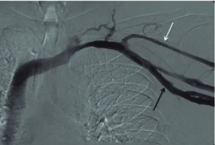
Figure 6. Smaller diameter of the cephalic vein (white arrow) with relation to the size of the catheter and its perpendicular insertion
into the axillary vein (black arrow).
primarily if there is no improvement in fever within 48 h of appropriate administration of antibiotics.28
Fungemia or bacteremia caused by the Bacillus species Corynebacterium jeikeium, Staphylococcus aureus, Pseudomonas aeruginosa or Stenotrophomonas maltophila and nontuberculous mycobacteria will very often persist despite administration of appropriate antibiotics, in which case the catheter must be removed. Catheter removal should also be considered when blood cultures remain positive more than 48 h after treatment with antibiotics; if no other site of infection can be identiied, or if bacteremia recurs after a course of antibiotics is completed.29
In our patient sample there were ive cases of catheter infection leading to removal because of the principal agents identiied: Klebsiella pneumoniae, Candida glabrata and Staphylococcus hominis. The results observed in our study are in line with the literature.
CONCLUSIONS
Placement of peripherally inserted central venous catheters under ultrasound guidance and with luoroscopy positioning offers a low incidence of complications and reduced infection rates and is safe and effective, particularly in cases with dificult vascular access, so these catheters are considered the irst-choice devices for central vascular access. Their maintenance requires rigorous training of the nursing team, who should only be responsible for preserving, caring for, and protecting the catheter, with the objective of minimizing complications caused by incorrect handling. The placement procedure should be performed by a trained physician who is able to manage and resolve possible complications related to insertion and use of the catheter.
REFERENCES
1. Cournand AF, Forssmann W, Richards DW. Werner Forssmann: biographical. Stockholm: The Nobel Foundation; 2014. http:// www.nobelprize.org/nobel_prizes/medicine/laureates/1956/ forssmann-bio.html. Acessado: 23/11/2016.
2. Freitas LCM, Raposo LCM, Finoquio RA. Instalação, manutenção e manuseio de cateteres venosos centrais de inserção periférica em pacientes submetidos a tratamento quimioterápico. Rev Bras Cancerol. 1999;45:19-29.
3. Vendramin P. Cateter central de inserção periférica (CCIP). In: Harada MJCS, Rego RC, editores. Manual de terapia intravenosa em pediatria. São Paulo: ELLU; 2005. cap. 7, p. 75-95.
4. Jesus VC, Secoli SR. Complicações acerca do cateter venoso central de inserção periférica (PICC). Cienc Cuid Saude. 2007;6(2):252-60.
5. Periard D, Monney P, Waeber G, et al. Randomized controlled trial of peripherally inserted central catheters vs. peripheral catheters for middle duration in-hospital intravenous therapy.
J Thromb Haemost. 2008;6(8):1281-8. PMid:18541001. http:// dx.doi.org/10.1111/j.1538-7836.2008.03053.x.
6. Safdar N, Maki DG. Risk of catheter-related bloodstream infection with peripherally inserted central venous catheters used in hospitalized patients. Chest. 2005;128(2):489-95. PMid:16100130. http://dx.doi.org/10.1378/chest.128.2.489.
7. Chemaly RF, Parres JB, Rehm SJ, et al. Venous thrombosis associated with peripherally inserted central catheters: a retrospective analysis of the Cleveland Clinic experience. Clin Infect Dis. 2002;34(9):1179-83. PMid:11941543. http://dx.doi.org/10.1086/339808.
8. Allen AW, Megargell JL, Brown DB, et al. Venous thrombosis associated with the placement of peripherally inserted central catheters. J Vasc Interv Radiol. 2000;11(10):1309-14. PMid:11099241.
9. Dawson RB. PICC Zone Insertion MethodTM (ZIMTM): a systematic
approach to determine the ideal insertion site for PICCs in the upper arm. J Assoc Vasc Access. 2011;16(3):156-60, 162-5. http:// dx.doi.org/10.2309/java.16-3-5.
10. Dariushnia SR, Wallace MJ, Siddiqi NH, et al. Quality Improvement Guidelines for Central Venous Access. J Vasc Interv Radiol. 2010;21(7):976-81. PMid:20610180. http://dx.doi.org/10.1016/j. jvir.2010.03.006.
11. Mehta N, Valesky WW, Guy A, Sinert R. Systematic review: is real-time ultrasonic-guided central line placement by ED physicians more successful than the traditional landmark approach? Emerg Med J. 2013;30(5):355-9. PMid:22736720. http://dx.doi.org/10.1136/ emermed-2012-201230.
12. Hockley SJ, Hamilton V, Young RJ, et al. Efficacy of the CathRite system to guide bedside placement of peripherally inserted central venous catheters in critically ill patients: a pilot study. Critical Care & Resuscitation. 2007;9(3):251-5. PMid:17767451.
13. Krstenic WJ, Brealey S, Gaikwad S, Maraveyas A. The effectiveness of nurse led 2-d ultrasound guided insertion of peripherally inserted central catheters in adult patients: a systematic review. J Assoc Vasc Access. 2008;13(3):120-5. http://dx.doi.org/10.2309/ java.13-3-4.
14. McMahon DD. Evaluating new technology to improve patient outcomes: a quality improvement. J Infus Nurs. 2002;25(4):250-5. PMid:12131507. http://dx.doi.org/10.1097/00129804-200207000-00008.
15. Simcock L. No going back: advantages of ultrasound-guided upper arm PICC placement. J Assoc Vasc Access. 2008;13(4):191-7. http:// dx.doi.org/10.2309/java.13-4-6.
16. Lobo BL, Vaidean G, Broyles J, Reaves AB, Shorr RI. Risk of venous thromboembolism in hospitalized patients with peripherally inserted central catheters. J Hosp Med. 2009;4(7):417-22. PMid:19753569. http://dx.doi.org/10.1002/jhm.442.
17. Links DJ, Crowe PJ. Horner’s syndrome after placement of a peripherally inserted central catheter. JPEN J Parenter Enteral Nutr. 2006;30(5):451-2. PMid:16931616. http://dx.doi.org/10.11 77/0148607106030005451.
18. Wu ET, Takeuchi M, Imanaka H, Higuchi T, Kagisaki K. Chylothorax as a complication of innominate vein thrombosis induced by a peripherally inserted central catheter. Anaesthesia. 2006;61(6):584-6. PMid:16704595. http://dx.doi.org/10.1111/j.1365-2044.2006.04640.x.
19. Liem TK, Yanit KE, Moseley SE, et al. Peripherally inserted central catheter usage patterns and associated symptomatic upper extremity venous thrombosis. J Vasc Surg. 2012;55(3):761-7. PMid:22370026. http://dx.doi.org/10.1016/j.jvs.2011.10.005.
21. Burns D. The Vanderbilt PICC Service: program, procedural, and patient outcomes successes. J Assoc Vasc Access. 2005;10(4):1-10. http://dx.doi.org/10.2309/java.10-4-10.
22. Hoffer EK, Bloch RD, Borsa JJ, Santulli P, Fontaine AB, Francoeur N. Peripherally inserted central catheters with distal versus proximal valves: prospective randomized trial. J Vasc Interv Radiol. 2001;12(10):1173-7. PMid:11585883. http://dx.doi.org/10.1016/ S1051-0443(07)61676-5.
23. Song L, Li H. Malposition of peripherally inserted central catheter: experience from 3.012 patients with cancer. Exp Ther Med. 2013;6(4):891-3. PMid:24137284.
24. Aladangady N, Roy R, Costeloe KL. The cobweb sign: percutaneous silastic long line tip placement in tributaries of superficial veins. J Perinatol. 2005;25(10):671-3. PMid:16193077. http://dx.doi. org/10.1038/sj.jp.7211355.
25. Baskin JL, Reiss U, Wilimas JA, et al. Thrombolytic therapy for central venous catheter occlusion. Haematologica. 2012;97(5):641-50. PMid:22180420. http://dx.doi.org/10.3324/haematol.2011.050492. 26. Maki DG, Kluger DM, Crnich CJ. The risk of bloodstream infection in adults with different intravascular devices: a systematic review of 200 published prospective studies. Mayo Clin Proc. 2006;81(9):1159-71. PMid:16970212. http://dx.doi.org/10.4065/81.9.1159.
27. Chopra V, Ratz D, Kuhn L, Lopus T, Chenoweth C, Krein S. PICC-associated bloodstream infections: prevalence, patterns, and predictors. Am J Med. 2014;127(4):319-28. PMid:24440542. http:// dx.doi.org/10.1016/j.amjmed.2014.01.001.
28. Sundriyal D, Shirsi N, Kapoor R, et al. Peripherally inserted central catheters: our experience from a cancer research centre. Indian J Surg Oncol. 2014;5(4):274-7. PMid:25767338. http://dx.doi. org/10.1007/s13193-014-0360-1.
29. Schiffer CA, Mangu PB, Wade JC, et al. Central venous catheter care for the patient with cancer. J Clin Oncol. 2013;31(10):1357-70. PMid:23460705. http://dx.doi.org/10.1200/JCO.2012.45.5733.
*
Correspondence
Marcelo Kalil Di Santo Rede D’or Hospital e Maternidade São Luiz – HMSL Itaim, Serviço de Cirurgia Vascular e Endovascular Rua Itapeva, 286, 6º andar, conjunto 64 – Bela Vista CEP 01332-000 - São Paulo (SP), Brazil Tel.: +55 (11) 4314-6900 E-mail: [email protected]
Author information
MKDS, DT and RGN - Assistant physicians, Serviço de Cirurgia Vascular e Endovascular, Rede D’or Hospital e Maternidade São Luiz (HMSL Itaim). AMN - Resident physician (General Surgery), Hospital da Beneicência Portuguesa de São Paulo. ES - Continuing Education Nurse, Chief of the Infusion herapy Team, HMSL. CTD - Medical student (5th year), Centro Universitário São Camilo (CUSC). MACJ - Physician, Coordinator, Grupo em Acesso Vascular, Serviço de Cirurgia Vascular e Endovascular, HMSL. JAK - Chief physician, Serviço de Cirurgia Vascular e Endovascular, HMSL.
Author contributions
Conception and design: MKDS Analysis and interpretation: MKDS, JAK, MACJ Data collection: MKDS, DT, RGN, AMN, ES Writing the article: MKDS, JAK, CTD Critical revision of the article: MKDS, JAK Final approval of the article*: MKDS, DT, RGN, AMN, ES, JAK, CTD, MACJ Statistical analysis: MKDS, JAK, MACJ Overall responsibility: MKDS, JAK