Floating thrombi in femoral veins
Trombo flutuante em veia femoral
Matheus Bertanha1, Rafael Elias Farres Pimenta1, Gustavo Muçouçah Sampaio Brandão1, Marcone Lima Sobreira1,
Regina Moura1, Rodrigo Gibin Jaldin1, Paula Angeleli Bueno de Camargo1, Winston Bonetti Yoshida1
Abstract
A loating venous thrombus in the femoral vein is a type of thrombus with a high potential for pulmonary embolization. However, the most appropriate management for these cases is still controversial. Clinical treatments, using anticoagulants or ibrinolytics, open thrombectomies, or thrombectomies by means of endovascular devices have all been used, although the criteria for indication of each are not yet deined. We present 3 clinical cases of loating thrombi in femoral veins with diferent etiologies and discuss their respective treatments and outcomes.
Keywords: venous thrombosis; thrombectomy; anticoagulants; ibrinolysis; pulmonary embolism.
Resumo
O trombo venoso lutuante em veia femoral é um tipo de trombo com alto potencial de embolização pulmonar. Entretanto, ainda é controversa a conduta mais apropriada nesses casos. Tratamentos clínicos com anticoagulantes ou ibrinolíticos e trombectomias abertas ou por meio de dispositivos endovasculares vêm sendo empregados ainda sem um critério de indicação bem deinido. Apresentamos três casos clínicos de trombos lutuantes em veia femoral, de etiologias distintas, cujos tratamentos e respectivas evoluções serão discutidos.
Palavras-chave: trombose venosa; trombectomia; anticoagulantes; ibrinólise; embolia pulmonar.
1 Universidade Estadual Paulista Júlio de Mesquita Filho – UNESP, Faculdade de Medicina de Botucatu, Departamento de Cirurgia e Ortopedia, Botucatu, SP, Brazil.
Financial support: None.
Conlicts of interest: No conlicts of interest declared concerning the publication of this article. Submitted: July 09, 2017. Accepted: October 03, 2017.
INTRODUCTION
A loating venous thrombus in the femoral vein is a type of thrombus with a high potential for pulmonary embolization.1-4 It is characterized by a thrombotic
mass that is not attached to the venous wall and advances through the lumen of a deep collector vein.5 According to Voet et al.,6 the prevalence of
loating thrombus in a study of 44 cases of proximal deep venous thrombosis (DVT) was 18%, and the most common site was the femoral vein (38% at the saphenofemoral junction), followed by the popliteal vein (26% at the junction with the small saphenous vein) and the external iliac vein (15%). In that patient sample, 87% of thrombi disappeared after 3 months of anticoagulant treatment according to follow-up duplex ultrasound (DUS), irrespective of site. Floating thrombi can also be seen in patients who exhibit only supericial venous thrombosis (SVT),7 in whom this
can extend to a deep vein,4 but it has also been reported
as a complication of endovenous laser treatment for incompetent saphenous veins.8 Chengelis et al.9
conducted a study in which 30 patients out of a total of 263 cases of SVT also had DVT and 12 of these patients had a loating thrombus in the femoral vein. Sobreira et al.10 observed a DVT rate of 21.7% and a
pulmonary embolism (PE) rate of 28.3% in a series of 60 cases of SVT.
There is no well-established consensus on the best treatment for these cases in order to prevent PE and recurrence of DVT. In this Therapeutic Challenge we present three cases of loating thrombi in femoral veins, their treatments and their outcomes.
PART I – CLINICAL SITUATIONS
Case 1 - A 69-year-old male patient presented with
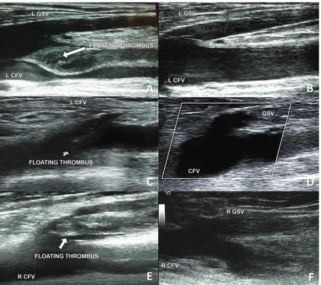
sudden onset dyspnea with chest pain and with vital signs within normal limits. The clinical suspicion of PE was conirmed with angiotomography. He had a history of arterial hypertension, ulcerative rectocolitis, and a previous DVT episode involving the left lower limb several years previously (he was unable to provide an exact date), on which occasion he had been put on anticoagulation with warfarin for 1 year, after which it had been withdrawn. Peripheral pulses were palpable. A venous DUS conducted to investigate the emboligenic source revealed a loating thrombus in the femoral vein, in addition to DVT involving the entire left femoral and popliteal veins (Figure 1A).
Case 2 - A 50-year-old male patient presented
complaining of edema of the left lower limb with onset 1 week previously after particularly intense physical effort (work-related). He did not exhibit
any other associated comorbidities. He reported that he did not smoke or abuse alcohol, had had no previous surgery, immobilization, trauma, obesity, restrictions to walking. He had no family history of PE, DVT, or SVT. On physical examination he was normotensive, with edema of the left lower limb (+++/4) and hyperemia, heat, and pain along the path of the great saphenous vein, from the proximal leg to the proximal thigh of the left lower limb, with no sign of severe chronic venous insuficiency, except for sparse non-communicating varicose veins. Peripheral pulses were palpable and there was muscle hardening in the region of the left thigh and calf. A venous DUS examination of the left lower limb revealed an incompressible great saphenous vein, with no blood low and with echogenic content (thrombus) in the lumen. The thrombus extended from the mid-leg to the saphenofemoral junction, where it “dived” into the lumen of the common femoral vein and appeared to be “loating” (Figure 1C; Video 1 of Supplementary Material), moving in time with arterial pulsations. Other supericial and deep veins were normal. The diagnostic conclusions of the examination were ascending thrombophlebitis of the great saphenous vein and loating thrombus of the common femoral vein, both in the left lower limb.
Case 3 - The patient was a 58-year-old female
with varicose veins and long-term chronic venous insuficiency and a venous ulcer of the right lower limb. The ulcer originated from a trauma in the region of the right medial malleolus, with consequent local infection, which was treated using antibiotics (cephalexin). The patient had an allergic reaction to the antibiotics and was obliged to remain at rest, treating the wound with localized dressings. She had intense pain at the ulcer site, worse when standing up. After 15 days of conservative treatment, she sought specialist medical care, at which point she presented with the additional symptoms of hyperemia, pain, and heat along the path of the right great saphenous vein. On clinical examination she was normotensive, distal arterial pulses were present, and she had an active venous ulcer, with granulation tissue, of 4 cm in diameter, on the medial surface of the right leg (CEAP 6). Venous DUS revealed SVT of the right great saphenous vein, extending along its entire path to the saphenofemoral junction, and a loating thrombus was observed at the limit of the saphenofemoral junction, moving with the heartbeat (Figure 1E).
Possible treatment options that were considered for these cases included: anticoagulant treatment3,11;
ibrinolytic treatment or thrombolysis12; venous
aspirative percutaneous thrombectomy11; and inferior
vena cava ilter (VCF).11
PART II – WHAT WAS DONE
Case 1 - The patient was treated with full
anticoagulation while in hospital, with a combination of unfractionated heparin and warfarin until the international normalized ratio had been brought within therapeutic levels (INR in the range of 2-3). In view of the PE episode and the loating thrombus, it was decided to implant a removable VCF. A DUS examination of the fourth day after hospital admission showed that the VCF was free from thrombi and well-located, with maintenance of the ultrasonographic status seen previously. He was discharged from hospital after
5 days with a prescription for 5 mg warfarin and instructions to attend routine outpatients follow-up to check anticoagulation levels. At 6 month follow-up, his clinical status was good and a control examination with venous DUS showed total remission from the loating thrombus, with signs of recanalization of the veins that had been involved in the DVT episode (Figure 1B). The VCF was still well-positioned and free from thrombi and was not removed.
Case 2 - The patient was given anticoagulation
treatment with enoxaparin sodium and warfarin. On the ifth day of treatment he returned for reassessment and exhibited considerable improvement of the edema (+/4) and inlammatory signs. Low molecular weight heparin was withdrawn and the 5 mg warfarin dose
was continued, with maintenance of therapeutic INR levels. The patient was prescribed elastic stockings and instructed to return to normal activities. The following week, he returned for another assessment and found to be asymptomatic, with no residual edema, with discretely heightened color along the path of the great saphenous vein in the proximal 1/3 of the leg and the distal thigh. Laboratory tests for thrombophilias were ordered. There were no intercurrent conditions of any type during the initial phase of anticoagulation or up to the time of writing (maintenance phase) and tests for secondary thrombophilias were negative (tests for silent cancer and antiphospholipid antibody syndrome). The venous DUS examination conducted 3 months after the clinical event showed total recanalization of the common femoral vein and the great saphenous vein and total remission of the loating thrombus (Figure 1D).
Case 3 - The patient was treated with a combination
of 1 mg/kg of enoxaparin every 12h (60 mg) and warfarin while in hospital. Tests for secondary thrombophilias were negative. During outpatients follow-up, therapeutic anticoagulation levels were maintained for 3 months and then anticoagulation was withdrawn. The follow-up venous DUS examination conducted after completion of anticoagulation treatment showed total disappearance of the loating thrombus, recanalization of the great saphenous vein and an absence of signs of DVT (Figure 1F).
DISCUSSION
In view of the potential autolysis of loating thrombi,6
anticoagulant treatment appears to be an attractive noninvasive option, particularly after SVT that has reached the saphenofemoral junction.13,14 Besides the obvious
antithrombotic effects, the anticoagulants, especially heparins, also have anti-inlammatory properties that amplify their beneits.15 In cases of SVT in isolation,
the American College of Chest Physicians (ACCP) 2012 consensus recommended using fondaparinux at prophylactic doses (2.5 mg/day) or low molecular weight heparin for 45 days.16 However, when DVT
is also present, treatment should be focused on this disease, increasing the duration of anticoagulation. The initial treatment should be provided in a hospital environment. Home treatment should be ruled out because of the high risk of complications related to the possibility of noncompliance with treatment.17
The advantages of surgical treatment are: faster treatment of symptoms and shorter hospital stays, which reduces costs.13 However, simple ligature
of the saphenous trunk does not impede passage of
thrombi via perforating and tributary vessels and does not attenuate any hypercoagulable state that may be present.13 During surgery, in addition to ligature, it is
also recommended that the head of the thrombus that is entering the lumen of the femoral vein should be removed (thrombectomy) by venotomy at the arch of the saphenous vein.1 The complications of this type
of procedure are hematoma and hemorrhages, the risk of embolizing the thrombus, and recurrence of local thrombosis.13 In general, anticoagulant treatment
during the postoperative period is recommended.13
Thrombolytic treatment effectively dissolves the thrombus and reduces postthrombotic syndrome (RR: 0.66; 95%CI: 0.47-0.94), but it is associated with a higher frequency of hemorrhagic complications.12
The ATTRACT study18,19 randomized 692 patients with
proximal DVT for ibrinolytic treatment or conventional treatment (anticoagulants and elastic stockings). After 2 years it was observed that ibrinolytic treatment did not prevent postthrombotic syndrome, but did reduce its severity in 25% of cases (18% vs. 24%).18
A VCF is indicated if anticoagulants are contraindicated or provoke complications, according to the ACCP consensus.11,16 There are no guidelines
for their use in cases of loating thrombi. However, in cases of extensive loating thrombi involving the iliac veins and even the vena cava, use of a VCF could be evaluated, because of the high risk of PE, especially when anticoagulants fail or are contraindicated.
CONCLUSIONS
There are no speciic treatment guidelines for cases of loating venous thrombosis with thrombi that extend into the femoral vein. Anticoagulant treatment appears to be an effective and less invasive option, and surgery can be associated with complications. While there is no consensus, in cases with more extensive loating thrombi, involving iliac veins or even the vena cava, using a VCF could be evaluated, because of the high risk of PE, particularly when anticoagulants are contraindicated.
REFERENCES
1. Casian D, Gutsu E, Culiuc V. Extraluminal venous interruption for free-floating thrombus in the deep veins of lower limbs. Chirurgia (Bucur). 2010;105(3):361-4. PMid:20726302.
2. Noel AA, Gloviczki P, Charboneau JW. Free-floating femoral vein thrombus in a patient with aspergillosis. Int Angiol. 2000;19(1):75-8. PMid:10853690.
4. Raulin S, Raulin C, Greve B. Free-floating thrombus in the femoral vein--a challenge in phlebologic diagnostics. Eur J Dermatol. 2001;11(6):564-8. PMid:11701410.
5. Leon L, Giannoukas AD, Dodd D, Chan P, Labropoulos N. Clinical significance of superficial vein thrombosis. Eur J Vasc Endovasc Surg. 2005;29(1):10-7. PMid:15570265. http://dx.doi.org/10.1016/j. ejvs.2004.09.021.
6. Voet D, Afschrift M. Floating thrombi: diagnosis and follow-up by duplex ultrasound. Br J Radiol. 1991;64(767):1010-4. PMid:1742580. http://dx.doi.org/10.1259/0007-1285-64-767-1010.
7. Jorgensen JO, Hanel KC, Morgan AM, Hunt JM. The incidence of deep venous thrombosis in patients with superficial thrombophlebitis of the lower limbs. J Vasc Surg. 1993;18(1):70-3. PMid:8326661. http://dx.doi.org/10.1067/mva.1993.42072.
8. Araujo WJB, Timi JRR, Erzinger FL, Caron FC. Trombose induzida pelo calor endovenoso: relato de dois casos tratados com rivaroxabana e revisão da literatura. J Vasc Bras. 2016;15(2):147-52. http://dx.doi. org/10.1590/1677-5449.009816.
9. Chengelis DL, Bendick PJ, Glover JL, Brown OW, Ranval TJ. Progression of superficial venous thrombosis to deep vein thrombosis. J Vasc Surg. 1996;24(5):745-9. PMid:8918318. http://dx.doi.org/10.1016/ S0741-5214(96)70007-1.
10. Sobreira ML, Maffei FH, Yoshida WB, et al. Prevalence of deep vein thrombosis and pulmonary embolism in superficial thrombophlebitis of the lower limbs: prospective study of 60 cases. Int Angiol. 2009;28(5):400-8. PMid:19935595.
11. Kearon C, Akl EA, Ornelas J, et al. Antithrombotic Therapy for VTE Disease: CHEST Guideline and Expert Panel Report. Chest. 2016;149(2):315-52. PMid:26867832. http://dx.doi.org/10.1016/j. chest.2015.11.026.
12. Watson L, Broderick C, Armon MP. Thrombolysis for acute deep vein thrombosis. Cochrane Database Syst Rev. 2016;11:CD002783. PMid:27830895.
13. Sobreira ML, Yoshida WB, Lastória S. Tromboflebite superficial: epidemiologia, fisiopatologia, diagnóstico e tratamento. J Vasc Bras. 2008;7(2):131-43. http://dx.doi.org/10.1590/S1677-54492008000200007.
14. Ascer E, Lorensen E, Pollina RM, Gennaro M. Preliminary results of a nonoperative approach to saphenofemoral junction thrombophlebitis. J Vasc Surg. 1995;22(5):616-21. PMid:7494365. http://dx.doi.org/10.1016/S0741-5214(95)70049-8.
15. Ranjbaran H, Wang Y, Manes TD, et al. Heparin displaces interferon-gamma-inducible chemokines (IP-10, I-TAC, and Mig) sequestered in the vasculature and inhibits the transendothelial migration and arterial recruitment of T cells. Circulation. 2006;114(12):1293-300. PMid:16940188. http://dx.doi.org/10.1161/ CIRCULATIONAHA.106.631457.
16. Kearon C, Akl EA, Comerota AJ, et al. Antithrombotic therapy for VTE disease: antithrombotic therapy and prevention of thrombosis,
9th ed: American College of Chest Physicians Evidence-Based Clinical Practice Guidelines. Chest. 2012;141:e419S-94S.
17. Maffei FHA, Rollo HA, Lastória S. Tratamento anticoagulante das tromboses venosas. In: Maffei FHA, et al. Doenças Vasculares Periféricas. 5. ed. Rio de Janeiro: GEN; 2016. p. 1813-26.
18. Society of Interventional Radiology. ATTRACT Trial results provide insight into DVT therapy. Washington, D.C.: SIR, 2017 [citado 2017 jul 09]. http://www.sirtoday.org/attract-study/.
19. Comerota AJ. Catheter-directed thrombolysis for iliofemoral deep vein thrombosis: helpful or hurtful? Expert Rev Hematol. 2015;8(2):131-3. PMid:25652095. http://dx.doi.org/10.1586/174 74086.2015.1007863.
*
Correspondence Matheus Bertanha Universidade Estadual Paulista Júlio de Mesquita Filho – UNESP, Faculdade de Medicina de Botucatu, Departamento de Cirurgia e Ortopedia Via Domingos Sartori, s/n - Distrito de Rubião Junior CEP 18607-621 - Botucatu (SP), Brazil Tel.: +55 (14) 3811-6305 E-mail: [email protected]
Author information MB, MLS and RM - Assistant professors, PhDs, Departamento de Cirurgia e Ortopedia, Faculdade de Medicina de Botucatu, Universidade Estadual Paulista Júlio de Mesquita Filho (UNESP). REFP, RGJ and PABC - Assistant physicians, Departamento de Cirurgia e Ortopedia, Faculdade de Medicina de Botucatu, Universidade Estadual Paulista Júlio de Mesquita Filho (UNESP). GMSB - Collaborating professor, Departamento de Cirurgia e Ortopedia, Faculdade de Medicina de Botucatu, Universidade Estadual Paulista Júlio de Mesquita Filho (UNESP). WBY - Full professor, Departamento de Cirurgia e Ortopedia, Faculdade de Medicina de Botucatu, Universidade Estadual Paulista Júlio de Mesquita Filho (UNESP), e editor-chefe do J Vasc Bras.
Author contributions Conception and design: WBY, MB Analysis and interpretation: WBY Data collection: GMSB, RFP, PABC, RGJ Writing the article: WBY, MB, MLS, RM Critical revision of the article: WBY, MB, MLS, RM Final approval of the article*: MB, RFP, GMSB, MLS, RM, RGJ, PABC, WBY Statistical analysis: N/A. Overall responsibility: WBY, MB
Supplementary Material
Supplementary material accompanies this paper.
Video 1. Venous duplex ultrasound of case 2 with loating thrombus at the right saphenofemoral junction.