159
REV. HOSP. CLÍN. FAC. MED. S. PAULO 56(5):159-162, 2001 SEPTEMBER-OCTOBER
From the Department of Vascular Surgery, Hospital das Clínicas, Faculty of Medicine, University of São Paulo.
PERSISTENCE OF THE EMBRYONIC LATERAL
MARGINAL VEIN:
REPORT OF TWO CASES
Raúl Rojas Martinez, Pedro Puech-Leão, Paulo Motta Guimarães and Baptista Muraco Netto
RHCFAP/3054
ROJAS MARTINEZ R et al. - Persistence of the embryonic lateral marginal vein: report of two cases. Rev. Hosp. Clín. Fac. Med.
S. Paulo 56(5):159-162, 2001.
Purpose: Congenital venous malformations of the lower limbs represent a particular challenge for the vascular surgeon.
Persistence of fetal veins is a rare malformation, and the most common is the persistence of the lateral marginal vein usually observed in patients with Klippel-Trenaunnay Syndrome. The persistence of this embryonic vein as an isolated venous malformation without the other characteristics of the Klippel-Trenaunnay Syndrome has not yet been reported. This paper describes two cases.
Methods: Two patients, a 17-year-old male patient and a 16-year-old female, have had since their birth a large venous trunk in
the lateral aspect of the right leg and thigh. The limbs underwent duplex scanning and phlebography. The surgical removal of the lateral marginal vein was performed.
Results: Surgical treatment resulted in very good functional and aesthetic results. Follow-up at 26 months showed no evidence of varicose vein recurrence.
Conclusions: To achieve good results, surgical intervention may be indicated in cases of orthopedic deformity, hemorrhage,
symptomatic, and unaesthetic lesions.
DESCRIPTORS: Klippel-Trenaunay Syndrome. Marginal vein. Congenital venous malformations. Angiodysplasias.
Varicosities.
Based on their anatomic and patho-logic structure, congenital vascular mal-formations can be divided into 5 groups: capillary, arterial or venous when they have only one component, venous malformations that have arterio-venous fistulas, and complex lesions when different combinations of the pre-vious 4 types occur. Congenital venous malformations of the lower limbs in-clude: valvular agenesis, extrinsic con-striction, hypoplasia, atresia or aplasia of veins, persistence of fetal veins, hy-pertrophy, tortuousities, and varicosi-ties1-3. Clinical findings of venous
mal-formations may vary significantly, from superficial lesions such as varicosities or circumscribed lesions to deep lesions
that involve muscle and bone and can cause limb hypertrophy and deformity. Venous anomaly may occur as an iso-lated lesion, or is most often associated with complex vascular malformations like the Klippel-Trenaunnay Syndrome (KTS). It is important to distinguish per-sistence of fetal veins from primary varicosities and from varicosities sec-ondary to arteriovenous fistula or deep venous thrombosis3,4.
Persistence of fetal veins is a rare congenital malformation. The most
common is the persistence of the lat-eral marginal vein, which is usually as-sociated with complex congenital venous malformations such as KTS1,4.
Its presence as an isolated lesion has not been previously reported. This ar-ticle presents two cases of persistent embryonic lateral marginal vein as an isolated venous malformation without being part of KTS, treated surgically. Literature review did not reveal any ci-tation of similar cases.
CASES REPORT
160
REV. HOSP. CLÍN. FAC. MED. S. PAULO 56(5):159-162, 2001 SEPTEMBER-OCTOBER
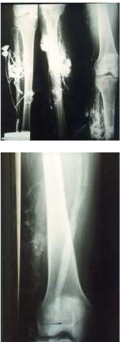
white woman, complaining of varicosi-ties, pain, and heaviness on the right lower limb. They had no history of venous thrombosis, and neither family had varicosity or venous malformations. Physical examination revealed a large venous trunk existing since birth in the lateral aspect of the right lower limb from the ankle to the upper thigh. In the lateral aspect of the upper leg below the knee, the marginal vein had developed substantial collateral vari-cosity. One patient presented a large incompetent perforator vein that devel-oped a venous aneurysm below the knee (Fig. 1). Limb pulse was palpable and normal with no audible bruit or palpable thrill.
Venous duplex scanning of the limbs showed a competent and normal deep venous system with competent sapheno-femoral and saphenopopliteal junctions. Phlebography demonstrated incompe-tence of the abnormal lateral vein com-municating to the common femoral vein, and to incompetent perforators veins below the knee. Reflux of saphenous junctions and perforator veins communi-cating to the saphenous system was not
detected. The deep venous system of the extremity was phlebographically normal (Figs. 2 and 3).
Surgical removal of the lateral mar-ginal vein and its tributaries was per-formed through small transverse inci-sions in the skin and suture of the per-forating veins behind the fascia with-out the use of the vein stripper. The ends of the lateral marginal vein in the ankle and thigh were tied (Fig. 4).
Ex-cept for the absence of the valves, the removed vein was macroscopically similar to primary varicosity, with a di-lated and thick wall.
Surgical removal of the lateral mar-ginal vein was performed with very good functional and aesthetic results. The patients recovered well and were discharged the day after surgery with elastic stockings and physiotherapy ex-ercises. They returned to normal activi-ties two weeks after surgery. The mean duration of follow-up was 26 months. There was no clinical or physical evi-dence of recurrence of varicose veins; the saphenous vein system remains normal.
DISCUSSION
In 1900, the French physicians Klippel and Trenaunnay described a clinical syndrome consisting of con-genital varicose veins, cutaneous he-mangiomas, and hypertrophy of the same extremity6. The
Klippel-Trenaunnay Syndrome (KTS) is the most common complex congenital venous malformation. Deep vein apla-Fig. 1 - Preoperative design on the skin of
persistent of the lateral marginal vein showing the typical feature of a large lateral venous channel.
Fig. 4 - Surgical removal of the lateral marginal vein through small excisions in the skin with suture of perforating veins.
161
REV. HOSP. CLÍN. FAC. MED. S. PAULO 56(5):159-162, 2001 SEPTEMBER-OCTOBER
sia or hypoplasia and a persistent valveless lateral channel are frequently involved. The precise etiology of con-genital venous malformations is un-known. The mechanism and time of embryonic damage are uncertain, and no hereditary factors have been con-firmed. De Takats and other authors have suggested that the anatomic-pathologic changes seen in angiodys-plasias can be explained by aberrations in vasculogenesis occurring in specific stages of embryonic development1,2,7,8.
The congenital vascular anomalies could be comprised essentially of just one type of vessel anomaly: capillary, arterial, or venous anomaly. In evalu-ating the venous dysplasias, we have to separate two basic features: phlebo-angioma and phlebectasia. Phlebo-angioma is essentially a venous hamar-toma with varying amounts of capillary and cavernous elements. These lesions are usually found in the subcutaneous tissue and muscles and can produce symptoms of heaviness or pain. Phle-bectasia, in contrast to phleboangioma, is a congenital dilatation of a large, su-perficial venous trunk that dilates pro-gressively over time. Usually, the mus-cular and deep veins are not dilated, but may be hypoplastic or aplastic. It is especially noted that in this disorder, the greater saphenous vein is often not involved in these congenital varicosi-ties2,3. More often, the varicosity is a
lateral vein running from the ankle up across the knee and thigh toward the greater trochanter and then into the in-guinal or gluteal area. This phlebec-tasia is related to the congenital persis-tence of the lateral marginal vein, also called a “lateral venous anomaly” by Dodd and Cockett or a “lateral mar-ginal vein of Servelle”5,9. This
relation-ship suggests the persistence of the embryonic dorsal or sciatic vein system that normally should have involuted around the tenth to twelfth week of intrauterine life2,3,12. In many cases, the
lateral marginal vein develops
incom-petent collateral and perforator veins that form a large varicose mass. This persistent vein, usually thick-walled, may or may not be valveless, and is not always visible. The termination of the lateral vein is variable, may be single or multiple, and is best determined by phlebography. Common sites of termi-nation are the common or profunda femoral vein, the external or internal iliac vein, popliteal vein, greater saphe-nous vein, and inferior vena cava1.
Baskerville et al. performed phlebog-raphy in 36 cases of KTS and demon-strated that the lateral marginal vein drained into the popliteal vein (11%), superficial femoral vein (17%), deep femoral vein (20%), external iliac vein (5%), greater saphenous vein (14%), and the internal iliac artery through the gluteal veins (33%)10. Our two patients
showed a large varicose trunk in the lateral aspect of the limb that began in the ankle and ran up to the upper thigh with a varicose mass and incompetent perforators below the knee without communication with the greater saphe-nous vein. In the two cases, the vein drained into the common femoral vein. This persistent vein is often associ-ated with hypoplasia or aplasia of some parts of the deep venous system of the extremity, and its presence should al-ways alert to this possibility; careful phlebography should be carried out before surgical removal of the lateral marginal vein is considered1,3,5,11,12. The
persistent vein can occur alone, or more often, is associated with KTS or with other angiodysplasias, such as the Servelle-Martorell syndrome or the Kasabach-Merrit Syndrome1,3,5,10,11.
There was no history suggestive of ge-netic transmission. The most common concomitant congenital anomalies were digital agenesis, congenital hip dislocation, coxa vara, atresia of the ear canal, spina bifida, syndactylia, and clinodactylia3,10-12. The two patients
un-derwent duplex scanning and phlebog-raphy that revealed a normal and
competent deep venous system. Nei-ther presented skin nevus, limb hyper-trophy, or another congenital anomaly. Servelle demonstrated on 768 pa-tients with KTS, who had undergone surgery the presence of varicose veins in 36%, the majority with persistence of the lateral marginal vein5. Gloviczki
et al. demonstrated in 40 patients with KTS varicosities in 25 cases, with per-sistent lateral marginal vein in 7 cases12. Baskerville et al. reported that
in 49 patients with KTS, the lateral vein was present in 68%, with incom-petent perforator veins below the knee in 45% of the cases10. In our institution,
in a series of 180 patients with KTS, 17 patients (9%) had persistence of the lateral marginal vein (not published).
Symptoms derived from the persis-tent lateral vein may be related to the resulting orthopedic deformity or to the changes due to venous stasis such as edema, pain, eczema, dermatitis, indu-ration of the skin and subcutaneous tis-sue, ulceration, and thrombophlebitis. Phleboliths can develop from repeated episodes of local thrombosis3. In a
study of 40 cases of KTS, the lateral marginal vein presented with hemor-rhage in 22% and thrombophlebitis in 16%10. Our two patients complained of
“burning” pain and heaviness on the right lower limb that was prominent with orthostatism. The persistent vein was prominent, painful, and dilated progressively over time.
162
REV. HOSP. CLÍN. FAC. MED. S. PAULO 56(5):159-162, 2001 SEPTEMBER-OCTOBER
removal of the lateral marginal vein is considered. Phlebography identifies anomalies of the deep vein system, and it helps to localize incompetent perforating veins. If aplasia or hypoplasia of the deep venous system is present, an extensive varicectomy should not be performed because these
varicose veins serve as substitute channels for the obstructed deep vein1,3,10,12.
The persistence of the embryonic lateral marginal vein may occur as an isolated venous malformation without association with other congenital venous syndromes and is very rare.
The best method of treatment in these cases depends on the extent of the le-sion, the severity of the symptoms, and the clinical condition of the patient. We suggest surgical intervention in cases of orthopedic deformity, hemorrhage, symptomatic, or unaesthetic lesions.
REFERENCES
1. MULLIKEN J & YOUNG A - Combined Vascular Malformations. In: MULLIKEN J. Vascular Birthmarks: Hemangiomas and Malformations. Philadelphia, Saunders, 1988. p. 159, 256-58. 2. MALAN & PUGLIONISI A - Congenital angiodysplasias of the
extremities. In: Generalities and classification, venous dysplasias. J Cardiovasc Surg, 1964; 5:87-130.
3. HOLLIER L, BERGAN J & YAO JST - Chapter 21: Surgical treatment of congenital venous malformations. In: BERGAN J. Surgery of the veins. Orlando, Grune, 1985. p. 275-283.
4. MYERS T & JANES J - Comprehensive surgical treatment of cavernous hemangiomas of the lower extremity. Surgery, 1955; 27(2): 184-197.
5. SERVELLE M - Klippel and Trénaunay’s Syndrome. Ann Surg, 1985; 201(3): 365-372.
6. KLIPPEL M & TRENAUNAY P - Du naevus variquex osteohypertrophique. Arch Gen Med (Paris) 1900; 3:641-672.
7. DETAKATS G - Vascular anomalies of the extremities: Report of 5 cases. SurgGynecol Obstet 1932; 55:227-237.
8. VOLLMAR J - Angiodysplasie und skeletsystem. Chirurg, 1976; 47(4): 205-213.
9. DODD H & COCKETT FB - The Pathology and Surgery of the Veins of the Lower Limbs. 2nd. ed. New York, Churchill, 1976. p. 187-190.
10. BASKERVILLE PA et al. - The Klippel-Trenaunay Syndrome: clinical, radiological and hemodynamic features and management. Br J Surg 1985;72: 232-236.
11. VOLLMAR J & VOSS EU - Vena marginalis lateralis persistens - die vergessene vene der angiologeu. VASA 1979; 8: 192-202. 12. GLOVICZKI P & HOLLIER LH - Surgical implications of
Klippel-Trenaunay Syndrome. Ann Surg 1983; 197(3): 353- 362. Received for publication on April 20, 2001.
RESUMO RHCFAP/3054
ROJAS MARTINEZ R e col. -Persistência da veia marginal do embrião: relato de dois casos. Rev. Hosp. Clín. Fac. Med. S. Paulo 56(5):159-162, 2001.
Objetivo: As anomalias venosas
congênitas representam um desafio es-pecial para o cirurgião vascular. A per-sistência de veias fetais é uma malformação rara, sendo o exemplo mais comum a persistência da veia marginal embrionária na face lateral do membro inferior em pacientes com hemangiodisplasias complexas,
parti-cularmente a Síndrome de Klippel-Trenaunnay. Seu aparecimento como defeito vascular congênito único é raríssimo. Mostramos 2 destes casos.
Método: Dois pacientes, com 17 e
16 anos sendo um do sexo masculino, apresentavam cordão varicoso, único e exuberante na face lateral da coxa e per-na direita, presentes desde o per- nascimen-to. Os membros foram avaliados com mapeamento Duplex e flebografia. Foi realizado tratamento cirúrgico com ressecção total da veia marginal.
Resultado: Ambos pacientes
evo-luíram satisfatoriamente. O
seguimen-to médio foi de 26 meses, sem evidên-cia de recorrênevidên-cia das varizes.
Conclusão: A terapêutica nestes
casos depende do estado clínico e ex-tensão da lesão. As indicações para o tratamento cirúrgico incluem: sangra-mento, lesões tróficas, dor, edema e deformidade estética, obtendo-se bons resultados.