Introduction
With increasing frequency, imaging tests are performed in elderly patients; this is due to a progressive increase in life expectancy, which in turn is due to improved living conditions and medical advances.(1,2) In the elderly, it is often difficult to establish what normality is, or rather, what changes are consistent with the aging process. This is due to the numerous anatomical and physiological changes that occur during the aging process. In clinical practice, the challenge
is to determine the extent to which the changes found in elderly individuals are due to the aging process.(3) The objective of the present review was to describe the most common aging-related chest imaging findings.
We conducted a systematic review of the medical literature on the subject, covering the period between 1950 and 2011 and including articles in Portuguese, English, French, Italian, and Spanish. We search the PubMed, LILACS,
The chest and aging: radiological findings*
O tórax e o envelhecimento: manifestações radiológicas
Bruno Hochhegger, Gustavo Souza Meirelles, Klaus Irion, Gláucia Zanetti, Eduardo Garcia, José Moreira, Edson Marchiori
Abstract
In the elderly (conventionally defined as individuals ≥ 60 years of age), it is often difficult to establish what normality is, because of the numerous anatomical and physiological modifications that occur during the aging process. As a result, the greatest challenge is to differentiate between the normal aging process and the onset of disease. Healthy elderly people commonly present borderline findings on chest imaging. We systematically reviewed the medical literature on the subject, covering the period between 1950 and 2011, including articles in Portuguese, English, French, Italian, and Spanish. We searched the PubMed, LILACS, and SciELO databases, using the search terms “age”, “aging”, “lung”, “thorax”, “chest”, “X-ray”, “radiography”, “pulmonary”, and “computed tomography”—as well as their corresponding translations—in various combinations. We included only original or review articles on aging-related chest imaging findings. In broad terms, aging results in physiological modifications that must be recognized so as not to be erroneously interpreted as pathological.
Keywords: Aging; Thorax; Lung; Diagnostic imaging.
Resumo
Nos idosos (convencionalmente definidos como indivíduos com idade ≥ de 60 anos), é muitas vezes difícil estabelecer o que é normal devido a inúmeras modificações anatômicas e fisiológicas que ocorrem durante o processo de envelhecimento. Como resultado, o principal problema consiste em diferenciar o ponto em que o envelhecimento é normal daquele no qual a doença começa. Os achados radiológicos do tórax de pessoas idosas sadias são comumente limítrofes. Revisamos sistematicamente a literatura médica sobre o assunto, abrangendo o período entre 1950 e 2011, incluindo artigos em português, inglês, francês, italiano e espanhol. A busca foi feita através das bases de dados PubMed, LILACS e SciELO, utilizando os seguintes termos: age, aging, lung, thorax, chest, X-ray, radiography, pulmonary, computed tomography e suas traduções correspondentes, em combinações variadas. Os critérios de inclusão foram artigos originais e de revisão de achados radiológicos no tórax relacionados ao envelhecimento. Em linhas gerais, o envelhecimento resulta em modificações fisiológicas que devem ser reconhecidas de forma a não serem erroneamente interpretadas como patologias.
Descritores: Envelhecimento; Tórax; Pulmão; Diagnóstico por imagem.
* Study carried out at the Pereira Filho Medical Center, Santa Casa Hospital Complex in Porto Alegre, Porto Alegre, Brazil. Correspondence to: Bruno Hochhegger. Rua João Alfredo, 558/301, Cidade Baixa, CEP 90050-230, Porto Alegre, RS, Brasil. Tel. 55 51 3314-3665 E-mail: [email protected]
Financial support: None.
studies have established the age at which this finding is first seen, this is known to be due to aging-related muscle mass loss, becoming more pronounced with age.(4-10) However, there are no objective criteria for the diagnosis of this imaging finding. Another common finding is costal cartilage calcification, which is seen as small islands of compact bone tissue or as nodules, being mistaken for solitary pulmonary nodules in some cases.(3)
The spinal column is another site where age-related degenerative changes are common. The main changes are osteoporosis and spondylosis. The term spondylosis refers to degenerative changes of the spinal column, including reduced intervertebral space, bone sclerosis adjacent to the intervertebral discs, and marginal vertebral osteophytes. In general, vertebral osteophytes are more commonly seen on the right side of the spinal column, which is due to the presence of the descending aorta on the left side. The association of pronounced dorsal kyphosis with a more convex sternum contributes to the so-called “barrel chest” deformity, a phenotypic configuration of the chest in elderly individuals. When combined, these parietal changes cause stiffening of the chest wall, having an unfavorable impact on respiratory mechanics.(11-13)
Barrel chest is an imaging finding that is typically (but not exclusively) seen in elderly individuals. The differential diagnosis should be made primarily with COPD. The diagnosis of COPD should be based on other findings, such as pulmonary emphysema, bronchial wall and SciELO databases for relevant references,
using the search terms “age”, “aging”, “lung”, “thorax”, “chest”, “X-ray”, “radiography”, “pulmonary”, and “computed tomography”— as well as their corresponding translations—in various combinations. In addition, review articles on the subject were hand searched for articles for inclusion in the present review, as were the references in all of the articles that were considered relevant. We included original or review articles on aging-related chest imaging findings. We found 152 articles. Of those, 12 were selected by reading the abstract. In addition to the articles on aging-related chest imaging findings, we included 39 articles on clinical, pathological, and functional aspects in order to underpin the discussion.
The imaging findings were didactically divided into three major groups: findings in the chest wall; findings in the mediastinum; and findings in the lung parenchyma.
Aging-related changes in
radiological findings in the chest
wall
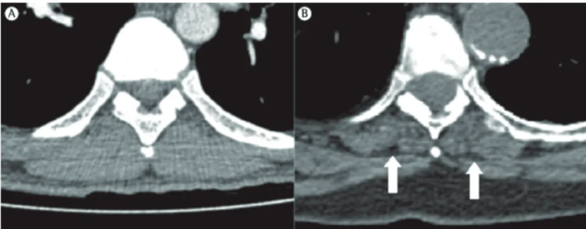
One of the most common imaging findings in the aging chest wall is a reduction in the thickness of the parietal muscles, particularly when compared with that of those in younger individuals, a change that can be easily seen on CT scans (Figure 1). This reduction is one of the major causes of increased pulmonary transparency on chest X-rays in the elderly. Although no
mass (due to hypertrophy) and age-related changes in the elastic properties of the myocardium.(18,19)
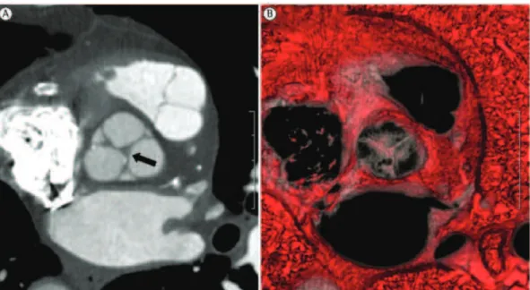
The principal radiological features of the “aging heart” include increased myocardial muscle mass and thickness (in particular, increased left ventricular muscle mass) due to myocyte hypertrophy and increased connective tissue matrix (Figure 2); marginal thickening of the heart valves (commonly the mitral and aortic valves) due to fat, collagen, and calcium salt deposition, causing wear of the valve annulus and, consequently, mild heart valve regurgitation in 90% of healthy patients over 80 years of age; and coronary sclerosis, possibly leading to changes in myocardial perfusion.(16,17,20-23) Although most of these changes have no clinical significance in healthy patients, they can contribute to decompensation in cases of cardiac overload due to external factors, such as infectious processes.
In most cases, the signs of right heart overload are due to increased pulmonary capillary resistance (as occurs in COPD and mitral valve dysfunction) and generally have a pathological basis; in contrast, the signs of left heart overload (left ventricular hypertrophy) are exclusively associated with cardiac aging in some cases.(22)
thickening, and bronchiectasis.(14,15) In patients with COPD, age is a factor that certainly contributes to disease progression. However, the individual roles of age and COPD in barrel chest cannot be determined by imaging tests.
Common imaging findings in the elderly include diaphragmatic bulging due to muscle hypertrophy and dyskinesia in some areas, particularly on the right side, probably caused by the effort of the hemidiaphragm to maintain the anatomical relationship between the lung and the liver.(11,13)
Aging-related changes in
radiological findings in the
mediastinum
Approximately 10% of the elderly population presents with changes that are exclusively related to cardiac aging. This select group of individuals is characterized by the exclusive presence of primary findings of cardiac aging, i.e., radiological findings that are not related to common comorbidities in this age group (arterial hypertension, COPD, atherosclerosis, diabetes, and renal failure).(16,17)
The most common physiological change related to cardiovascular aging is diastolic dysfunction, which is due to increased left ventricular muscle
collagen becomes more stable because of an increase in the number of intermolecular cross-links. The most widely accepted hypothesis is that lung elastic recoil is lost because of changes in the spatial arrangement of the network of collagen fibers or because of a protein known as pseudoelastin.(31)
Studies of the senescence-accelerated mouse have shown remarkably increased alveolar duct size during the aging process. Enlarged terminal air spaces have also been reported, having been characterized as a relatively homogeneous destruction, with few cellular infiltrates in the alveoli, suggesting that, unlike what occurs in emphysema, air space enlargement was not due to inflammation of the lung parenchyma.(36,37)
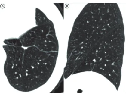
Turner et al.(34) found that the ratio of lung weight to body weight does not decrease with age in individuals in the 20-60 year age bracket, a finding that suggests little or no lung destruction, or tissue replacement.(34,36,37) During the aging process, the alveolar ducts increase in diameter and the alveoli become larger and shallower. After the fourth decade of life, part of the elastic fibers in the respiratory bronchioles and alveoli degenerate, their complacency therefore decreasing.(38) These changes are more pronounced around the alveolar ducts. Consequently, there is alveolar duct dilatation, followed by air space enlargement.(32) This enlargement is remarkably homogeneous, unlike the irregular distribution of air space enlargement in emphysema (Figures 3 and 4).
Morphometric studies have shown a progressive increase in the average distance between air space walls, as well as a decrease in the surface area of air space wall per unit of lung volume. (34,39,40) These changes begin in the third decade
of life and progress linearly and continuously, resulting in a 25-30% decrease in the surface area of air space wall per unit of lung volume in nonagenarians.(39,40) Although these changes are histologically different from those seen in pulmonary emphysema, in which there is destruction of the alveolar walls, they result in similar changes in lung compliance. As occurs with pulmonary emphysema, these changes cause a reduction in the supporting tissues around the airways, where there is a trend toward collapse of small (< 2-mm) airways and, consequently, changes in airflow. These morphostructural changes in the lung parenchyma constitute an aging-Changes in the aorta include elongation and
dilation, which are the major factors responsible for the chest X-ray finding of upper mediastinal enlargement in the elderly. In most patients, parietal calcifications of the aorta are most commonly seen in the aortic arch and in the descending portion of the aorta, constituting nonspecific radiological findings. However, in elderly individuals, calcification of the thoracic aorta, heart valves, and coronary arteries indicates a higher risk of cardiovascular diseases.(3,22,23)
Aging-related changes in
radiological findings in the lung
parenchyma
During the first two decades of life, the lungs grow and mature. The maximum number of alveoli is reached at approximately 10-12 years of age, and maturation of the respiratory system occurs at approximately 20 years of age in females and at approximately 25 years of age in males.
Decreased lung function is due to imbalances triggered by changes in the ventilation/perfusion ratio in patients at a more advanced age; however, together, these changes account for less than 3% of the total cardiac output, leading to a minimal (6-mmHg) decrease in PaO2 and having no clinical significance unless pulmonary function is affected by an underlying disease.(24-30)
One of the major aging-related physiological changes is decreased lung compliance. The two components of the elastic properties of the lung are surface and tissue forces.(31) There is no evidence that the surface-active lining of terminal respiratory units changes its basic mechanical behavior with age. No changes in the quality or quantity of alveolar surfactant have been described, and there is no evidence of changes in type II pneumocyte function.(32) However, changes in the lung parenchyma and chest wall are functionally significant.
Chest wall compliance decreases with age, which is principally due to musculoskeletal limitations, such as vertebral fractures, spondylosis, and progressive loss of respiratory muscle strength. (31,32) Lung parenchymal compliance normally
qualitative changes in collagen and in lung compliance.(11,13) The extent of the influence of each of these factors has yet to be determined. The initial pathophysiological consequence of these changes is air trapping due distal airway closure, with a progressive increase in RV. This related phenomenon that has been designated
“senile emphysema” (Figure 3).(38)
Aging-related parenchymal changes are caused by reduced blood flow from the systemic circulation through the bronchial arteries, as well as by the aforementioned quantitative/
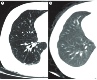
Figure 3 - Three-dimensional CT scan showing (in A) areas of increased small airway volume, known as senile emphysema (blue areas), in an 85-year-old patient. Note the homogeneous distribution of emphysematous areas. In B, three-dimensional CT scan of a healthy 23-year-old patient. Note that there are no areas suggestive of pulmonary emphysema.
pulmonary hypertension (a clinical manifestation of mild vascular sclerosis) that can redistribute pulmonary flow cranially and be mistaken for early signs of cardiac decompensation.(41-43)
The major determinants of static lung volumes are chest wall compliance and lung parenchymal compliance. Loss of lung parenchymal compliance and, to a lesser degree, decreased respiratory muscle strength result in an increase in RV (air mechanism is analogous to that of pulmonary
emphysema, with no signs of inflammation and no significant increase in TLC. At the same time, the ventilation/perfusion ratio changes because of a reduction in the number of alveoli with normal gas exchange; this has two pathophysiological consequences: increased physiological dead space and the shunt effect, both of which lead to a decrease in PaO2.(39,41) In addition, there is mild
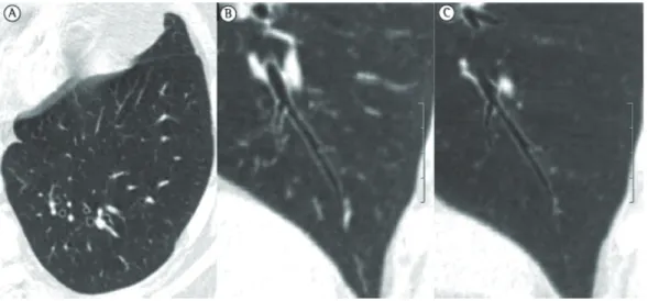
Figure 5 - Asymptomatic 87-year-old female patient. In A, axial CT scan of the left lower lobe showing laminar atelectasis at the lung bases. In B, sagittal reconstruction of this finding.
Figure 6 - Asymptomatic 83-year-old female patient. Axial CT scan taken in the prone position, showing subpleural linear septal thickening (arrows). These radiological findings are indistinguishable from those seen in patients with interstitial lung disease due to other causes.
Likewise, correlation with pulmonary function test results (particularly DLCO) can demonstrate how gas exchange is occurring and guide a conservative approach. Another fact that should be taken into consideration in the elderly is life expectancy and the metabolic need for gas exchange, given that patients whose activity is limited by extrathoracic disease have lower physiological needs. This brings us to the well-known Hippocratic principle of primum non nocere (above all, do no harm), which is increasingly true today, given the various choices of procedures and the increase in survival of the population.
References
1. Bonomo L, Larici AR, Maggi F, Schiavon F, Berletti R. Aging and the respiratory system. Radiol Clin North Am. 2008;46(4):685-702, v-vi. PMid:18922288. http:// dx.doi.org/10.1016/j.rcl.2008.04.012
2. Maggi S, Marzari C, Crepaldi G. Epidemiologia dell’invecchiamento. In: Guglielmi G, Schiavon F, Cammarota T, editors. Radiologia geriatrica. Milano: Springer; 2006. p. 13-20. http://dx.doi. org/10.1007/88-470-0486-1_3
3. Schiavon F, Nardini S, Favat M, Manfrin P, Chioatto P. Radiology of normal chest structures in the elderly patient [Article in Italian]. Radiol Med. 1993;86(4):418-31. 4. Barreto SM. O envelhecimento e a função pulmonar. J
Pneumol. 1983;9(3):160-5.
5. Bassey EJ, Harries UJ. Normal values for handgrip strength in 920 men and women aged over 65 years,
trapping), which increases by approximately 50% between ages 20 and 70 years. Conversely, VC progressively decreases to approximately 75% of optimal values. Therefore, TLC remains constant throughout life.(44) The closing volume of the small airways (volume at which the small airways begin to close during exhalation) increases with age. This premature closure begins to exceed functional residual capacity at age 44 years and exceeds it at age 65 years,(45) being closely related to the loss of supporting tissues around the airways. This is one of the theories for the aging-related decrease in the ventilation/perfusion ratio.(46-49)
A CT scan of the lung parenchyma shows findings that are quite common in the elderly, and it is speculated that these findings are related to collagen changes. These findings are laminar atelectasis, mostly posterior and basal, located in the dependent regions of the lungs (Figure 5); subpleural linear thickening (Figure 6); areas of air trapping (Figure 7); bronchial thickening and ectasia (Figure 8); and lung cysts.(41,50-53)
The differential diagnosis between normal, aging-related imaging findings and imaging findings secondary to disease is quite difficult, and it is often impossible to distinguish between the two types of findings using imaging tests alone. Monitoring these lesions is often necessary, and comparison with previous tests is indispensable.
22. Di Guglielmo L, Dore R, Raisaro A, Pallavicini D. Diagnostic imaging in the study of heart aging. Is the “senile heart” a fact? [Article in Italian]. Radiol Med. 1999;97(6):449-60. 23. McLaughlin MA. The aging heart. State-of-the-art
prevention and management of cardiac disease. Geriatrics. 2001;56(6):45-9; quiz 50. PMid:11417374. 24. Krumpe PE, Knudson RJ, Parsons G, Reiser K. The aging
respiratory system. Clin Geriatr Med. 1985;1(1):143-75. PMid:3913497.
25. Murray JF. Aging. In: Murray JF, editors. The normal lung: the basis for diagnosis and treatment of pulmonary disease. Philadelphia: Saunders; 1986. p. 339-60. 26. Janssens JP. Aging of the respiratory system: impact on
pulmonary function tests and adaptation to exertion. Clin Chest Med. 2005;26(3):469-84, vi-vii. PMid:16140139. http://dx.doi.org/10.1016/j.ccm.2005.05.004 27. Wagner PD, Laravuso RB, Uhl RR, West JB. Continuous
distributions of ventilation-perfusion ratios in normal subjects breathing air and 100 per cent O2. J Clin Invest. 1974;54(1):54-68. PMid:4601004 PMCid:301524. http://dx.doi.org/10.1172/JCI107750
28. Wagner PD, Saltzman HA, West JB. Measurement of continuous distributions of ventilation-perfusion ratios: theory. J Appl Physiol. 1974;36(5):588-99. PMid:4826323. 29. Leblanc P, Ruff F, Milic-Emili J. Effects of age and
body position on “airway closure” in man. J Appl Physiol. 1970;28(4):448-51. PMid:5437433.
30. Delclaux B, Orcel B, Housset B, Whitelaw WA, Derenne JP. Arterial blood gases in elderly persons with chronic obstructive pulmonary disease (COPD). Eur Respir J. 1994;7(5):856-61. PMid:8050540.
31. Well DS, Meier JM, Mahne A, Houseni M, Hernandez-Pampaloni M, Mong A, et al. Detection of age-related changes in thoracic structure and function by computed tomography, magnetic resonance imaging, and positron emission tomography. Semin Nucl Med. 2007;37(2):103-19. PMid:17289458. http://dx.doi.org/10.1053/j. semnuclmed.2006.10.004
32. Edge JR, Millard FJ, Reid L, Simon G. The radiographic appearances of the chest in persons of advanced age. Br J Radiol. 1964;37:769-74. Niewohner D, Kleinerman J, Liotta L. Elastic behavior of post-mortem human lungs: effects of aging and mild emphysema. J Appl Physiol 1975; 38(7):943-9.
33. Niewohner D, Kleinerman J, Liotta L. Elastic behavior of post-mortem human lungs: effects of aging and mild emphysema. J Appl Physiol 1975; 38(7):943-9. 34. Turner JM, Mead J, Wohl ME. Elasticity of human lungs
in relation to age. J Appl Physiol. 1968;25(6):664-71. PMid:5727191.
35. Lang MR, Fiaux GW, Gillooly M, Stewart JA, Hulmes DJ, Lamb D. Collagen content of alveolar wall tissue in emphysematous and non-emphysematous lungs. Thorax. 1994;49(4):319-26. PMid:8202900 PMCid:475363. http://dx.doi.org/10.1136/thx.49.4.319
36. Kurozumi M, Matsushita T, Hosokawa M, Takeda T. Age-related changes in lung structure and function in the senescence-accelerated mouse (SAM): SAM-P/1 as a new murine model of senile hyperinflation of lung. Am J Respir Crit Care Med. 1994;149(3 Pt 1):776-82. PMid:8118649.
37. Teramoto S, Fukuchi Y, Uejima Y, Teramoto K, Oka T, Orimo H. A novel model of senile lung: senescence-accelerated mouse (SAM). Am J Respir Crit Care Med. 1994;150(1):238-44. PMid:8025756.
and longitudinal changes over 4 years in 620 survivors. Clin Sci (Lond). 1993;84(3):331-7.
6. Carmeli E, Reznick AZ. The physiology and biochemistry of skeletal muscle atrophy as a function of age. Proc Soc Exp Biol Med. 1994;206(2):103-13. PMid:8208732. 7. Irion KL, Marchiori E, Hochhegger B, Porto Nda S, Moreira
Jda S, Anselmi CE, et al. CT quantification of emphysema in young subjects with no recognizable chest disease. AJR Am J Roentgenol. 2009;192(3):W90-6. PMid:19234245. http://dx.doi.org/10.2214/AJR.07.3502
8. Booth FW, Weeden SH, Tseng BS. Effect of aging on human skeletal muscle and motor function. Med Sci Sports Exerc. 1994;26(5):556-60. http://dx.doi. org/10.1249/00005768-199405000-00006
9. Baumgartner RN, Stauber PM, McHugh D, Koehler KM, Garry PJ. Cross-sectional age differences in body composition in persons 60+ years of age. J Gerontol A Biol Sci Med Sci. 1995;50(6):M307-16. http://dx.doi. org/10.1093/gerona/50A.6.M307
10. Newman AB, Haggerty CL, Goodpaster B, Harris T, Kritchevsky S, Nevitt M, et al. Strength and muscle quality in a well-functioning cohort of older adults: the Health, Aging and Body Composition Study. J Am Geriatr Soc. 2003;51(3):323-30. PMid:12588575. http:// dx.doi.org/10.1046/j.1532-5415.2003.51105.x 11. Bernadac P. Le poumon du troisième age. Encycl Med Chir
(Paris), Radiodiagnostic VI. 1991;4-02-05:324980-10. 12. Muiesan G, Sorbini CA, Grassi V. Respiratory function in the
aged. Bull Physiopathol Respir (Nancy). 1971;7(5):973-1009. 13. Zeleznik J. Normative aging of the respiratory system. Clin
Geriatr Med. 2003;19(1):1-18. http://dx.doi.org/10.1016/ S0749-0690(02)00063-0
14. Bafadhel M, Umar I, Gupta S, Raj JV, Vara DD, Entwisle JJ, et al. The role of CT scanning in multidimensional phenotyping of COPD. Chest. 2011;140(3):634-42. PMid:21454400 PMCid:3168858. http://dx.doi. org/10.1378/chest.10-3007
15. Irion KL, Hochhegger B, Marchiori E, Porto Nda S, Baldisserotto Sde V, Santana PR. Chest X-ray and computed tomography in the evaluation of pulmonary emphysema. J Bras Pneumol. 2007;33(6):720-32. PMid:18200374. http://dx.doi.org/10.1590/S1806-37132007000600017 16. Badano L, Carratino L, Giunta L, Calisi P, Lucatti
A. Age-induced changes in the cardiovascular system in normal subjects [Article in Italian]. G Ital Cardiol. 1992;22(9):1023-34. PMid:1291420. 17. Midiri M. Thoracic examination in cardiac aging [Article
in Italian]. Radiol Med. 2003;106(3 Suppl 1):50-3. 18. Boogers MJ, van Werkhoven JM, Schuijf JD, Delgado V,
El-Naggar HM, Boersma E, et al. Feasibility of diastolic function assessment with cardiac CT: feasibility study in comparison with tissue Doppler imaging. JACC Cardiovasc Imaging. 2011;4(3):246-56. PMid:21414572. http:// dx.doi.org/10.1016/j.jcmg.2010.11.017
19. Mandinov L, Eberli FR, Seiler C, Hess OM. Diastolic heart failure. Cardiovasc Res. 2000;45(4):813-25. http://dx.doi. org/10.1016/S0008-6363(99)00399-5
20. Badano L, Carratino L, Giunta L, Calisi P, Lucatti A. Age-induced changes in the cardiovascular system in normal subjects [Article in Italian]. G Ital Cardiol. 1992;22(9):1023-34. PMid:1291420.
47. Quanjer P. Standardized lung function testing: report working party ‘‘Standardization of lung function tests’’. European Community for Coal and Steel, Luxembourg. Bull Eur Physiopathol Respir 1983;19(Suppl 5):1-95. PMid:6616097.
48. Kerstjens HA, Rijcken B, Schouten JP, Postma DS. Decline of FEV1 by age and smoking status: facts, figures, and fallacies. Thorax. 1997;52(9):820-7. PMid:9371217 PMCid:1758654. http://dx.doi.org/10.1136/thx.52.9.820 49. Dockery DW, Ware JH, Ferris BG Jr, Glicksberg DS, Fay
ME, Spiro A 3rd, et al. Distribution of forced expiratory volume in one second and forced vital capacity in healthy, white, adult never-smokers in six U.S. cities. Am Rev Respir Dis. 1985;131(4):511-20. PMid:3873193. 50. Hansell DM. Thin-section CT of the lungs: the Hinterland
of normal. Radiology. 2010;256(3):695-711. Erratum in: Radiology. 2010;257(3):897. PMid:20720066. http:// dx.doi.org/10.1148/radiol.10092307
51. Copley SJ, Wells AU, Hawtin KE, Gibson DJ, Hodson JM, Jacques AE, et al. Lung morphology in the elderly: comparative CT study of subjects over 75 years old versus those under 55 years old. Radiology. 2009;251(2):566-73. PMid:19401580. http://dx.doi.org/10.1148/ radiol.2512081242
52. Hochhegger B, Irion K, Bello R, Marchiori E, Moreira J, Porto Nda S, et al. Understanding the classification, physiopathology and the diagnostic radiology of bronchiectasis [Article in Portuguese]. Rev Port Pneumol. 2010;16(4):627-39. PMid:20700560. 53. Irion KL, Marchiori E, Hochhegger B. Tomographic
diagnosis of pulmonary emphysema. J Bras Pneumol. 2009;35(9):821-3. http://dx.doi.org/10.1590/ S1806-37132009000900001
38. Verbeken EK, Cauberghs M, Mertens I, Clement J, Lauweryns JM, Van de Woestijne KP. The senile lung. Comparison with normal and emphysematous lungs. 1. Structural aspects. Chest. 1992;101(3):793-9. PMid:1541148. http:// dx.doi.org/10.1378/chest.101.3.793
39. Gillooly M, Lamb D. Airspace size in lungs of lifelong non-smokers: effect of age and sex. Thorax. 1993;48(1):39-43. PMid:8434351 PMCid:464237. http://dx.doi.org/10.1136/thx.48.1.39
40. Thurlbeck WM. The internal surface area of nonemphysematous lungs. Am Rev Respir Dis. 1967;95(5):765-73. PMid:6023510.
41. Sharma G, Goodwin J. Effect of aging on respiratory system physiology and immunology. Clin Interv Aging. 2006;1(3):253-60. PMid:18046878 PMCid:2695176. http://dx.doi.org/10.2147/ciia.2006.1.3.253
42. Freundlich IM. Redistribution of pulmonary blood flow. AJR Am J Roentgenol. 1985;145(6):1315-6. PMid:3877444. 43. Levin DL, Buxton RB, Spiess JP, Arai T, Balouch J,
Hopkins SR. Effects of age on pulmonary perfusion heterogeneity measured by magnetic resonance imaging. J Appl Physiol. 2007;102(5):2064-70. PMid:17303711. http://dx.doi.org/10.1152/japplphysiol.00512.2006 44. Crapo RO. The aging lung. In: Mahler DA, editor.
Pulmonary disease in the elderly patient. New York: Marcel Dekker; 1993. p. 1-21.
45. Tockman M. Aging of the respiratory system. In: Hazzard WR, Blass JP, Halter JB, Ouslander JG, Tinetti ME, editors. Principles of geriatric medicine and gerontology. New York: McGraw-Hill; 1994. p. 555-64.
About the authors
Bruno Hochhegger
Thoracic Radiologist. Department of Pulmonology, Santa Casa Hospital Complex in Porto Alegre; and Professor of Radiology. Universidade Federal de Ciências da Saúde de Porto Alegre – UFSCPA, Federal University of Health Sciences of Porto Alegre – Porto Alegre, Brazil.
Gustavo Souza Meirelles
Radiologist. Fleury Medicina Diagnóstica, São Paulo, Brazil. Klaus Irion
Consultant Radiologist. Liverpool Heart and Chest Hospital, Liverpool, United Kingdom. Gláucia Zanetti
Professor of Pulmonology. Petrópolis School of Medicine, Petrópolis, Brazil. Eduardo Garcia
Professor of Pulmonology and Geriatrics. Universidade Federal de Ciências da Saúde de Porto Alegre – UFSCPA, Federal University of Health Sciences of Porto Alegre – Porto Alegre, Brazil.
José Moreira
Professor of Pulmonology. Universidade Federal do Rio Grande do Sul – UFRGS, Federal University of Rio Grande do Sul – School of Medicine, Porto Alegre, Brazil.
Edson Marchiori
Professor of Radiology. Federal University of Rio de Janeiro, Rio de Janeiro, Brazil.
Erratum
Manuscript: The chest and aging: radiological findings
Publication: Jornal Brasileiro de Pneumologia. 2012;38(5):656-65
DOI:10.1590/S1806-37132012000500016
On page 656 of the original publication, in the third line, where is written “Gustavo Pontes de Meireles” should be read “Gustavo Souza Meirelles.”
On pages 658, 660, 662, and 664 of the original publication, in the header, where is written “Meireles GP” should be read “Meirelles GS.”