Arq Neuropsiquiatr 2006;64(1):128-131
Hospital São José, Complexo Hospitalar Santa Casa de Porto Alegre, Porto Alegre RS, Brazil:1Postgraduate Professor at the Medical
School - Fundação Faculdade Federal de Ciências Médicas de Porto Alegre (FFFCMPA), Neuro s u rgeon at Hospital São José - Complexo Hospitalar Santa Casa (HSJ/CHSC); 2Medical - Residents in Neurosurgery at HSJ/CHSC.
Received 6 June 2005, received in final form 24 August 2005. Accepted 17 October 2005.
Dr. Jorge Luiz Kraemer - Rua Padre Chagas 415 / 702 - 90570-080 Porto Alegre RS - Brasil. E-mail: [email protected]
VERTEBROBASILAR DOLICHOECTASIA
AS A CAUSE OF TRIGEMINAL NEURALGIA
THE ROLE OF MICROVASCULAR DECOMPRESSION
Case report
Jorge Luiz Kraemer
1, Arthur de Azambuja Pereira Filho
2,
Gustavo de David
2, Mario de Barros Faria
2ABSTRACT - Our purpose is to re p o rt a case of trigeminal neuralgia caused by vert e b robasilar dolichoec-tasia treated with microvascular decompression. A 63-year-old man sought treatment for a recurrent lan-cinating left facial pain in V2 and V3 trigeminal territories. The computed tomography angiography re v e a l e d a mechanical compression of the left trigeminal nerve due to vertebrobasilar dolichoectasia. The patient
was submitted to a left suboccipital craniotomy. Shredded Teflon®was introduced in the conflicting
neu-rovascular area, achieving a satisfactory decompression. The patient’s pain resolved immediately. Ve rt e b robasilar dolichoectasia is a rare cause of trigeminal neuralgia and a successful outcome can be achieved with microvascular decompression.
KEY WORDS: trigeminal neuralgia, vertebrobasilar dolichoectasia, microvascular decompression.
Dolicoectasia vertebrobasilar como causa de neuralgia trigeminal: o papel da descompressão microvascular. Relato de caso
RESUMO - O objetivo desse estudo é relatar um caso de neuralgia trigeminal causado por dolicoectasia vertebrobasilar tratado com descompressão microvascular. Um homem (63 anos) consultou por neuralgia trigeminal re c o rrente na hemiface esquerda (territórios V2 e V3). A angiotomografia cerebral revelou com-pressão mecânica do nervo trigêmio esquerdo devido à dolicoectasia vertebrobasilar. O paciente foi sub-metido à craniotomia suboccipital esquerda. Introduziu-se Te f l o n®na área de conflito neuro v a s c u l a r,
obten-do-se uma descompressão satisfatória. O paciente apresentou remissão da dor imediatamente. A dolicoec-tasia vert e b robasilar é uma causa rara de neuralgia trigeminal e uma excelente evolução pode ser alcança-da com a descompressão microvascular.
PALAVRAS-CHAVE: neuralgia trigeminal, dolicoectasia vertebrobasilar, descompressão microvascular.
Trigeminal neuralgia is a common facial pain syn-d rome which usually affects misyn-dsyn-dle-agesyn-d ansyn-d elsyn-der- elder-ly people. The syndrome consists of paroxysms of lan-cinating pain, usually in the distribution of the mandibular and maxillary divisions of the trigeminal n e rve. Patients often involuntarily wince when expe-riencing this severe pain, providing the derivation of the termtic douloure x1. The most common cause of
idiopathic trigeminal neuralgia is microvascular com-pression of the nerve2. A compressing vessel is
iden-tified for most patients who undergo micro s u rg i c a l d e c o m p ression, being the superior cerebellar art e ry responsible for 75% of cases3. Other arteries, such as
the anteroinferior cerebellar art e ry (10%), postero i n-ferior cerebellar art e ry (1%), vertebral art e ry (2%), basilar art e ry (1%), and primitive trigeminal art e ry or its variants, have also been identified as the cause of this condition4,5. Tumors, aneurysms and vascular
m a l f o rmations are observed in only a few cases3.
Arq Neuropsiquiatr 2006;64(1) 129
of trigeminal neuralgia3 , 6. Many surgical or
nonsur-gical modalities of treatment have been pro p o s e d for trigeminal neuralgia. Microvascular decompre s-sion is the most effective surgical modality available. It is nondestructive, mortality and morbidity rates a re low when properly perf o rmed, and it confers the best short and long-term quality of life to the patients7.
The purpose of this study is to re p o rt and discuss a rare case of trigeminal neuralgia due to vert e-b roe-basilar dolichoectasia successfully treated with m i c rovascular decompression and documented by computed tomography angiography (CTA).
CASE
A 63-year-old man with a past medical history of hyper-tension sought tr eatment after experienci ng a re c u rre n t lancinating left facial pain in trigeminal territories (V2 and V3) for almost five years. The pain was described as sharp and electrical and was exacerbated by talking, chewing and sometimes was spontaneously triggered. These symptoms resolved by October 2001, after a percutaneous surg i c a l p ro c e d u re(radiofrequency lesioning of the gasserian gan-glion). After a pain-free period of almost 4 years, the pain re c u rred with the same characteristics. High doses of car-bamazepine and amitriptyline did not relieve the pain ade-q u a t e l y. The patient was re f e rred with clinically intractable symptoms and subsequently considered for micro s u rg i c a l decompression after neurological reinvestigation.
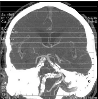
The patient’s neurological examination revealed hyper-esthesia in the V2 and V3 distribution of the trigeminal n e rve on the left side. All the others aspects of the neuro-logical examination were normal. The CTA revealed a
mechanical compression at the left trigeminal nerve due to vert e b robasilar dolichoectasia (Figs 1 and 2). Surg e ry was indicated.
The patient was placed in the prone oblique( p a r k bench)position, and a left suboccipital craniotomy was per-f o rmed. The dura was opened, and cere b rospinal per-fluid was released at the cisterna magna to provide a capacious work-ing environment. Arachnoid dissection revealed a large vas-cular stru c t u re, later identified as the basilar dolichoectat-ic art e ry, dislocating and compressing the left trigeminal n e rve at its root entry zone (Fig 3). Shredded Te f l o n®w a s
i n t roduced in the conflicting neurovascular area (between the artery and the trigeminal nerve), achieving a satisfac-tory decompression. There was no other vascular or nerve microsurgical manipulation.
Fig 1. Standard CTA (coronal view) showing the dolichobasilar ectasia.
Fig 2. Reconstruction 3D CTA showing the dolichobasilar ecta -sia and its relations to skull base.
130 Arq Neuropsiquiatr 2006;64(1)
The patient’s lancinating facial pain resolved immedi-ately after surg e ry. He initially presented with mild dise-quilibrium, but it was completely resolved at a 3-month follow-up examination.
DISCUSSION
Ve rt e b robasilar dolichoectasia is an uncommon vasculopathy of unclear etiology which affects the a rterial wall of vertebral and/or basilar art e r i e s8.
Tr a d i t i o n a l l y, vert e b robasilar dolichoectasia has been re g a rded as athero s c l e rotic in nature, although recently Mizutani and Aruga have suggested that some cases re p resent a dissecting pro c e s s9 , 1 0. This
dis-ease causes arterial elongation and enlarg e m e n t , with subsequent haemodynamic and haemostatic changes, which, in turn, lead to thrombosis, micro -embolisation, and brainstem compression, with or without aneurysm form a t i o n1 1. A variety of clinical
s y n d romes have been associated with ectatic vert e-b roe-basilar arteries. These include a nume-ber of isolat-ed or combinisolat-ed brainstem/cranial nerve syndro m e s , c e rv i c o m e d u l l a ryjunction compression, transient or p e rmanent motor deficits, cerebellar dysfunction, central sleep apnea, hydrocephalus and ischemic stroke11-13.
D i rect compression by vert e b robasilar dolichoec-tasia is an uncommon cause for trigeminal neural-gia. The incidence, as estimated in previous re p o rt s , ranges from 0.9% to 5.7%1 4. Piatt et al.1 5re p o rted 2
cases in a series of 105 patients. Bederson et al.1 6re l a
t-ed 4 cases in a group of 256 operatt-ed cases. Klun et a l .1 7re p o rted 2 cases in a group of 220 operated
patients. Vascular compression usually occurs at or near the root entry zone (REZ) of the trigeminal n e rve, as re p o rted by some authors. Hamlyn1 8
o b s e rved that 42 out of 46 patients who underw e n t posterior fossa surg e ry for treatment of trigeminal neuralgia had a vessel in contact with the nerve. Of those, 28 had a vessel in contact at the REZ, 12 had a vessel in contact lateral to the REZ (the point of contact with the nerve was more than one-half of the vessel’s diameter away from the brainstem), and 2 had a vessel in contact at the REZ as well as later-al to it. Sindou et later-al.1 9o b s e rved the presence of a
contacting vessel in 97% of 579 patients with idio-pathic trigeminal neuralgia. The site of contact was at the REZ in 52% of cases, in the mid-third of the n e rve in 54%, and at the exit of the nerve fro m M e c k e l ’s cave in 10%. In the present case, the con-flicting neurovascular area was located at the REZ.
Several operative treatments for trigeminal neu-ralgia are in current use, including radiofre q u e n c y
gasserian rhizotomy, glycerol postgasserian rhizoly-sis, balloon compression of the gasserian ganglion, and microvascular decompression of the trigeminal root. When cranial nerve dysfunction, especially trigeminal neuralgia, is caused by anomalies of cal-i b e r, length, and tortuoscal-ity of the vert e b ro b a s cal-i l a r a rteries, alternative techniques, such as re p o s i t i o n-ing of the tortuous vert e b robasilar art e ry by pulln-ing it toward the nearby dura mater2 0and encirc l i n g
method of trigeminal nerve decompre s s i o n1 4h a v e
been re p o rted re c e n t l y. In the present case case, the authors thought that these techniques would not bring advantages over the microvascular decompre s-sion20.
M i c rovascular decompression for hyperactive dys-function of cranial nerves was initially developed by G a rdener and Miklos2 1and Gardner and Sava2 2a n d
was perfected and popularized by Jannetta2 3 - 2 5a f t e r
the introduction of the micro s u rgical technique under an operative micro s c o p e2 6. Microvascular decompre
s-sion for trigeminal neuralgia has proven to be a high-ly effective and safe surgical pro c e d u rein alleviating the effects of neurovascular compre s s i o n2 7. Compare d
to alternative treatments, microvascular decompre s-sion offers significant advantages for trigeminal neu-r a l g i a2 8. There is a growing body of evidence
sug-gesting microvascular decompression as the best sur-gical modality for trigeminal neuralgia7. The rates of
success (free of pain, without medication) are supe-rior or at least equal to those of other re p o rted tre a t-ments, with substantially lower rates of facial numb-ness28.
The majority of the series in the literature re p o rt s a percentage of pain relief between 63% and 94%7
with well-defined follow-ups (mean time2 years). H o w e v e r, the incidence of re c u rrence has been re p o rted to range from 3 to 30%2 9. Long-term
fol-low-up studies revealed that most postoperative re c u rrences of trigeminal neuralgia occurred in the first 2 years after surg e ry2 9. Mendoza and
I l l i n g w o rt h3 0 re p o rted that 90% of re c u rre n c e s
o c c u rred within 2 years. The annual rate of re c u r-rence for trigeminal neuralgia decreases below 2% within 5 years after surg e ry and below 1% within 10 years after surg e ry2 9. Twenty-year follow-up data
demonstrated that 30% of successfully tre a t e d patients experienced trigeminal neuralgia re c u r-rences29.
recur-Arq Neuropsiquiatr 2006;64(1) 131
rence. Preoperative sensory deficits, a history of a trigeminal ablative pro c e d u re, and the number of trigeminal divisions affected by trigeminal neuralgia were not significant predictors29.
In the present case, the patient’s lancinating facial pain resolved immediately after surgery. He initially presented with mild disequilibrium, but it was com-pletely resolved at a 3-month follow-up examination. We attribute it to the manipulation of the vestibu-lar nerve, and the complete resolution of this symp-tom after a 3-month follow-up re i n f o rces this suspi-cion. The follow-up period in the present case is cer-tainly short, but the patient does not present any of the previously re p o rted predicting re c u rrence fac-tors, so we strongly believe that the cure with m i c rovascular decompression in this case is very like-ly to be obtained.
In conclusion, vert e b robasilar dolichoectasia is a r a re cause of trigeminal neuralgia and a successful outcome can be achieved with microvascular decom-pression.
REFERENCES
1. Ali MJ, Gebarski S, Thompson BG. Transient magnetic resonance imag-ing signal alterations in the brainstem after microvascular decompre s-sion for trigeminal neuralgia: case report. Neuro s u rgery 2004; 55: E1023-1026.
2. Deshmukh VR, Hott JS, Tabrizi P, Nakaji P, Feiz-Erfan I, Spetzler R. Cavernous malformation of the trigeminal nerve manifesting with trigeminal neuralgia: case report. Neurosurgery 2005;56:E623. 3. Du R, Binder DK, Halbach V, Fischbein N, Barbaro N. Trigeminal
neu-ralgia in a patient with a dural arteriovenous fistula in Meckel’s cave: case report. Neurosurgery 2003;53:216-221.
4. Barker FGII, Jannetta PJ, Bissonette DJ, Larkins MV, Jho HD. The long-term outcome of microvascular decompression for trigeminal neural-gia. N Engl J Med 1996;334:1077-1083.
5. Morita A, Fukushima T, Miyazaki S, Shimizu T, Atsuchi M. Tic dou-l o u reux caused by primitive trigeminadou-l artery or its variant. J Neuro s u rg 1989;70:415-419.
6. Kirsch E, Hausmann O, Kaim A, Gratzl O, Steinbrich W, Radu EW. Magnetic resonance imaging of vertebrobasilar ectasia in trigeminal neuralgia. Acta Neurochir (Wien) 1996;138:1295-1299.
7. Ashkan K, Marsh H. Microvascular decompression for trigeminal neu-ralgia in the elderly: a review of the safety and eff i c a c y. Neuro s u rg e r y 2004;55:840-850.
8. Ubogu EE, Chase CM, Ve r rees MA, et al. Cervicomedullary junction c o m p ression caused by vertebral artery dolichoectasia and re q u i r i n g surgical treatment: case report. J Neurosurg 2002;96:140-143. 9. Craig R, Barnwell S. Catastrophic subarachnoid hemorrhage re s u l t i n g
f rom ru p t u red vertebrobasilar dolichoectasia: case report. Neuro s u rg e r y 1998;42:379-382.
10. Mizutani T, A ruga T. Dolichoectatic intracranial vertebrobasilar dissec-ting aneurysm. Neurosurgery 1992;31:765-773.
11. Ubogu EE, Zaidat OO. Ve r t e b robasilar dolichoectasia diagnosed by magnetic resonance angiography and risk of stroke and death: a cohort study. J Neurol Neurosurg Psychiatry 2004;75:22-26.
12. Yu YL, Moseley IF, Pullicino P, et al. The clinical picture of ectasia of the i n t r a c e rebral arteries. J Neurol Neuro s u rg Psychiatry 1982;45:29-36. 13. M i l a n d re L, Bonnefori B, Pestre P, et al. Ve r t e b robasilar arterial
dolicho-ectasia: complications and prognosis. Rev Neurol (Paris) 1991;147:714-722.
14. Yoshimoto Y, Noguchi M, Tsutsumi Y. Encircling method of trigemnal nerve decompression for neuralgia caused by tortuous vertebro b a s i-lar artery: technical note. Surg Neurol 1995;43:151-153.
15. Piatt J, Wilkins RH. Treatment of tic douloureux and hemifacial spasm by posterior fossa exploration: therapeutic implications of various neu-rovascular relationships. Neurosurgery 1984;14:462-471.
16. Bederson JB, Wilson CB. Evaluation of microvascular decompre s s i o n and partial sensory rhizotomy in 252 cases of trigeminal neuralgia. J Neurosurg 1989;71:359-367.
17. Klun B. Microvascular decompression and partial sensory rhizotomy in the treatment of trigeminal neuralgia: personal experience with 220 patients. Neurosurgery 1992;30:49-52.
18. Hamlyn PJ. Neurovascular relationships in the posterior cranial fossa, with special re f e rence to trigeminal neuralgia: Part I. Review of the lit-e r a t u rlit-e and dlit-evlit-elopmlit-ent of a nlit-ew mlit-ethod of vascular injlit-ection-filling in cadaveric controls. Clin Anat 1997;10:371-379.
19. Sindou M, Howeidy T, Acevedo G. Anatomical observations during m i c rovascular decompression for idiopathic trigeminal neuralgia (with c o r relations between topography of pain and site of the neuro v a s c u-lar conflict): Prospective study in a series of 579 patients. Acta Neuro c h i r (Wien) 2002;144:1-13.
20. Tomasello F, Alafaci C, Salpietro FM, Longo M. Bulbar compression by an ectatic vertebral artery: a novel neurovascular construct relieved by microsurgical decompression. Operative Neurosurg 2005;56:117-124. 21. Gardner WJ, Miklos MV. Response of trigeminal neuralgia to
decom-p ression of sensory root: discussion of cause of trigeminal neuralgia. JAMA 1959;170:1773-1776.
22. G a rdner WJ, Sava GA. Hemifacial spasm: a reversible pathophysio-logic state. J Neurosurg 1962;19:240-247.
23. Jannetta PJ. Neurovascular compression in cranial nerve and systemic disease. Ann Surg 1980;192:518-525.
24. Jannetta PJ. Treatment of trigeminal neuralgia by microoperative decom-p ression. Youmans JR (Ed). Neurological surg e r y, ed 2. Philadeldecom-phia: WB Saunders, 1982:3589-3603.
25. Jannetta PJ. Microsurgical management of trigeminal neuralgia. Arch Neurol 1985;42:800.
26. Kondo A. Follow-up results of microvascular decompression in trigem-inal neuralgia and hemifacial spasm. Neurosurgery 1997;40:46-52. 27. K u reshi SA, Wilkins R. Posterior fossa reexploration for persistent or
re c u r rent trigeminal neuralgia or hemifacial spasm: surgical findings and therapeutic implications. Neurosurgery 1998;43:1111-1116. 28. Kalkanis SN, Eskander EN, Carter BS, Barker FG. Microvascular
decom-p ression surgery in the United States, 1996 to 2000: mortality rates, and the effects of hospital and surgeon volumes. Neuro s u rgery 2003;52: 1251-1262.
29. Lee SH, Levy EI, Scarrow AM, Kassam A, Jannetta PJ. Recurrent trigem-inal neuralgia attributable to veins after microvascular decompre s s i o n . Neurosurgery 2000;46: 356.