The
bovine
tuberculosis
burden
in
cattle
herds
in
zones
with
low
dose
radiation
pollution
in
Ukraine
Richard
Weller
(1),
Artem
Skrypnyk
(2),
Andriy
Zavgorodniy
(2),
Borys
Stegniy
(2),
Anton
Gerilovych
(2),
Oleksandr
Kutsan
(2),
Svitlana
Pozmogova
(2)&
Svitlana
Sapko
(2)Summary
The authors describe a study of the tuberculosis (TB) incidence in cattle exposed to low doses of radiation resulting from the Chernobyl (pronounced ‘Chornobyl’ in Ukrainian) nuclear plant catastrophe in 1986. The purpose of the study was to determine if ionising radiation influences the number of outbreaks of bovine TB and their severity on farms in the Kyiv, Cherkasy and Chernigiv regions of Ukraine. These farms are all located within a 200 km radius of Chernobyl and have had low‐dose radiation pollution. Pathological and blood samples were taken from cattle in those regions that had positive TB skin tests.
Mycobacterium spp. were isolated, differentiated by PCR, analysed and tested in guinea‐pigs and rabbits. Species differentiation showed a significant percentage of atypical mycobacteria, which resulted in the allergic reactions to tuberculin antigen in the skin test. Mixed infection of M. bovis and M. avium subsp.
hominissuis was found in three cases. The results concluded that low‐dose radiation plays a major role in the occurrence of bovine TB in regions affected by the Chernobyl nuclear disaster.
Keywords
Bovine tuberculosis, Cattle, Chernobyl, Chornobyl, Low dose, Mycobacteria,
Mycobacterium bovis, Radiation, Tuberculosis, Ukraine.
Il
peso
della
tubercolosi
bovina
nelle
mandrie
site
nelle
zone
a
basso
inquinamento
radioattivo
in
Ucraina
Riassunto
Gli autori descrivono uno studio sull’incidenza della tubercolosi (TB) condotto sul bestiame esposto a bassi livelli di radiazioni provocate dal disastro nucleare di Chernobyl (‘Chornobyl’ in ucraino) nel 1986. Scopo dello studio era stabilire se le radiazioni ionizzanti influenzano il numero di focolai di TB bovina e la loro gravità nelle fattorie delle regioni di Kiev, Cherkasy e Chernigiv in Ucraina. Tutte queste fattorie si trovano entro un raggio di 200 km da Chernobyl e sono state esposte a un basso livello di inquinamento radioattivo. I campioni patologici ed ematici sono stati raccolti dai capi di bestiame di queste regioni che avevano risultati positivi ai test cutanei della TB. È stata accertata la presenza di Mycobacterium spp., che è
stato isolato, differenziato mediante PCR, analiz‐ zato e testato su cavie e conigli. La differenziazione delle specie ha evidenziato una percentuale significativa di micobatteri atipici, con reazioni allergiche all’antigene della tubercolina al test cutaneo. In tre casi è stata riscontrata un’infezione mista da M. bovis e M. avium subsp. hominissuis.
Sulla base dei risultati è stato concluso che le radiazioni a basso livello giocano un ruolo importante nell’insorgenza della TB bovina nelle regioni colpite dal disastro nucleare di Chernobyl.
(1) Biological Sciences Division, Pacific Northwest National Laboratory, P.O. Box 999, K9-81, Richland, Washington 99354, United States of America
dick.weller@pnl.gov
Parole chiave
Basso livello, Bestiame, Chernobyl, Chornobyl, Micobatteri, Mycobacterium bovis, Radiazione, Tubercolosi, Tubercolosi bovina, Ucraina.
Introduction
Bovine tuberculosis is a chronic bacterial disease of animals and humans caused by Mycobacterium bovis. In many countries, bovine tuberculosis is a major infectious disease of cattle, other domesticated animals and certain wildlife populations (35).
Bovine tuberculosis constitutes a public health problem as M. bovis is a zoonotic pathogenic agent, i.e. it can be transmitted from animals to humans; it has been identified in humans in many countries. M. bovis causes a disease clinically indistinguishable from tuberculosis (TB) infection caused by M. tuberculosis (4, 35). M. bovis infection has a significant economic
impact with worldwide annual losses to
agriculture of US$3 billion (8). This fact
supports the worldwide TB eradication
programmes which prescribe the slaughter of cattle showing positive reactions to the intradermal tuberculin skin test. This test remains the standard method for ante‐mortem diagnosis of bovine TB and forms the basis of measures aimed at TB eradication in Ukraine.
In Ukraine, TB diagnosis is conducted using a
combination of epidemiological, allergic
(e.g. skin test), pathological and bacteriological methods. Ante‐mortem TB diagnosis is based solely on the skin test. All cattle in Ukraine must be skin tested with the mammalian tuberculin antigen (made from M. bovis) twice a year and all reactors must be removed from the herd immediately and slaughtered. In addition, the entire complex of veterinary,
sanitary and organisational measures,
specified in the current instruction, must be performed (16, 17, 18, 19, 36).
However, skin test results are influenced by a number of variables such as the stage and severity of disease, prevalence of cross‐ reacting organisms and a host of other factors (27, 32). On many farms, the comparative skin test, utilising both tuberculin for mammals and
allergen prepared from М. scrofulaceum and M. intracellulare (allergen from atypical mycobacteria: AAM), is used to discriminate between animals infected with M. bovis and those sensitised to tuberculin due to exposure to non‐pathogenic mycobacterial organisms of environmental origin (20).
The Chernobyl (pronounced ‘Chornobyl’ in
Ukrainian) nuclear plant catastrophe in 1986 changed the ecological situation in Ukraine dramatically. It caused radioactive nuclide pollution of over 35 million hectares. The
human population in this area exceeded
5 million. In the 30‐km zone affected by the disaster, as well as in the adjacent territories, there was a broad spectrum of radionuclides of different forms, half‐life periods and energies of radiation. In the polluted territory, small doses of long‐term radiation by isotopes 134Сs, 137Cs and 90Sr were observed (29).
After the Chernobyl disaster, TB‐infected cattle were held for extended periods because the
pollution of their meat by radioactive
substances had resulted in them exceeding maximum permissible indices for slaughter. As a result, in the Narovlyansk district of the
Gomel region, tuberculosis lesions were
detected in 516 carcasses for four months in 1987 and in 570 carcasses in 1988 (14, 25).
The lack of veterinary specialists and technical workers on the TB‐affected farms caused the delayed and poor‐quality implementation of
organisational, economic, veterinary and
hygiene measures. The high radioactive
pollution resulted in the spread of the TB infectious agent and increased the number of animals that reacted to the tuberculin skin test (13).
At present, a large cattle population is reared on a territory that was polluted by a low‐dose
of radiation, i.e. the Kyiv, Cherkasy,
Zhytomyr, Rivne and Chernigiv regions.
during the past 24 years, in both ‘clean’ and polluted areas, there are regions (Crimea, Rivne) where no outbreaks of TB occurred (31). Ionising radiation can influence both the infectious disease agent and the host. The level of such influence depends on the characteristics of radiation, the dose, dose rate and many other factors (29).
The state of health of the host plays an important role in susceptibility to TB. In humans who lived in areas polluted by radioactivity; the form of TB observed was usually acute and it developed in a short period, with massive bacterial excretion of the fibro‐cavernous forms of the agent. Further‐ more, there was a large increase in the primary tuberculosis reactivation frequency (1, 33). This was conditioned by over‐exertion of
compensatory‐adaptive mechanisms in the
immune system which led to atrophy of the thymus and immune deficiency (33).
It was reported, that radiation, above the background level, leads to a decrease of the pool of circulating lymphocytes in the blood. This results in the development of a secondary
immunodeficiency and the initiation of
autoimmune processes in the animal (3). On the other hand, mycobacteria isolated from humans exposed to a low radiation dose (0.03‐ 0.09 Sv) were more virulent in the mice model and the agent had a virulence index of 1.01±0.034 while the control group (myco‐ bacteria isolated from humans who were not exposed to the radiation) had a virulence index of 0.85±0.041. At the same time, other authors noted that the ability of ‘radioactive’ mycobacteria to form the cell‐wall deficient variants had increased (12).
Low doses of gamma radiation (0.00645‐
0.0654 C/kg) contribute to the acceleration of growth on nutrient media of both reference and field strains of the tuberculosis agents, as well as atypical mycobacteria. The mechanism of the small dose gamma radiation stimulating effect is due to the more rapid lipid metabolism in the chain of lipid peroxidation in the membranes of mycobacteria. However,
under natural conditions in a polluted
environment, the radiation doses do not reach such a ‘stimulating’ level (15).
In the radioactive polluted zones of the Semipalatinsk region, extensive changes in mycobacterial biological properties (pathogenic, atypical, L‐forms) were detected (6, 7, 28).
It was observed that 76.5% of coccoid cultures had high virulence which the authors believe is evidence of an increase in ‘virulent characteristics’ of the M. tuberculosis isolated in the area polluted by radiation (9). Further‐ more, the authors concur with the findings of Golyshevskaya et al. who believe that the affect of ionising radiation on micro‐ and macro‐ organisms is insufficiently known and requires further study.
The aim of our study was to analyse the impact of bovine tuberculosis on farms located in the territories where low dose radiation pollution was recorded, as a consequence of the Chernobyl catastrophe.
Materials
and
methods
Skin tests were performed using the purified protein derivative (PPD) tuberculin prepared from M. bovis in the Sumy Biological Factory. A 0.1 ml dose of the PPD was injected intradermally in the middle of the left neck with a needleless injection device; the hair on the site had been clipped and the site was treated with 70% alcohol. The reaction to the tuberculin was observed and recorded 72 h after the injection.
A total of 81 pathological samples and 28 blood samples were taken from cattle at slaughter that had revealed positive skin tests. Samples were collected from animals in the Kyiv, Cherkasy and Chernigiv regions.
The following tissues, from slaughter cattle, were collected for pathological examination:
submaxillary, retropharyngeal, prescapular,
upper udder and mesenteric lymph nodes as well as lung, liver and spleen. Before inoculation for bacterial isolation the material was treated with 3‐5% H2SO4.
45°С to obtain enough bacterial mass for the biochemical tests. The following physiological
characteristics of mycobacteria were
investigated (5, 19):
rate and characteristics of culture growth morphology and pigmentation of colonies catalase, nicotinamidase and pyrazinamidase
activities
ability to hydrolyse Tween‐80 and urea tolerance to sodium chloride
reduction of tellurite and the ability to take up iron.
The sensitisation and pathogenic characteristics of Mycobacterium cultures isolated were studied by experimental inoculation of guinea‐ pigs, rabbits and chickens.
Mycobacteria were differentiated to the species level according to the results of biochemical tests, experimental infection in laboratory animals and polymerase chain reaction (PCR). The latter was conducted in the following way: 1‐5 μl of isolated DNA was added to a master
mix which contained 5 μl PCR‐buffer (10×) with MgCl2 (1.5 mM), 2 μl dNTP (2 mМ), 1 μl
forward and 1 μl reverse primers (20 pМ), 0.2 μl Taq‐polymerase (5 U/μl), PCR‐grade water and 2 μl betaine (5 М) for reaction
optimisation. Amplified loci, annealing
temperature, number of cycles and references are presented in Table I.
Results
Several farms were selected for skin test investigation based on their TB history. TB screening was performed on two farms located in the Kyiv region, three in the Cherkasy region and two in the Chernigiv region. In total, 7 134 head of cattle were skin tested and 359 (5.03%) animals gave positive results (Fig. 1).
Gross pathological examinations at slaughter of the animals that had reacted to the skin test revealed 72 animals with TB lesions (20.05% of
Table I
Differentiation of Mycobacterium species and species complexes using the polymerase chain reaction
Mycobacterium species Amplified locus Primers Annealing (°С) No. of cycles Reference
Genus Mycobacterium 16S rRNA Mycgen F
Mycgen R
60 35 34
M. tuberculosis complex IS 6110 INS-1
INS-2
65 25 23
M. bovis RvD1-Rv2031c JB 21
JB 22
65 25 26
M. tuberculosis mtр40 PT 1
PT 2
65 25 23
M. avium complex M. kansasii
16S rDNA Mycgen F
MycAv R
65 25 34
M. intracellulare 16S rDNA Mycint F
Mycgen R
60 40 34
M. a. hominissuis M. a. paratuberculosis
FR 300 FR 300 P1
FR 300 P2
65 25 22
M. a. avium M. a. hominissuis M. a. silvaticum
IS 1245 A 1245
B 1245
65 25 11
M. a. avium M. a. silvaticum
IS 901 IS 901 P1
IS 901 P2
65 25 22
M. a avium M. a. silvaticum
IS 902 P102fµ
P103r
65 30 24
M. a. paratuberculosis IS 900 IS900/P3N
IS900/P4N
Figure 1
Response to intradermal administration of purified protein derivative-tuberculin
for mammals in cattle infected with Mycobacterium bovis
the total number of reactors). Severe lesions were found in submaxillary, retropharyngeal, prescapular and mesenteric lymph nodes and also in the lungs, liver and spleen (Fig. 2).
Figure 2
Tuberculosis lesions in lymph node of a cow
infected with Mycobacterium bovis
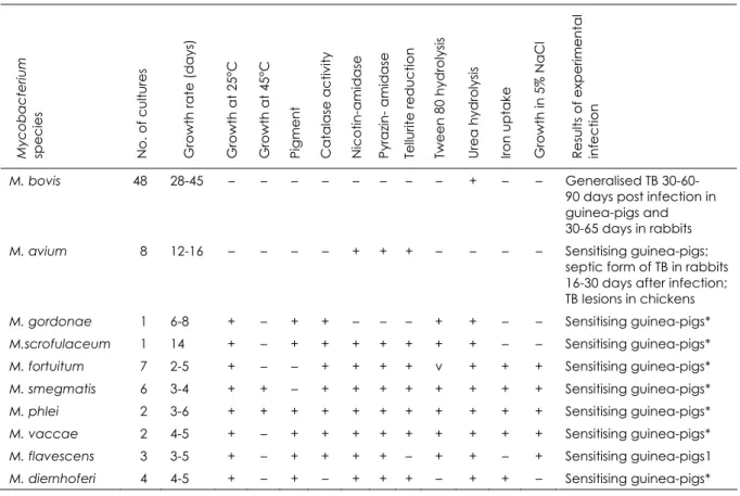
A total of 82 Mycobacterium cultures were isolated from 109 tissue and blood samples taken from 72 reactor cattle. The cultures exhibited different morphology, growth rate and pigmentation. Forty‐eight cultures were referred to as pathogenic and 34 as atypical mycobacteria. The atypical mycobacteria were
classified as follows using the Runyon
classification:
two belonged to group II eight to group III
twenty‐four to group IV.
Physiological features of isolated mycobacteria
and species differentiation based on
biochemical test results are presented in Table II.
All samples were positive in PCR when tested with the Mycobacterium genus‐specific primers. When the Mycobacterium‐positive samples were tested with IS 6110‐specific PCR primers, 48 samples (58.54%) were positive and thus were identified as M. tuberculosis complex. All IS 6110‐positive samples were also positive in PCR with M. bovis‐specific primers to the gene RvD1‐Rv2031c.
To determine if the PCR‐positive samples were also mixed with M. tuberculosis, all of those that reacted to the IS 6110 and to RvD1‐ Rv2031c primers were also tested using specific PCR primers for M. tuberculosis. All samples gave negative results.
Table II
Species identification of mycobacteria isolated from cattle positive in skin tests
Results are based on the biochemical tests shown
Myc o bact eriu m spe c ie s
No. of cultur
es
Growth rat
e
(d
ays)
Growth at 25°C Growth at 45°C Pigme
n t Catalase activi ty Nicotin-ami d ase
Pyrazin- amidase Te
llurite re ducti o n Twe e n 80 hy droly sis Ure a hy droly sis Iron uptak e
Growth in 5%
NaCl
Re
sults of e
xpe ri me ntal infe ction
M. bovis 48 28-45 – – – – – – – – + – – Generalised TB
30-60-90 days post infection in guinea-pigs and 30-65 days in rabbits
M. avium 8 12-16 – – – – + + + – – – – Sensitising guinea-pigs;
septic form of TB in rabbits 16-30 days after infection; TB lesions in chickens
M. gordonae 1 6-8 + – + + – – – + + – – Sensitising guinea-pigs*
M.scrofulaceum 1 14 + – + + + + + + + – – Sensitising guinea-pigs*
M. fortuitum 7 2-5 + – – + + + + v + + + Sensitising guinea-pigs*
M. smegmatis 6 3-4 + + – + + + + + + + + Sensitising guinea-pigs*
M. phlei 2 3-6 + + + + + + + + + + + Sensitising guinea-pigs*
M. vaccae 2 4-5 + – + + + + + + + + + Sensitising guinea-pigs*
M. flavescens 3 3-5 + – + + + + – + + – + Sensitising guinea-pigs1
M. diernhoferi 4 4-5 + – + – + + + – + + – Sensitising guinea-pigs*
TB tuberculosis – negative reaction + positive reaction v variable reaction
* sensitising guinea-pigs to tuberculin for mammals and to allergen from atypical mycobacteria
M. avium complex. Eight samples (9.75%) of the 34 remaining atypical mycobacteria were PCR‐positive using these primers. M. avium subspecies identification was performed using PCRs with primers specific to insertion elements IS1245, IS901, IS902, genes 16S rDNA and FR300. The results were as follows:
DNA of M. avium subsp. avium was identified in three (3.66%) samples
DNA of M. avium subsp. hominissuis in 5 (6.1%) samples
DNA of M. intracellulare or M. avium subsp. paratuberculosis was not found in any sample. Taking into account growth rates and different colony morphology, three M. bovis cultures
were suspected of being ‘mixed’. These
cultures were examined using PCR primers specific to the M. avium complex and gave positive results. Further PCR typing revealed that these three cultures were mixed with M. avium subsp. hominissuis.
Discussion
Data concerning species differentiation of mycobacteria cultures isolated from cattle that had reacted positively to the skin test are summarised in Figure 3. Species differentiation
was based on the results of culture,
morphological and biochemical methods and the results of the PCR tests.
The high percentage (41.5%) of atypical mycobacteria isolated from the 72 cattle that had reacted to the skin test is significant. The percentage of M. avium complex was 9.8%; M. fortuitum was 8.5% and M. smegmatis was
7.3%. The isolation of other atypical
Mycobacterium species from cattle is relatively rare (Fig. 3).
M. fortuitum
8.5% M. bovis
58.5% M. scrofulaceum
1.2% M. avium subsp.
hominissuis 6.1%
M. gordonae 1.2% M. diernhoferi
4.9% M. flavescens
3.7% M. vaccae
2.4%
M. phlei 2.4% M. smegmatis
7.3%
M. avium subsp. avium
3.7%
Figure 3
Results of species differentiation of Mycobacterium cultures isolated from cattle that gave positive
results to the skin test (n = 72)
mycobacteria which may have stimulated the skin reactions to tuberculin. These results confirm conclusions of our previous work (10, 30). Inappropriate poor sanitary conditions of animal keeping (10, 21, 36) may have provided the atypical mycobacteria the opportunity to
invade and multiply in the extremely
weakened animals.
The TB situation in the territories polluted with low doses of radiation remains difficult and the burden of cattle tuberculosis is still heavy. As reported previously, the dynamics of improvement of the TB status of cattle herds in these territories is remarkably lower than in other regions of the country and the impact of TB is appreciably higher (31). This analysis has been confirmed by the isolation of M. bovis strains with high virulence from this region;
these strains caused generalised disease in guinea‐pigs 38‐64 days after experimental injection.
Ante‐mortem diagnosis of TB becomes more complicated as a large percentage (41.5%) of tuberculin reactions are caused by atypical mycobacteria.
Results of this study showed the negative influence of low doses of radiation on the TB status of cattle herds. This has increased the impact of cattle TB in regions affected by the Chernobyl catastrophe.
Grant
support
This work was supported by the United States
Department of Energy, Initiatives for
Proliferation Prevention Program (Project P160).
References
1. Abramovskaya A.K., Gurevich G.L., Surkova L.K., Shpakovskaya N.S. & Yushko V.A. 1994. Rationale
for detection and clinical aspects of tuberculosis in areas of unfavourable radiological environment
[in Russian]. Probl Tuberk, 2, 18-19.
2. Bartos M., Hlozek P. & Pavlik I. 2006. Identification of members of Mycobacterium avium species by
Accu-Probes, serotyping, and single IS900, IS901, IS1245 and IS901-flanking region PCR with internal
3. Byaletskiy S.A. 1997. Specificity of anti-epizootic measures in the farms, located on the territory,
contaminated by radioactive nuclides [in Russian]. InProc. International Conference,
24-26 September, Kharkiv. IECVM, Kharkiv, 89 pp.
4. Cataldi A. & Romano M.I. 2007. Tuberculosis caused by other members of the M. tuberculosis
complex. In Tuberculosis 2007: from basic science to patient care, First Ed. (J.C. Palomino, S.C. Leão
& V. Ritacco, eds), 283-314 (www.tuberculosistextbook.com/tuberculosis2007.pdf accessed on 11 May 2009).
5. Dostal S., Richter E. & Harmsen D. 2003. Concise guide to mycobacteria and their molecular
differentiation. Ridom Press, Würzburg, 206 pp.
6. Fedoseev V.S. 1983. Changeability of tuberculosis agent and its connection with epizootic state [in
Russian]. Bull All-Russian Inst ExpVete Y.R. Kovalenko, Moscow, 51, 28.
7. Fedoseev V.S., Rubtsova I.N., Kirilenko N.G. & Baygazanov A.N. 1989. Methodical recommendations.
Isolation of L-forms of tuberculosis mycobacteria from animals. [in Russian]. Developed by the Department of microbiology and virology of Semipalatinsk Zoo-Veterinary Institute. Approved by the section of veterinary of scientific-research board of State Agrarian Industry of Kazakhstan. Protocol N4 of 23.12.1988. Alma-Ata, Kaynar, 19 pp.
8. Garnier T., Eiglmeier K., Camus J.-C., Medina N., Mansoor H., Pryor M., Duthoy S., Grondin S.,
Lacroix C., Monsempe C., Simon S., Harris H., Atkin R., Doggett J., Mayes R., Keating L., Wheeler P.R., Parkhill J., Barrell B.G., Cole S.T., Gordon S.T. & Hewinson R.G. 2003. The complete genome sequence
of Mycobacterium bovis. Proc Natl Acad Sci USA, 100, 7877-7882.
9. Golyshevskaya V.I., Cheshek V.I. & Shevchenko N.I. 1993. Microbiological diagnostics of tuberculosis
in the zones of strict radioactive control [in Russian]. Probl Tuberk, 6, 30-32.
10. Gorzheyev V.M. 2005. Epizootological monitoring and improvement of the system of cattle
tuberculosis control in the farms of Ukraine [in Ukrainian]. PhD thesis, 16.00.08. Kharkiv, 126 pp.
11. Guerrero S., Bernasconi C., Burki D., Bodmer T. & Telenti A. 1995. A novel insertion element from
Mycobacterium avium, IS 1245, is a specific target for analysis for strain relatedness. J Clin Microbiol,
33, 304-307.
12. Gurevich G.L., Abramovskaya A.K., Dorozhkova I.R. & Surkova L.K. 1993. Characteristics of biological
properties of the causative agent in patients with pulmonary tuberculosis exposed to prolonged
low-dose radiation [in Russian]. Probl Tuberk, 2, 46-48.
13. Illin B.N. 1990. If there is adaptation to the influence of ionizing radiation [in Russian]. Radiation
hygiene, Leningrad, 47-65.
14. Kassich V.Yu. 1991. Diagnostics of cattle tuberculosis in the conditions of ionizing radiation [in
Russian]. Abstract of PhD thesis. 16.00.03, Moscow, 21 pp.
15. Kassich V.Yu. 2004. Changeability of mycobacteria, epizootiological monitoring, methods of animal
tuberculosis control in the conditions of radioactive influence [in Ukrainian]. Abstract of doctoral thesis: 16.00.03. UAAS, Institute of Experimental and Clinical Veterinary Medicine, Kharkiv, 42 pp.
16. Kassich V.Yu., Fuks P. & Zavgorodniy A. 1999. Scientific and practical achievements in the
application of the method of allergic diagnostics of cattle tuberculosis [in Ukrainian]. Vet Med
Ukraine, 9, 18.
17. Kassich Yu., Zavgorodniy A., Kassich V., Verbitsky P., Gorzheev V., Kucheryavenko O., Lyolya V.,
Yakovlev V. & Negiba S. 2001. Determination of reaction on tuberculin in cattle by the method of
application of simultaneous allergic test [in Ukrainian]. Vet Med Ukraine, 5, 14-15.
18. Kassich Yu., Zavgorodniy A., Verbitsky P., Gorzheev V., Kucheryavenko O., Ignatov M., Lyolya V.,
Chasnyk M., Minasyan G., Getmansky V. & Ignatenko L. 2002. Rehabilitation measures in the farms
with cattle tuberculosis [in Ukrainian]. Vet Med Ukraine, 1, 13-14.
19. Kassich Yu.Ya., Babkin A.F., Zavgorodniy A.I., Gorzheev V.M., Borzyak A.T., Pavlenko M.S. &
Manchenko V.M. 1997. Methodical recommendations on determination of reaction on tuberculin in cattle from the safe concerning tuberculosis farms and determination of specific belonging of
mycobacteria cultures [in Ukrainian]. Vet Med Ukraine, 2, 32-38.
20. Kassich Yu.Ya., Zavgorodniy A.I., Kassich V.Yu, Gorzheyev V.M., Pavlenko M.S. & Lyolya V.V. 2004.
Achievements of science and practice in control of animal tuberculosis in Ukrainian farms [in
Russsian]. Vet Pathol, 1- 2 (9), 38-41.
21. Kolos Yu., Stets’ V. & Titarenko V. 2006. The problem of animal tuberculosis [in Ukrainian]. Vet Med
Ukraine, 11, 10-12.
22. Kunze Z.M., Portaels F. & McFadden J.J. 1992. Biologically distinct subtypes of Mycobacterium avium
23. Lie’bana E., Aranaz A., Francis B. & Cousins D. 1996. Assessment of genetic markers for species
differentiation within the Mycobacterium tuberculosis complex. J Clin Microbiol, 34 (4), 933-938.
24. Moss M.T., Malik Z.P., Tizard M.L., Green E.P., Sanderson J.D. & Hermon-Taylor J. 1992. IS902, an
insertion element of the chronic-enteritis-causing Mycobacterium avium subsp. silvaticum. J Gen
Microbiol, 138 (1), 139-145.
25. Ovdienko N.P., Sypin V.D. & Kassich V.Yu. 2002. Monitoring of tuberculosis in the zone of radioactive
pollution [in Russian]. Veterinaria, 3, 5-10.
26. Rodriguez J.G., Mejia G.A., Del Portillo P., Patarroyo M.E. & Murillo L.A. 1995. Species-specific
identification of Mycobacterium bovis by PCR. Microbiology, 141, 2131-2138.
27. Rua-Domenech de la R., Goodchild A.T., Vordermeier H.M., Hewinson R.G., Christiansen K.H. &
Clifton-Hadley R.S. 2006. Ante-mortem diagnosis of tuberculosis in cattle: a review of the tuberculin
tests, gamma-interferon assay and other ancillary diagnostic techniques. Res Vet Sci, 81 (2), 190-210.
28. Rubtsova I.N. 1983. Change of mycobacteria form in animal organism and their meaning in
bacteriological diagnostics of tuberculosis [in Russian]. Bulletin ARIEVYa R. Kovalenko, 51, 32.
29. Shabliy V.Yu., Barkevich V.A., Voronenko V.R., Ostal’tsev V.F. & Ryzhenko V.V. 1995. Influence of
small doses of ionizing radiation on infectious and epidemiological process [in Russian]. In Proc.
International Conference on common epizootiology, immunology, ecology and methodical problems, 20-22 September, Kharkiv, Institute of Experimental and Clinical Veterinary Medicine, Kharkiv, 571-572.
30. Skrypnyk A.V. 2007. Molecular-and-genetic differentiation of mycobacteria, isolated in Ukraine and
their phylogenetic correlations [in Ukrainian]. PhD thesis, 16.00.03. Kharkiv, 182 pp.
31. Skrypnyk A., Zavgorodniy A., Stegniy B., Skrypnyk V. & Gerilovych A. 2008. Study of Chernobyl
catastrophe influence on bovine TB epizootic process intensity in radioactive polluted and ‘clean’
territories of Ukraine in 1983-2007. In Proc. 18th European Congress of clinical microbiology and
infectious diseases, 19-22 April, Barcelona. Blackwell Publishing/Wiley, San Francisco, European
Society of Clinical Microbiology and Infectious Diseases (ESCMID), Abstract No. P1567
(www.blackwellpublishing.com/eccmid18/abstract.asp?id=69465 accessed on 18 May 2009).
32. Snider D.E. 1982. The tuberculin skin test. Am Rev Resp Dis, 125 (3), 108-118.
33. Surkova L.K. & Shtil’man M.-Y. 1993. Characteristics of thanatogenesis and pathomorphology of
tuberculosis in the Gomel region in relation to the Chernobyl AES accident [in Russian]. Probl Tuberk,
2, 20-24.
34. Wilton S. & Cousins D. 1992. Detection and identification of multiple mycobacterial pathogens by
DNA amplification in a single tube. PCR Methods Appl, 1 (4), 269-273.
35. World Organisation for Animal Health (Office International des Épizooties: OIE) 2008. Bovine
tuberculosis, Chapter 2.4.7. In Manual of diagnostic tests and vaccines for terrestrial animals, 2008
OIE, Paris, 683-697 (www.oie.int/eng/normes/mmanual/2008/pdf/2.04.07_BOVINE_TB.pdf accessed on 18 May 2009).
36. Zelinsky M. 2000. Cattle tuberculosis. Causes of emergence and factors which prevent the