Early Mouse Heart Development
Svetlana Gavrilov
1, Richard P. Harvey
2,3, Virginia E. Papaioannou
1*1 Department of Genetics and Development, Columbia University Medical Center, New York, New York, United States of America, 2 Developmental and Stem Cell Biology Laboratory, Victor Chang Cardiac Research Institute, Darlinghurst, New South Wales, Australia, 3 St. Vincent's Hospital Clinical School, University of New South Wales, Kensington, New South Wales, Australia
Abstract
Members of the T-box family of transcription factors are important regulators orchestrating the complex regionalization of the developing mammalian heart. Individual mutations in Tbx20 and Tbx3 cause distinct congenital heart abnormalities in the mouse: Tbx20 mutations result in failure of heart looping, developmental arrest and lack of chamber differentiation, while hearts of Tbx3 mutants progress further, loop normally but show atrioventricular convergence and outflow tract defects. The two genes have overlapping areas of expression in the atrioventricular canal and outflow tract of the heart but their potential genetic interaction has not been previously investigated. In this study we produced compound mutants to investigate potential genetic interactions at the earliest stages of heart development. We find that Tbx20; Tbx3 double heterozygous mice are viable and fertile with no apparent abnormalities, while double homozygous mutants are embryonic lethal by midgestation. Double homozygous mutant embryos display abnormal cardiac morphogenesis, lack of heart looping, expression patterns of cardiac genes and time of death that are indistinguishable from Tbx20 homozygous mutants. Prior to death, the double homozygotes show an overall developmental delay similar to Tbx3 homozygous mutants. Thus the effects of Tbx20 are epistatic to
Tbx3 in the heart but Tbx3 is epistatic to Tbx20 with respect to developmental delay.
Citation: Gavrilov S, Harvey RP, Papaioannou VE (2013) Lack of Genetic Interaction between Tbx20 and Tbx3 in Early Mouse Heart Development. PLoS ONE 8(7): e70149. doi:10.1371/journal.pone.0070149
Editor: Reiner Albert Veitia, Institut Jacques Monod, France
Received May 2, 2013; Accepted June 20, 2013; Published July 25, 2013
Copyright: © 2013 Gavrilov et al. This is an open-access article distributed under the terms of the Creative Commons Attribution License, which permits unrestricted use, distribution, and reproduction in any medium, provided the original author and source are credited.
Funding: Richard P. Harvey holds a National Health and Medical Research Council (NHMRC) Australia Fellowship (573705). This work was funded by grants from the NHMRC (573732) and National Heart Foundation of Australia (Richard P. Harvey) and the National Institutes of Health, Eunice Kennedy Shriver National Institute of Child Health and Human Development (NIH/NICHD), Grant Number 4R37HD033082 (Virginia E. Papaioannou). The content is solely the responsibility of the authors and does not necessarily represent the official views of the funders. The funders had no role in study design, data collection and analysis, decision to publish, or preparation of the manuscript.
Competing interests: The authors have declared that no competing interests exist. * E-mail: [email protected]
Introduction
The four-chambered mammalian heart develops from a simple linear heart tube by polar elongation, myocardial differentiation and morphogenesis. This complex regionalization of the developing heart is orchestrated by multiple signaling modules and transcriptional circuits. Members of the T-box family of transcription factors are particularly important regulators of myocardial proliferation and patterning. T-box family members Tbx1, Tbx2, Tbx3, Tbx5, Tbx18 and Tbx20 are expressed in the heart and regulate various aspects of embryonic heart development. The importance of unperturbed T-box gene function is highlighted by mutations in mice and human, which are associated with congenital heart disease [1–3]. Patients heterozygous for mutations in the coding region of TBX20 suffer from diverse cardiac defects, including ventricular septal defects, aberrant valvulogenesis, tetralogy of Fallot and cardiomyopathy [4–7].
Mutations in TBX3 result in the ulnar-mammary syndrome in which a subset of patients has ventricular septal defects [8] or conduction defects [9].
with both cell-autonomous and non-cell-autonomous defects of this process. Chamber myocardial genes are not activated in Tbx20 deficient hearts while Tbx2, which is normally restricted to the atrioventricular canal (AVC) and OFT, is ectopically expressed throughout the cardiac crescent and linear heart tube [3,13–15]. Tbx20 heterozygous mice survive postnatally but show dilated cardiomyopathy, which phenocopies at least a subset of human TBX20 mutant defects [3].
In the mouse heart, Tbx3 expression is first detected in the inflow tract at the onset of heart looping. Its expression delineates the developing cardiac conduction system, endocardial cushions in the AVC, and the mesenchyme of the OFT [16,17]. Tbx3 enables development of the cardiac conduction system by restricting cell division and repressing the chamber-specific gene expression program. Tbx3 deficient embryos are developmentally retarded and die at midgestation apparently due to yolk sac deficiencies although Tbx3 mutant hearts are also abnormal [18,19]. Tbx3 deficiency results in variable heart defects including increased cell division in the atrioventricular canal, incomplete ventricular septation, double outlet right ventricle, and delayed aortic arch formation. Homozygous mutants also show a failure of atrioventricular convergence, the process by which the inflow region is displaced dorsally to the ventricular segment during heart looping [19,20].
Thus, when individually mutated, Tbx20 and Tbx3 cause distinct congenital heart abnormalities, but their overlapping expression in the AVC and outflow tract in the developing heart raises the question of whether they act independently or through transcriptional regulation of common target genes. Tbx20 expression appears ectopically in the apex of the ventricular septum of Tbx3 mutant hearts at E12.5 [10] and Tbx3 expression is markedly down regulated at E9.5 in the AVC of embryos with a conditional deletion of Tbx20 [21]. However, the potential genetic interaction between these two genes has not been investigated with respect to phenotypic effects. In this study, we investigated the phenotype of Tbx20; Tbx3 double homozygous mutants to explore possible genetic interaction between these two genes based on their overlapping expression in the AVC and OFT. With respect to heart development, the double homozygous phenotype was indistinguishable from that of the Tbx20 single homozygous mutant in that heart looping and development was arrested early in development. In addition, a general developmental delay characteristic of Tbx3 homozygous mutants was apparent in the double homozygous mutants, apparently independent of the cardiac phenotype.
Materials and Methods
Mice
All experiments were carried out in strict accordance with the recommendations in the Guide for the Care and Use of Laboratory Animals of the National Institutes of Health under protocols approved by the Columbia University Institutional Animal Care and Use Committee (protocol No. AC-AAAD3302). Mutant alleles were maintained on a mixed genetic background derived from 129, C57BL/6Tac and ICR
mice. Mice heterozygous for a null mutation of the Tbx20 locus, Tbx20lacZ [3], hereafter designated as Tbx20-, or a null mutation
of the Tbx3 locus, Tbx3tm1Pa [18], hereafter designated as
Tbx3-, were intercrossed to produce double heterozygotes,
which were intercrossed to generate double homozygotes as well as other genetic combinations. Embryos and weanling mice were genotyped for Tbx20 and Tbx3 by PCR from lysates of yolk sac or tail tip, respectively. The mutant allele of Tbx20 was genotyped with primers: Primer 1 (LoxP F2): GACTGGAGAGGCCATCAAAA-3’ and Primer 2 (LacZR2): 5’-GTTTTCCCAGTCACGACGTT- 3’; and the wild type allele was genotyped with primers: T20 wt sense: 5”-CCCAAGGAGAAGGAGGCAGCAGAGAAC-3’ and T20 wt antisense: 5’- CGCAAGTATAAAATGGGGGTTCCTGACC-3’. PCR conditions were 3 minutes at 94oC, 30 cycles (30 seconds at 94oC, 30 seconds at 61oC, 60 seconds at 72oC), and 5 minutes at 72oC. The primers and PCR conditions for Tbx3 were as previously described [18].
Embryo collection and in situ hybridization
For timed pregnancies, females were placed with males overnight and checked the following morning for the presence of vaginal plugs. Noon on the day of the plug was designated E0.5. Embryos between E8.5 and E9.25 were dissected out of the uteri and their extra-embryonic membranes in phosphate buffered saline (PBS) with 0.2% bovine serum albumin and were fixed in 4% paraformaldehyde in PBS overnight. Standard procedures were used for whole mount in situ hybridization [22].
Statistics
Statistical analyses were performed using the Chi-square distribution, Mann Whitney U test and notched box plot analysis (Excel, Graph Pad Prism and R http://www.wessa.net/ rwasp_notchedbox1.wasp).
Results
Generation of Tbx20-/-; Tbx3-/- double homozygous
mutants
Mice heterozygous for Tbx20- or Tbx3- were intercrossed to
generate compound heterozygotes, Tbx20+/-; Tbx3+/-, which
were viable and fertile and present at the expected Mendelian frequency at weaning (Χ2=4.08; p>0.10; Table 1). No apparent abnormalities were observed. Compound heterozygotes were intercrossed and embryos of all possible genotypes were recovered at midgestation at the expected Mendelian frequency (Χ2 = 14.38; p>0.05; Table 1).
Embryonic lethality and abnormal cardiac
morphogenesis in Tbx20; Tbx3 compound mutant embryos
heterozygotes to single heterozygotes. All embryos homozygous mutant for Tbx20, including Tbx20-/-; Tbx3+/+ (n=27), Tbx20-/-; Tbx3+/- (n=29), as well as double homozygous mutant embryos (n=14), were morphologically indistinguishable from one another at any given developmental stage: Starting at the heart looping stage, heart tube formation was retarded, hearts were small, and heart looping failed, resulting in two vertically oriented chamber-like swellings with a characteristic hour-glass shape by E9.25 (Figure 1A–C and Tables 2 & 3), as previously described for Tbx20-/- hearts [3,13–15]. Embryos
that were Tbx20+/-; Tbx3-/-, on the other hand, initiated heart looping normally but some displayed atrioventricular
Table 1. Number of offspring of each genotype from intercrosses of Tbx20 and Tbx3 heterozygotes, recovered at weaning, and from crosses of mice double heterozygotes for both genes, recovered at midgestation.
Cross Genotypes Total
Tbx20 +/+ +/- +/+ +/- -/- -/- +/+ +/- -/-
Tbx3 +/+ +/+ +/- +/- +/+ +/- -/- -/- -/- Tbx20+/- x Tbx3+/- 28 23 18 16 85*
Tbx20+/-; Tbx3+/- x
Tbx20+/-; Tbx3+/- 11 15 13 30 12 21 6 6 12 126**
*. Recovered and genotyped at weaning. **. Recovered and genotyped E8.5 - E9.25.
convergence defects by E9.25 (2/4) similar to Tbx3-/- embryos (4/4) (Figure 1E–G and Table 3), as previously described [19]. Wild type and single or double heterozygotes had normal heart morphology (n=120).
Limb abnormalities in Tbx3-/- embryos were not observed
between E8.5 and E9.25, which is in agreement with previous work [18]. The only abnormality seen in yolks sac or gross placental morphology was a reduction in yolk sac vasculature in 1 of 4 Tbx20+/-; Tbx3-/- embryos at E8.5 and 1 of 6 double
homozygous mutant embryos at E9.25 (Table 3). This resembled the reduced vasculature reported for Tbx3-/- and
Tbx20-/- embryos at E9.5 [3,18].
Developmental delay in Tbx3-/- and Tbx20-/-; Tbx3
-/-embryos
Among the embryos from Tbx20+/-; Tbx3+/- double
heterozygous crosses at E8.5, some single or double homozygous embryos showed developmental delay, but over 90% were living (65/71) (Table 2); by E9.25, however, 37% (7/19) were dead (Table 3). Thus, to assess the extent of developmental delay associated with each genotype prior to death, we scored living embryos at E8.5 (n=160) using somite number to determine developmental stage. To account for between-litter variation, the ratio of the number of somites for an embryo to the mean number of somites in its litter was calculated for each embryo. A Mann Whitney U test showed that there is no significant difference in this ratio between Tbx20-/- homozygous mutants and Tbx20-/-; Tbx3+/- compound
Figure 1. Tbx20-/-; Tbx3-/- double homozygotes show heart defects similar to Tbx20-/- embryos. Whole mount ventral (A–F)
and left side (G, H) views of the heart (E9.25; 15-20 somite embryos) of Tbx20; Tbx3 single, compound and double mutants compared with wild type (D, H). All Tbx20-/- embryos have an unlooped, hourglass-shaped heart (A-C, outlined in A), whereas Tbx3
-/-embryos either wild type or heterozygous for Tbx20 (E, F, G) have normally looped hearts but show atrioventricular convergence defects (G, arrows) compared with wild type (H). lv, left ventricle; rv, right ventricle; t, telencephalon.
mutant embryos, nor between Tbx3-/- homozygous mutants and
Tbx20+/-; Tbx3-/- compound mutants. Thus, single and
compound mutants were combined and the distributions of ratios for embryos of each group were compared using notched box plots (Figure 2). The analysis showed that both Tbx20-/-;
Tbx3-/- double homozygotes and Tbx3-/- mutants are significantly delayed developmentally compared to wild type or Tbx20-/- embryos, but are similar to one another. Thus, with respect to developmental delay, Tbx20-/-; Tbx3-/- embryos resemble Tbx3-/- embryos (Figure 2).
Cardiac gene expression in Tbx20-/-; Tbx3-/- embryos
Molecular markers of cardiac development were used to further characterize the phenotype of compound and double homozygous mutant embryonic hearts. Tbx2 is normally expressed in the non-chamber myocardium, most prominently in the AVC. As previously reported [14], Tbx2 expression was
not altered in Tbx20-/- hearts at E8.75 (data not shown) but was
upregulated and ectopically expressed throughout the entire Tbx20-/- mutant heart by E9.25. Similar upregulation and ectopic expression of Tbx2 was seen in Tbx20-/-; Tbx3-/- double
homozygous mutant hearts, whereas expression in Tbx3
-/-hearts was normal (Figure 3A–D).
Expression of Nppa, a marker of chamber myocardium and a target of both Tbx20 and Tbx3, was severely down regulated in both Tbx20-/- and double homozygous mutant hearts from
E8.5-E9.25, demonstrating the lack of chamber differentiation. In contrast, precocious expression of Nppa in atrial myocardium was observed in Tbx3-/- embryos, as previously described [19] (Figure 3E–H), as well as in Tbx20+/-; Tbx3-/- compound
mutants (data not shown).
We observed no change in expression of Pitx2, which is important for cardiac remodeling and asymmetric development, in the left inflow tract (IFT) of Tbx20-/-, Tbx3-/- or Tbx20-/-; Tbx3 -/-embryos compared to stage-matched wild type controls (Figure
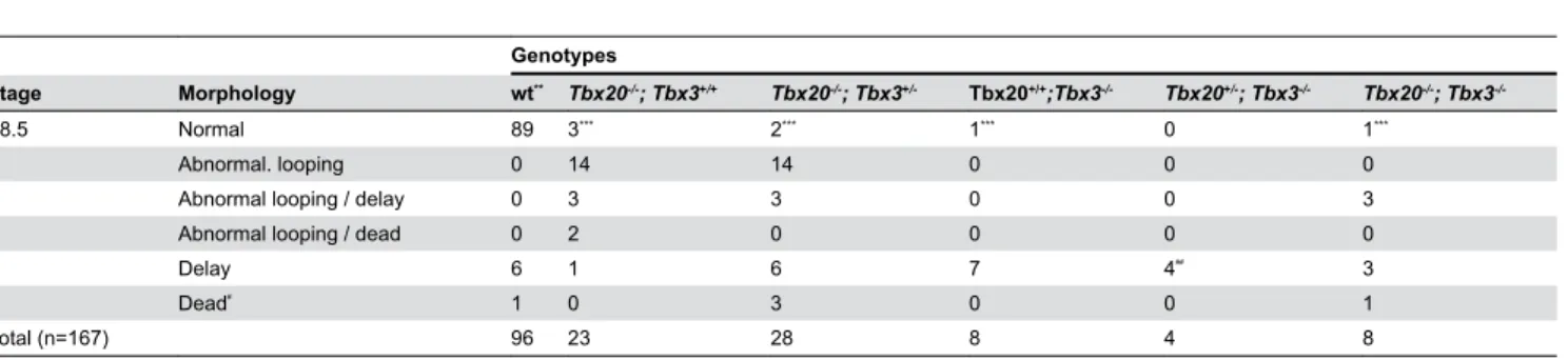
Table 2. Frequency of different morphological defects in embryos at E8.5 from matings of Tbx20+/-; Tbx3+/- double
heterozygotes*
.
Genotypes
Stage Morphology wt** Tbx20-/-; Tbx3+/+ Tbx20-/-; Tbx3+/- Tbx20+/+;Tbx3-/- Tbx20+/-; Tbx3-/- Tbx20-/-; Tbx3
-/-E8.5 Normal 89 3*** 2*** 1*** 0 1***
Abnormal. looping 0 14 14 0 0 0
Abnormal looping / delay 0 3 3 0 0 3
Abnormal looping / dead 0 2 0 0 0 0
Delay 6 1 6 7 4## 3
Dead# 1 0 3 0 0 1
Total (n=167) 96 23 28 8 4 8
*. Double heterozygotes were either intercrossed or crossed to single heterozygotes. **. wt, wild type; this group includes wild type and heterozygotes for one or both alleles. ***. Embryos were at the cardiac crescent stage, prior to heart looping.
#. Approximate time of death between E8.0 and E8.5; too degenerate to score for heart phenotype. ##. One of these embryos had a yolk sac defect.
Table 3. Frequency of different morphological defects in embryos at E9.25 from matings of Tbx20+/-; Tbx3+/- double
heterozygotes*.
Genotypes
Stage Morphology wt** Tbx20-/-; Tbx3+/+ Tbx20-/-; Tbx3+/- Tbx20+/+;Tbx3-/- Tbx20+/-; Tbx3-/- Tbx20-/-; Tbx3
-/-E9.25 Normal 23 0 0 0 0 0
Abnormal looping 0 0 0 0 0 1
Abnormal looping / delay 0 1 1 0 0 4***
Abnormal convergence 0 0 0 1 0 0
Abnormal convergence /delay 0 0 0 2 1 0
Abnormal convergence / dead 0 0 0 1 1 0
Delay 0 0 0 0 1 0
Dead# 1 3 0 0 1 1
Total (n=43) 24 4 1 4 4 6
*. Double heterozygotes were either intercrossed or crossed to single heterozygotes. **. wt, wild type; this group includes wild type and heterozygotes for one or both alleles. ***. One of these embryos had a yolk sac defect.
3I–L). Tbx5, which is expressed in the atrioventricular and atrial progenitors in an antero-posterior gradient, was slightly reduced in both Tbx20-/- and Tbx20-/-; Tbx3-/- embryos at E8.5 but no difference in expression was evident among the genotypes by E9.25 (Figure 3M–O).
Discussion
The DNA binding domain of T-box proteins, as well as T-box binding motifs identified in the promoters of downstream target genes, are highly conserved. This leads to the possibility that these transcription factors have targets in common and could compete or cooperate in transcriptional regulation of specific targets in areas of overlapping gene expression. Furthermore, some T-box proteins can bind DNA as dimers, raising the possibility of heterodimerization. In the developing heart at least seven T-box genes are expressed [1,23] and although each has a specific pattern of expression, there are extensive areas of expression overlap. Genetic studies have shown that some of these genes do indeed interact during heart development. Tbx1, Tbx2 and Tbx3 all interact in OFT development with Tbx1 upstream of Tbx2 and Tbx3. Double homozygous mutants for Tbx1 with either Tbx2 or Tbx3 have heart failure more severe than either single mutant alone [24]. At the level of gene regulation, Tbx2 and Tbx5 have been shown to repress or activate, respectively the expression of
Figure 2. Tbx20-/-; Tbx3-/- double homozygotes are
developmentally delayed as are Tbx3-/- embryos. Notched
box plots showing the distributions of the ratio of somite number in an embryo / the mean somite number for the litter in four different groups: wild type, Tbx20-/- homozygous mutants;
Tbx3-/- homozygous mutants and double homozygotes. Compound and single mutants were combined for this comparison as single mutants were not different from the compounds (Mann Whitney U test). Whiskers of the notched box plots represent 10th and 90th percentiles, boxes include the 25th through 75th percentile, and outliers are individually plotted. If the notches of two plots do not overlap, this indicates that the medians differ between the two. With respect to developmental stage, Tbx20-/-; Tbx3-/- double homozygotes
resemble Tbx3-/- embryos and both groups are delayed
compared to wild type or Tbx20-/- embryos. doi: 10.1371/journal.pone.0070149.g002
Nppa through competing interactions with the transcriptional co-factor Nkx2-5 [25–27]. Tbx3 and Tbx20 have also been shown capable of interacting with Nkx2-5 [12,17]. T-box genes might also regulate one another and in this respect, Tbx20 has been shown to repress Tbx2 expression in the developing heart, as in its absence, Tbx2 is upregulated throughout the chamber myocardium of the heart tube [3].
Tbx20 and Tbx3 have areas of overlapping expression in the OFT and AVC of the developing heart and thus have the potential to interact either by affecting common downstream target genes or by cross regulation. A potential example of cross regulation was noted in Tbx3 mutant hearts at E13.5-14.5 where Tbx20, which is normally expressed in the base of the developing interventricular septum, appears to be ectopically expressed in the developing conduction system at the apex of the septum where Tbx3 would normally be expressed [10]. However, whether this is genuine ectopic expression or simply the lack of Tbx3-expressing tissue in the abnormally developing septum is not clear. On the other hand, Tbx20 regulates Tbx3 in the AVC as deletion of Tbx20 specifically in the AVC leads to a downregulation of Tbx3 expression at E9.5 [21].
To investigate the phenotypic consequences of possible interactions between the two genes at the earliest stages of heart development, we produced double mutants using mutant alleles that ablate function and compared the morphological and molecular phenotypes of the single, compound and double mutants. We found no evidence for a genetic interaction in double heterozygotes, which were viable and fertile, or in double homozygotes, which displayed an embryonic heart phenotype indistinguishable from Tbx20 single homozygous mutants: morphologically the double homozygous mutant hearts displayed a vertically oriented hourglass shaped, two-chambered heart with no looping. Molecularly, Tbx2 was upregulated throughout the heart tube and Nppa was severely down regulated in the double homozygous mutants, similar to Tbx20 single mutants, whereas Pitx2 and Tbx5 were unchanged. This phenotype is indistinguishable from the Tbx20-/- phenotype where cardiac development is arrested and
mutant embryos are delayed in their overall development compared with littermates [18]. We observed a similar developmental delay in Tbx20-/-; Tbx3-/- double homozygous
embryos that was not seen in Tbx20 single mutants. Thus, developmental delay is associated with the Tbx3 mutant genotype, even though Tbx20 mutants have a more severe
Figure 3. Cardiac marker gene expression suggests that Tbx20-/-; Tbx3-/- double homozygous mutants show a Tbx20
-/-phenotype. Whole mount ISH left (A–H) or ventral (I–O) views of the head and heart of stage-matched wild type, single and double
mutant embryos showing expression of Tbx2 (E9.25; A-D), Nppa (E9.25; E-H), Pitx2 (E8.5; I-L) and Tbx5 (E9.25; M-O). Tbx2 is normally expressed in the AVC in wild type and Tbx3-/- hearts but is ectopically expressed in Tbx20-/- and double homozygotes. In Tbx20-/- and double homozygotes, Nppa expression is severely down regulated in ventricular myocardium, but in Tbx3-/- hearts it is precociously upregulated in atrial myocardium. There is no change in Pitx2 expression in the left inflow tract in Tbx20-/-, Tbx3-/- or double homozygous embryos when compared to wild type. Tbx5 is expressed in the atrioventricular and atrial progenitors in an antero-posterior gradient. There is no change in Tbx5 expression in either Tbx20-/- or double homozygotes compared to wild type at E9.25. at, atrium; AVC, atrioventricular canal; h, heart; IFT, inflow tract; lt IFT, left inflow tract; t, telencephalon; v, ventricle.
and earlier heart abnormality. It was previously postulated that vascular defects and apoptosis in the Tbx3 mutant yolk sac were responsible for this developmental delay [18], even though the subsequent discovery of a heart phenotype put this interpretation into question [19]. Both Tbx3 and Tbx20 are expressed in the yolk sac, Tbx20 in the mesoderm layer [3] and Tbx3 in both endoderm and mesoderm [29], and vascular deficiency has been reported in both single mutants at E9.5 [3,18]. In this study of E8.5-9.25 embryos, the incidence of abnormal yolk sac vasculature was very low in Tbx3 mutants and was not exacerbated in double mutants. Thus the developmental delay associated with the Tbx3 mutant genotype does not appear to be associated with an early yolk sac vasculature phenotype and remains to be elucidated.
Acknowledgements
We thank members of the Papaioannou laboratory for helpful discussions.
Author Contributions
Conceived and designed the experiments: RPH VEP SG. Performed the experiments: VEP SG. Analyzed the data: RPH VEP SG. Contributed reagents/materials/analysis tools: RPH VEP SG. Wrote the manuscript: VEP SG. Approved and edited the manuscript: RPH VEP SG.
References
1. Naiche LA, Harrelson Z, Kelly RG, Papaioannou VE (2005) T-box genes in vertebrate development. Annu Rev Genet 39: 219-239. doi: 10.1146/annurev.genet.39.073003.105925. PubMed: 16285859. 2. Greulich F, Rudat C, Kispert A (2011) Mechanisms of T-box gene
function in the developing heart. Cardiovasc Res 91: 212-222. doi: 10.1093/cvr/cvr112. PubMed: 21498422.
3. Stennard FA, Costa MW, Lai D, Biben C, Furtado MB et al. (2005) Murine T-box transcription factor Tbx20 acts as a repressor during heart development and is essential for adult heart integrity, function and adaption. Development 132: 2451-2462. doi:10.1242/dev.01799. PubMed: 15843414.
4. Kirk EP, Sunde M, Costa MW, Rankin SA, Wolstein O et al. (2007) Mutations in cardiac T-box factor gene TBX20 are associated with diverse cardiac pathologies, including defects of septation and valvulogenesis and cardiomyopathy. Am J Med Genet 81: 280-291. PubMed: 17668378.
5. Liu C, Shen A, Li X, Jiao W, Zhang X et al. (2008) T-box transcription factor TBX20 mutations in Chinese patients with congenital heart disease. Eur J Med Genet 51: 580-587. doi:10.1016/j.ejmg. 2008.09.001. PubMed: 18834961.
6. Hammer S, Toenjes M, Lange M, Fischer JJ, Dunkel I et al. (2008) Characterization of TBX20 in human hearts and its regulation by TFAP2. J Cell Biochem 104: 1022-1033. doi:10.1002/jcb.21686. PubMed: 18275040.
7. Qian L, Mohapatra B, Akasaka T, Liu J, Ocorr K et al. (2008) Transcription factor neuromancer/TBX20 is required for cardiac function in Drosophila with implications for human heart disease. Proc Natl Acad Sci U S A 105: 19833-19838. doi:10.1073/pnas.0808705105. PubMed: 19074289.
8. Meneghini V, Odent S, Platonova N, Egeo A, Merlo GR (2006) Novel
TBX3 mutation data in families with Ulnar-Mammary syndrome indicate a genotype-phenotype relationship: mutations that do not disrupt the T-domain are associated with less severe limb defects. Eur J Med Genet 49: 151-158. doi:10.1016/j.ejmg.2005.04.021. PubMed: 16530712. 9. Linden H, Williams R, King J, Blair E, Kini U (2009) Ulnar Mammary
syndrome and TBX3: expanding the phenotype. Am J Med Genet A 149A: 2809-2812. doi:10.1002/ajmg.a.33096. PubMed: 19938096. 10. Bakker ML, Boukens BJ, Mommersteeg MT, Brons JF, Wakker V et al.
(2008) Transcription factor Tbx3 is required for the specification of the atrioventricular conduction system. Circ Res 102: 1340-1349. doi: 10.1161/CIRCRESAHA.107.169565. PubMed: 18467625.
11. Kraus F, Haenig B, Kispert A (2001) Cloning and expression analysis of the mouse T-box gene Tbx20. Mech Dev 100: 89-91. PubMed: 11118890.
12. Stennard FA, Costa MW, Elliott DA, Rankin S, Haast SJP et al. (2003) Cardiac T-box factor Tbx20 directly interacts with Nkx2-5, GATA4, and GATA5 in regulation of gene expression in the developing heart. Dev Biol 262: 206-224. doi:10.1016/S0012-1606(03)00385-3. PubMed: 14550786.
13. Takeuchi JK, Mileikovskaia M, Koshiba-Takeuchi K, Heidt AB, Mori AD et al. (2005) Tbx20 dose-dependently regulates transcription factor networks required for mouse heart and motoneuron development. Development 132: 2463-2474. doi:10.1242/dev.01827. PubMed: 15843409.
14. Cai C-L, Zhou W, Yang L, Bu L, Qyang Y et al. (2005) T-box genes coordinate regional rates of proliferation and regional specification
during cardiogenesis. Development 132: 2475-2487. doi:10.1242/dev. 01832. PubMed: 15843407.
15. Singh MK, Christoffels VM, Dias JM, Trowe M-O, Petry M et al. (2005)
Tbx20 is essential for cardiac chamber differentiation and repression of
Tbx2. Development 132: 2697-2707. doi:10.1242/dev.01854. PubMed: 15901664.
16. Washkowitz AJ, Gavrilov S, Begum S, Papaioannou VE (2012) Diverse functional networks of Tbx3 in development and disease. Wires Syst Biol Med 4: 273-283. doi:10.1002/wsbm.1162. PubMed: 22334480. 17. Hoogaars WMH, Tessari A, Moorman AFM, de Boer PAJ, Hagoort J et
al. (2004) The transcriptional repressor Tbx3 delineates the developing central conduction system of the heart. Cardiovasc Res 62: 489-499. doi:10.1016/j.cardiores.2004.01.030. PubMed: 15158141.
18. Davenport TG, Jerome-Majewska LA, Papaioannou VE (2003) Mammary gland, limb, and yolk sac defects in mice lacking Tbx3, the gene mutated in human ulnar mammary syndrome. Development 130: 2263-2273. doi:10.1242/dev.00431. PubMed: 12668638.
19. Mesbah K, Harrelson Z, Théveniau-Ruissy M, Papaioannou VE, Kelly RG (2008) Tbx3 is required for outflow tract development. Circ Res 103: 743-750. doi:10.1161/CIRCRESAHA.108.172858. PubMed: 18723448.
20. Ribeiro I, Kawakami Y, Büscher D, Raya A, Rodríguez-León J et al. (2007) Tbx2 and Tbx3 regulate the dynamics of cell proliferation during heart remodeling. PLOS ONE, 2: e398. PubMed: 17460765.
21. Cai X, Nomura-Kitabayashi A, Cai W, Yan J, Christoffels VM et al. (2011) Myocardial Tbx20 regulates early atrioventricular canal formation and endocardial epithelial-mesenchymal transition via Bmp2. Dev Biol 360: 381-390. doi:10.1016/j.ydbio.2011.09.023. PubMed: 21983003.
22. Wilkinson DG (1992) Whole mount in situ hybridization of vertebrate embryos. Oxford: IRL Press.
23. Hurlin PJ, Steingrìmsson E, Copeland NG, Jenkins NA, Eisenman RN (1999) Mga, a dual-specificity transcription factor that interacts with Max and contains a T-domain DNA-binding motif. Eur Molecular Biol Organization J 18: 7019-7028. PubMed: 10601024.
24. Mesbah K, Rana MS, Francou A, van Duijvenboden K, Papaioannou VE et al. (2012) Identification of a Tbx1/Tbx2/Tbx3 genetic pathway governing pharyngeal and arterial pole morphogenesis. Hum Mol Genet 21: 1217-1229. doi:10.1093/hmg/ddr553. PubMed: 22116936. 25. Hiroi Y, Kudoh S, Monzen K, Ikeda Y, Yazaki Y et al. (2001) Tbx5
associates with Nkx2-5 and synergistically promotes cardiomyocyte differentiation. Nat Genet 28: 276-280. doi:10.1038/90123. PubMed: 11431700.
26. Bruneau BG, Nemer G, Schmitt JP, Charron F, Robitaille L et al. (2001) A murine model of Holt-Oram syndrome defines roles of the T-box transcription factor Tbx5 in cardiogenesis and disease. Cell 106: 709-721. doi:10.1016/S0092-8674(01)00493-7. PubMed: 11572777. 27. Habets PEMH, Moorman AFM, Clout DEW, van Roon MA, Lingbeek M
et al. (2002) Cooperative action of Tbx2 and Nkx2.5 inhibits ANF expression in the atrioventricular canal: implications for cardiac chamber formation. Genes Dev 16: 1234-1246. doi:10.1101/gad. 222902. PubMed: 12023302.
PNAS 109: E154-E163. doi:10.1073/pnas.1115165109. PubMed: