and Heart Failure
Alexander Lukasz1,2., Gernot Beutel3., Philipp Ku¨mpers2, Agnieszka Denecke1,4, Mechthild Westhoff-Bleck4, Bernhard Schieffer4, Johann Bauersachs4, Jan T. Kielstein1, Oktay Tutarel4*
1Department of Nephrology and Hypertension, Hannover Medical School, Hannover, Germany,2Department of Medicine D, Division of General Internal Medicine, Nephrology, and Rheumatology, University Hospital Muenster, Muenster, Germany,3Department of Hematology and Oncology, Hannover Medical School, Hannover, Germany,4Department of Cardiology and Angiology, Hannover Medical School, Hannover, Germany
Abstract
Background:Chronic heart failure is an important cause for morbidity and mortality in adults with congenital heart disease (ACHD). While NT-proBNP is an established biomarker for heart failure of non-congenital origin, its application in ACHD has limitations. The angiogenic factors Angiopoietin-1 and -2 (Ang-1, Ang-2), vascular endothelial growth factor (VEGF), and soluble receptor tyrosine kinase of the Tie family (sTie2) correlate with disease severity in heart failure of non-congenital origin. Their role in ACHD has not been studied.
Methods:In 91 patients Ang-2 and NT-proBNP were measured and related to New York Heart Association class, systemic ventricular function and parameters of cardiopulmonary exercise testing. Ang-1, VEGF, and sTie2 were also measured.
Results:Ang-2 correlates with NYHA class and ventricular dysfunction comparable to NT-proBNP. Further, Ang-2 showed a good correlation with parameters of cardiopulmonary exercise testing. Both, Ang-2 and NT-proBNP identified patients with severely limited cardiopulmonary exercise capacity. Additionally, Ang-2 is elevated in patients with a single ventricle physiology in contrast to NT-proBNP. VEGF, Ang-1, and sTie2 were not correlated with any clinical parameter.
Conclusion:The performance of Ang-2 as a biomarker for heart failure in ACHD is comparable to NT-proBNP. Its significant elevation in patients with single ventricle physiology indicates potential in this patient group and warrants further studies.
Citation:Lukasz A, Beutel G, Ku¨mpers P, Denecke A, Westhoff-Bleck M, et al. (2013) Angiopoietin-2 in Adults with Congenital Heart Disease and Heart Failure. PLoS ONE 8(6): e66861. doi:10.1371/journal.pone.0066861
Editor:Rajesh Gopalrao Katare, University of Otago, New Zealand
ReceivedDecember 28, 2012;AcceptedMay 10, 2013;PublishedJune 24, 2013
Copyright:ß2013 Lukasz et al. This is an open-access article distributed under the terms of the Creative Commons Attribution License, which permits unrestricted use, distribution, and reproduction in any medium, provided the original author and source are credited.
Funding:The article processing charge was funded by means of the DFG-Project ‘‘Open Access Publishing‘‘ by the German Research Council. The funders had no role in study design, data collection and analysis, decision to publish, or preparation of the manuscript.
Competing Interests:The authors have declared that no competing interests exist. * E-mail: otutarel@hotmail.com
.These authors contributed equally to this work.
Introduction
Chronic heart failure (CHF) is an important cause for morbidity and mortality in adults with congenital heart disease (ACHD) [1]. Heart failure symptoms may not always correlate with objective measures like systemic ventricular function or parameters of cardiopulmonary exercise testing [2,3]. The rarity of individual malformations and the complex anatomy and physiology make assessing the cardiac function difficult [4]. Therefore, the prevalence of heart failure in these patients is underappreciated [5,6]. A simple investigation like a blood test to detect early stages of heart failure and predict those at risk of deterioration would be valuable [4]. B-type natriuretic peptide (BNP) and N-terminal pro-BNP (NT-propro-BNP) are established biomarkers for diagnosis and management of heart failure due to acquired heart disease [7]. Unfortunately, the clinical use of those markers in adults with congenital heart disease is limited [8,9]. Therefore diagnosis and treatment monitoring is frequently based on cardiopulmonary exercise testing [4,6,10,11], which is time consuming and not feasible in special patient groups.
vessel integrity, vascular leakage and expression of inflammatory genes [16,19]. Ang-2 is stored in the endothelium and rapidly released upon different activators e.g. hypoxemia [20]. Recently published studies report a significant increase of the soluble Tie2 receptor, Ang-2 and VEGF in patients with CHF due to acquired heart disease when compared with healthy controls [21,22]. Serum Ang-2 correlates with an impaired exercise capacity and reduced ventilatory capacity in CHF patients [22]. The role of these circulating endothelial factors in ACHD has not been studied before. Therefore, the aim of this study was to elucidate the potential diagnostic value of Ang-1, Ang-2, soluble Tie2 and VEGF for heart failure in adults with congenital heart disease.
Materials and Methods
Ethics Statement
The study was approved by the local Ethics Committee of Hannover Medical School, Germany. All patients gave written informed consent.
The patients were recruited during a routine outpatient visit at the Adult Congenital Heart Disease Clinic of the Hannover Medical School. All patients in whom a venous blood sampling was feasible were eligible for this study.
A clinical workup including medical history, physical examina-tion, 12-lead electrocardiogram, transthoracic echocardiography, and cardiopulmonary exercise testing was performed.
The severity of the congenital heart defect was graded according to complexity as proposed by guidelines [23]. The patients were further classified according to their symptoms of heart failure using the New York Heart Association (NYHA) functional classification which is based on patient’s symptoms and the limitations to normal physical activities [7].
Laboratory Methods
Blood samples for measurement of plasma Ang-1, Ang-2, VEGF, sTie2 and NT-proBNP, and routine biochemistry were drawn. Blood samples were immediately cooled on ice, centrifuged at 1,500 g and 4uC for 10 min. Supernatants were stored in 1 ml aliquots at –80uC until further use. Plasma concentrations of Ang-1 and Ang-2 were measured by an in-house immunoluminometric assay as was previously reported in detail [24]. In brief the assays had detection limits of 0.12 ng/mL 1) and 0.2 ng/mL (Ang-2). Inter- and intra-assay imprecision was#8.8 and 3.7% for Ang-1 and was#4.6 and 5.2% for Ang-2, respectively. Plasma VEGF (biologically active VEGF-A121 and VEGF-A165) and sTie2 were measured using commercially available sandwich ELISA kits (R&D Systems, Minneapolis, MN, USA) according to the manufacturer’s instructions. All other measurements were done with routine laboratory tests using certified assay methods.
Echocardiography
A standard 2D-Doppler transthoracic echocardiogram was performed according to the recommendations for the assessment of ventricular function and valvular heart disease issued by the American Society of Echocardiography [25]. Systemic ventricular systolic function was assessed qualitatively (i.e. normal, moderately or severely impaired).
Cardiopulmonary Exercise Studies
Cardiopulmonary exercise studies were performed on a bicycle in sitting position, starting with 25 W, increasing further 25 W every 2 min. All patients exercised to the end of their tolerance. A 12-lead ECG was recorded throughout the exercise test to determine heart rate and heart rate response. Systolic blood pressure and blood pressure response, as well as work rate (W/kg) were measured. Ventilation, oxygen uptake (VO2), and carbon
dioxide production (VCO2), were measured continuously by a
breath-by-breath method. Subjects breathed through a fitted mask and a hot-wire anemometer (Oxycon Delta, Ja¨ger, Hoechberg, Germany) measuring inspired and expired flow continuously.
Statistical Analysis
Continuous data are presented as mean6standard deviation. Categorical data are presented as counts and proportions. Between group comparisons were examined using Student’sttest for continuous and Mann-Whitney-U test for categorical variables. One-way ANOVA was used if more than two groups were compared. The least significant difference (LSD) method was used as a post hoc-test.
For the parameters of cardiopulmonary exercise testing cut off values representing patients with limitations of their cardiopulmo-nary exercise capacity were defined as previously described [14]: peak oxygen uptake (peak VO2) ,20 ml/min/kg, ventilatory
equivalent for carbon dioxide (EQCO2) .34, ventilatory
equiv-alent for oxygen (EQO2) .34, oxygen pulse if female ,9 ml/
heartbeat, if male,12 ml/heartbeat. A group of patients that was severely affected was defined by a peakVO2,20 ml/min/kg or an
EQCO2.34 or a combination of both. The cut-offs were chosen
according to published data and clinical experience [2,3,26]. Receiver-operating characteristic curves (ROC curve) for these parameters were drawn and the areas under the curves calculated. All tests were two-sided and significance was accepted at p,0.05. Data analysis was performed using SPSS (SPSS Inc., Chicago, IL, USA). Figures were prepared using GraphPad Prism (GraphPad Prism Software Inc., San Diego, CA, USA).
Table 1.Clinical characteristics of study population.
Age(yrs) 30.4610.7
Sex
female 37 (40.7)
male 54 (59.3)
BMI(kg/m2) 23.564.2
Complexity of congenital heart disease
simple 18 (19.8)
moderate 36 (39.6)
severe 37 (40.7)
Systemic ventricle
left 65 (71.4)
right 12 (13.2)
single ventricle 14 (15.4)
Systemic ventricular function
normal 52 (57.1)
moderately impaired 32 (35.2) severely impaired 7 (7.7)
NYHA class
NYHA I 55 (60.4)
NYHA II 21 (23.1)
NYHA III 15 (16.5)
Results
Ninety-one patients were included in this cross-sectional study. Ang-1, VEGF and Tie2 could only be analyzed in 80 patients.
Table 1andTable 2show the clinical characteristics of the study population. Cardiopulmonary exercise testing was performed in 70 patients. Five of these performed a submaximal exercise test (respiratory exchange ratio ,1.05) and were excluded from analysis. Nine patients were cyanotic. Out of these three had an exercise test. The values for EQCO2 were not used in these
patients since EQCO2 is not a marker of prognosis in cyanotic
patients.
Ventricular Function
Ang-1, VEGF and Tie2 were not statistically different in patients with normal ventricular function compared to patients with moderate or severe ventricular dysfunction. (Table 3). Ang-2 did reach a statistically significant difference in patients with normal ventricular function compared to patients with severe
ventricular dysfunction (3.5364.19 ng/ml vs. 7.4867.57 ng/ml, p,0.05), but not to those with moderate ventricular dysfunction (5.365.18, p = 0.11). There was also no significant difference between the last two. (Table 3andFigure 1A).
NT-proBNP reached a statistically significant difference in patients with normal ventricular function compared to those with severe ventricular dysfunction (1426177 pg/ml vs. 115661540 pg/ml, p,0.0001) and between patients with normal ventricular function and moderate ventricular dysfunction (1426177 pg/ml vs. 3946477 pg/ml, p,0.05) as well as between patients with moderate and severe ventricular dysfunction (3946477 pg/ml vs. 115661540 pg/ml, p,0.001). (Table 3
andFigure 1B).
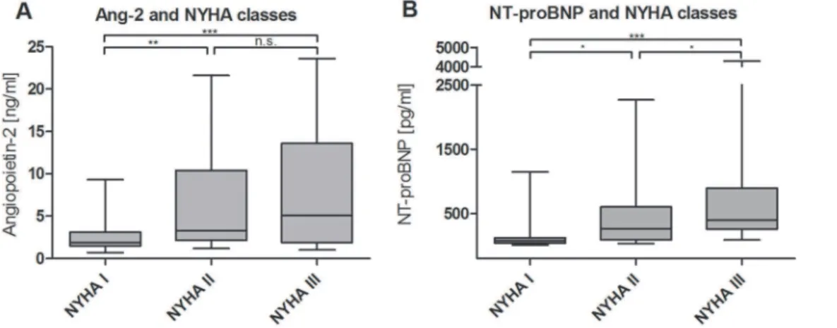
NYHA Class
Ang-2 differentiated between patients in NYHA class I (2.5261.6 ng/ml) and NYHA class II (6.8366.56 ng/ml, p,0.0001), as well as those in NYHA class III (8.2366.88 ng/ ml, p,0.0001), but not between patients in NYHA class II and III
Table 2.Type of congenital heart defect grouped according to ventricular function.
Ventricular function
Congenital heart defect All Normal Moderately impaired Severely impaired
TGA and CCTGA 11 (12.1) 2 (18.2) 5 (45.5) 4 (36.4)
Tetralogy of Fallot 12 (13.2) 6 (50) 6 (50) 0
Coarctation of the aorta 10 (11.0) 9 (90) 1 (10) 0
ASD or VSD 11 (12.1) 9 (81.8) 2 (18.2) 0
AVSD 5 (5.5) 5 (100) 0 0
Marfan syndrome 7 (7.7) 3 (42.9) 4 (57.1) 0
Congenital AS or PS 11 (12.1) 9 (81.8) 2 (18.2) 0
Single ventricle physiology 13 (14.3) 4 (30.8) 6 (46.2) 3 (23.1)
Miscellaneous 11 (12.1) 5 (45.5) 6 (54.5) 0
Data are expressed as counts (percentage); TGA = transposition of the great arteries; CCTGA = congenital corrected transposition of the great arteries; ASD = atrial septal defect; VSD = ventricular septal defect; AVSD = atrioventricular septal defect; AS = aortic valve stenosis; PS = pulmonary valve stenosis; miscellaneous: Ebstein’s anomaly, subaortic stenosis, supravalvular aortic stenosis, pulmonary atresia, Eisenmenger.
doi:10.1371/journal.pone.0066861.t002
Figure 1. Levels of biomarkers in comparison between normal, moderate and severe impairment of ventricular function.Ang-2 (A) and NT-proBNP (B) (*p,0.05, **p,0.001, ***p,0.0001).
(p = 0.34). (Table 3and Figure 2A) The results for Ang-2 as a function of NYHA class in each ventricular function subset can be found inTable 4. The highest values were found in the subgroup of patients in NYHA class III and severe ventricular function, while patients in NYHA class I and normal ventricular function had the lowest values. It seems that Ang-2 could mark NYHA class within a given ventricular function better than marking ventricular function within a given NYHA class, but the sample size in each group is too small to provide conclusive answers.
NT-proBNP was significantly different in patients in NYHA class I (1286202 pg/ml) compared to patients in NYHA class III (79661081 pg/ml, p,0.0001) and to patients in NYHA class II (4326517 pg/ml, p,0.05) as well as between the last two
(p,0.05). (Table 3 and Figure 2B). Ang-1, VEGF and Tie2 were not different between NYHA classes. (Table 3).
Cardiopulmonary Exercise Testing
Peak VO2was significantly higher in patients in NYHA class I
(28.667.4 ml/min/kg) vs. patients in NYHA class II (23.364.5 ml/min/kg, p = 0.01) and patients in NYHA class III (14.165.2 ml/min/kg, p,0.001) and also in comparison between NYHA class II and III (p = 0.001). Patients with a normal ventricular function had a significantly higher peak VO2
(28.567.6 ml/min/kg) compared to patients with a moderate (20.667.2 ml/min/kg, p,0.001) or severe ventricular dysfunction (21.068.4 ml/min/kg, p = 0.018). There was no significant difference between the last two (p = 0.917).
Table 3.Ang-1, Ang-2, VEGF, Tie2 and NT-proBNP levels according to NYHA class, ventricular function and systemic ventricle physiology.
Ang-1 (ng/ml) Ang-2 (ng/ml VEGF (pg/ml) Tie2 (ng/ml) NT-proBNP (pg/ml) NYHA Class
I 5.863.8 2.561.6 111.4662.5 19.963.3 1286200
II 6.565.7 6.866.6a 119.2
6114.4 23.165.7 4326517a
III 4.563.2 8.266.9b 156.7
6142.7 21.063.9 79661081b,c Ventricular function
normal 5.163.2 3.564.2 117.7684.8 20.564.2 1426177 moderately impaired 6.765.5 5.365.1 128.66111.1 21.064.1 3946477d
severely impaired 5.763.8 7.567.6e 122.2
6110.8 23.264.4 115661540f Systemic ventricle
right 7.567.3 4.965.2 117685.5 21.664.2 6426758 left 5.663.4 2.962.8 116.9681.6 20.163.7 1806250g
single 4.663.0 11.266.9h 144.5
6145.9 23.165.4 61761091i
Values are expressed as mean6SD;
ap
,0.01 to NYHA I - Ang-2 and p,0.05 for NT-proBNP;
bp,0.001 to NYHA I - Ang-2 and NT-proBNP; cp,0.05 for NT-proBNP and NYHA II; dp
,0.01 vs. normal ventricular function;
ep,0.05 vs. normal ventricular function;
fp,0.01 vs. moderate ventricular dysfunction and p,0.001 vs. normal ventricular function; gp
,0.01 vs. systemic right ventricle;
hp
,0.001 vs. systemic left and right ventricle;
ip,0.01 vs. systemic left ventricle.
doi:10.1371/journal.pone.0066861.t003
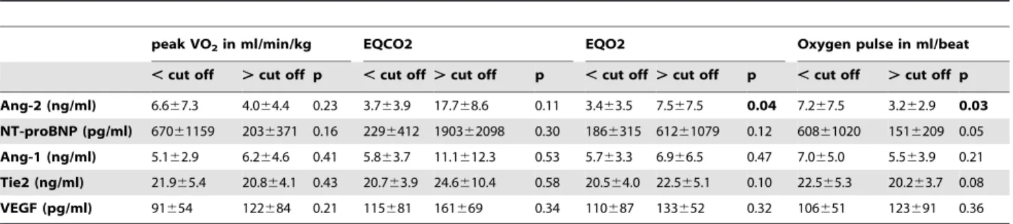
Ang-2 was elevated in patients with limited cardiopulmonary exercise compared to their peers. Significant differences were observed for EQO2(p = 0.04), and oxygen pulse (p = 0.03), but not
for peak VO2(p = 0.23) and EQCO2(p = 0.11). NT-proBNP was
not elevated in patients with limited cardiopulmonary exercise capacity. There was no significant difference for peak VO2
(p = 0.16), EQCO2(p = 0.30), EQO2(p = 0.12), and oxygen pulse
(p = 0.05). (Table 5).
Severely Limited Exercise Capacity
There were 16 patients with severely limited exercise capacity in our cohort of 65 patients with cardiopulmonary exercise testing (defined as peakVO2,20 ml/min/kg or an EQCO2.34 or a
combination of both). The areas under the receiver-operating characteristic (ROC) curves for identifying patients with severely limited cardiopulmonary exercise capacity were 0.784 for NT-proBNP and 0.656 for Ang-2.
Ventricular Physiology
Ang-2 was significantly elevated in patients with a single ventricle physiology (11.2166.94 ng/ml) in comparison with patients with a systemic left (2.9362.75 ng/ml, p,0.0001) or right ventricle (4.8665.22 ng/ml, p,0.0001), but not between the last two. (Table 3andFigure 3A) NT-proBNP was elevated in patients with a single ventricle (61761091 pg/ml) in comparison with a systemic left ventricle (1806250 pg/ml, p,0.001) but not with a systemic right ventricle (6426758 pg/ml). (Table 3and
Figure 3B).
Discussion
This is the first study to evaluate the role of circulating endothelial factors in adults with congenital heart disease. Ang-2
correlated with parameters of heart failure like NYHA classes and ventricular function. Furthermore, there was a good correlation between Ang-2 and parameters of cardiopulmonary exercise testing. Interestingly, elevated Ang-2 levels were found in patients with a single ventricle physiology.
Two recent studies elucidated the role of circulating endothelial factors in CHF due to acquired heart disease. Both showed an increase of circulating Ang-2 in patients with CHF [21,22]. Furthermore, Eleuteri et al. reported a stepwise increase of Ang-2 in CHF with increasing NYHA class. We also found elevated Ang-2 levels with increasing NYHA class in our patients. This is of importance, since Norozi et al. showed that the risk of heart failure increases with NYHA class in ACHD [3]. They found an odds ratio for patients in NYHA II compared to patients in NYHA I of 3.4 and for patients in NYHA III of 11.6 [3]. Therefore, Ang-2 might act as a surrogate marker for heart failure in ACHD.
In our study population there was no difference in Ang-1 levels according to NYHA class or ventricular function. This is in accordance with recent studies that reported stable Ang-1 levels in NYHA class I-IV with a trend to lower values in NYHA class III in CHF patients [21,22]. (Table 3).
We observed an increase of Ang-2 levels with worsening ventricular function. There was also a good correlation between ventricular function and NT-proBNP concentrations. This is in contrast to the finding of Larsson and colleagues [4]. In their study, the association of BNP/NT-proBNP with ventricular function was weak and only statistically significant when BNP and NT-proBNP data were combined [4]. It appeared that BNP/ NT-proBNP had especially poor value in differentiating between patients with no or mild ventricular impairment, which suggested a limited ability of BNP/NT-proBNP to diagnose heart failure at the initial stages [4].
Table 4.Ang-2 as a function of NYHA class in each ventricular function subset.
NYHA I NYHA II NYHA III
Ventricular function n Ang-2 n Ang-2 n Ang-2
normal 40 2.261.4 7 8.167.7a 5 7.6
66.3b moderately impaired 15 3.361.8c 11 6.6
66.9a 6 7.8 66.3a,d severely impaired 0 n/a 3 4.761.9 4 9.669.9a,d
Values are expressed as mean6SD;
ap,0.01 to NYHA I – normal; b
p,0.05 to NYHA I – normal;
cp
,0.05 to NYHA II – normal;
dp,0.05 to NYHA I – moderate. All other comparisons were not significant.
doi:10.1371/journal.pone.0066861.t004
Table 5.Association of endothelial factors and NT-proBNP with parameters of cardiopulmonary exercise testing.
peak VO2in ml/min/kg EQCO2 EQO2 Oxygen pulse in ml/beat
,cut off .cut off p ,cut off .cut off p ,cut off .cut off p ,cut off .cut off p Ang-2 (ng/ml) 6.667.3 4.064.4 0.23 3.763.9 17.768.6 0.11 3.463.5 7.567.5 0.04 7.267.5 3.262.9 0.03 NT-proBNP (pg/ml) 67061159 2036371 0.16 2296412 190362098 0.30 1866315 61261079 0.12 60861020 1516209 0.05
Ang-1 (ng/ml) 5.162.9 6.264.6 0.41 5.863.7 11.1612.3 0.53 5.763.3 6.966.5 0.47 7.065.0 5.563.9 0.21
Tie2 (ng/ml) 21.965.4 20.864.1 0.43 20.763.9 24.6610.4 0.58 20.564.0 22.565.1 0.10 22.565.3 20.263.7 0.08
VEGF (pg/ml) 91654 122684 0.21 115681 161669 0.34 110687 133652 0.32 106651 123691 0.36
In patients with CHF an association of elevated circulating Ang-2 and parameters of impaired exercise capacity was recently reported. In 87 patients with heart failure of non-congenital origin circulating Ang-2 levels were associated with lower peak oxygen consumption (peak VO2), increased VE/VCO2slope and shorter
exercise duration [22]. This is in accordance with our results that a limited cardiopulmonary exercise capacity in ACHD patients is associated with elevated Ang-2 levels. Further, Ang-2 was able to distinguish patients with an especially limited exercise capacity demonstrated by lower peak VO2and increased EQCO2. It has
been shown that poor exercise capacity identifies ACHD at risk for hospitalization or death [2]. Peak VO2predicted hospitalization or
death and was related to the frequency and duration of hospitalization in a large cohort of ACHD [2].
Furthermore, Ang-2 levels were elevated in patients with a single ventricle physiology. This is an interesting finding, because NT-proBNP might have its limitations in this subgroup. It was not elevated in patients with a single ventricle physiology in our study and also showed mixed results regarding its association with a variety of different parameters of heart failure in this population in a recent systematic review [9]. NT-proBNP and BNP are released from cardiomyocytes in response to increased myocardial wall stress due to volume- or pressure-overload states [27]. But in adults with congenital heart disease increased myocardial wall stress due to volume- or pressure-overload states is often not the main pathophysiologic mechanism of heart failure. This is especially true for patients with a single ventricle after the Fontan palliation in whom the leading problem is a limitation of preload [28]. This might explain the restrictions of NT-proBNP as a biomarker for heart failure in this patient group. Further, in patients who underwent the Fontan palliation elevated pulmonary vascular resistance can be found [28]. It was already demonstrated that Ang-2 is elevated and a promising biomarker in patients with elevated pulmonary vascular resistance in the context of idiopathic pulmonary arterial hypertension [29]. Therefore, one possible explanation for the elevated Ang-2 levels in patients with a Fontan circulation could be that elevated pulmonary vascular resistance leads to elevated Ang-2 levels in this subgroup. Interestingly, one study showed that BNP is only elevated in patients with a single ventricle physiology when the systemic ventricle fails but not when there is a failure of the Fontan connection [30]. Thus, Ang-2 may be a valuable biomarker for failure of the Fontan connection in this patient group.
The reason of elevated VEGF and Ang-2 levels in CHF and ACHD remain unclear, because higher levels of these factors have not been translated into clinically significant new vessel formation, as in cancer growth for example [31]. An increase of these angiogenic factors is amongst others possibly triggered by tissue hypoxia [21]. It is hypothesised that elevation of Ang-2 and VEGF in heart failure promotes endothelial repair mechanisms but does not lead to angiogenesis [31,32]. Although there is a proangio-genic milieu with elevated Ang-2 and VEGF additional endothe-lial nitric oxidase synthase (eNOS) is needed for the formation of new blood vessels [31]. ENOS is the downstream mediator for VEGF and is lacking in CHF. Current data of our group shows significantly elevated levels of ADMA, the most potent inhibitor of eNOS, in patients with ACHD and heart failure [14]. This could explain why despite elevated angiogenic factors their presence may not necessarily translate into angiogenesis. Therefore, these abnormal levels of angiogenic factors in ACHD may simply play a role in repair and maintenance of a dysfunctional or damaged endothelium [31]. However endothelial repair mechanisms involved remain to be determined.
A limitation of our study is its cross sectional design. A longitudinal design would be needed to elucidate the predictive value of circulating angiogenic factors in patients with ACHD. A long-term follow up study of the patients that participated in this study is planned. This would be of great interest since previous studies suggest that the predictive value of NT-proBNP may be limited in patients with heart failure in ACHD and its true prognostic value is unclear [4,9] Further, the number of patients enrolled especially those with a single ventricle was small. Therefore, our results have a hypothesis-generating character.
In conclusion, Ang2 could have potential as a biomarker of heart failure in ACHD. It shows especially promising results for patients with a Fontan circulation. A prospective study with a larger patient size is warranted to evaluate the diagnostic and prognostic potential of Ang-2 in this patient group.
Author Contributions
Conceived and designed the experiments: AL JTK OT. Performed the experiments: AL PK AD MWB. Analyzed the data: AL GB JTK OT. Contributed reagents/materials/analysis tools: BS JB. Wrote the paper: AL GB JB JTK OT.
References
1. Shaddy RE, Webb G (2008) Applying heart failure guidelines to adult congenital heart disease patients. Expert Rev Cardiovasc Ther 6: 165–174.
2. Diller GP, Dimopoulos K, Okonko D, Li W, Babu-Narayan SV, et al. (2005) Exercise intolerance in adult congenital heart disease: comparative severity, correlates, and prognostic implication. Circulation 112: 828–835.
3. Norozi K, Wessel A, Buchhorn R, Alpers V, Arnhold JO, et al. (2007) Is the Ability index superior to the NYHA classification for assessing heart failure?: comparison of two classification scales in adolescents and adults with operated congenital heart defects. Clin Res Cardiol 96: 542–547.
4. Larsson DA, Meurling CJ, Holmqvist F, Waktare JE, Thilen UJ (2007) The diagnostic and prognostic value of brain natriuretic peptides in adults with a systemic morphologically right ventricle or Fontan-type circulation. Int J Cardiol 114: 345–351.
5. Bolger AP, Coats AJ, Gatzoulis MA (2003) Congenital heart disease: the original heart failure syndrome. Eur Heart J 24: 970–976.
6. Bolger AP, Sharma R, Li W, Leenarts M, Kalra PR, et al. (2002) Neurohormonal activation and the chronic heart failure syndrome in adults with congenital heart disease. Circulation 106: 92–99.
7. Dickstein K, Cohen-Solal A, Filippatos G, McMurray JJ, Ponikowski P, et al. (2008) ESC Guidelines for the diagnosis and treatment of acute and chronic heart failure 2008: the Task Force for the Diagnosis and Treatment of Acute and Chronic Heart Failure 2008 of the European Society of Cardiology. Developed in collaboration with the Heart Failure Association of the ESC (HFA) and endorsed by the European Society of Intensive Care Medicine (ESICM). Eur Heart J 29: 2388–2442.
8. Eindhoven JA, van den Bosch AE, Boersma E, Roos-Hesselink JW (2012) The usefulness of brain natriuretic peptide in simple congenital heart disease - a systematic review. Cardiol Young: 1–10.
9. Eindhoven JA, van den Bosch AE, Jansen PR, Boersma E, Roos-Hesselink JW (2012) The usefulness of brain natriuretic Peptide in complex congenital heart disease: a systematic review. J Am Coll Cardiol 60: 2140–2149.
10. Garg R, Raman SV, Hoffman TM, Hayes J, Daniels CJ (2008) Serum markers of systemic right ventricular function and exercise performance. Pediatr Cardiol 29: 641–648.
11. Giannakoulas G, Dimopoulos K, Bolger AP, Tay EL, Inuzuka R, et al. (2010) Usefulness of natriuretic Peptide levels to predict mortality in adults with congenital heart disease. Am J Cardiol 105: 869–873.
12. Tousoulis D, Charakida M, Stefanadis C (2005) Inflammation and endothelial dysfunction as therapeutic targets in patients with heart failure. Int J Cardiol 100: 347–353.
13. Diller GP, van Eijl S, Okonko DO, Howard LS, Ali O, et al. (2008) Circulating endothelial progenitor cells in patients with Eisenmenger syndrome and idiopathic pulmonary arterial hypertension. Circulation 117: 3020–3030. 14. Tutarel O, Denecke A, Bode-Boger SM, Martens-Lobenhoffer J, Lovric S, et al.
(2012) Asymmetrical dimethylarginine–more sensitive than NT-proBNP to diagnose heart failure in adults with congenital heart disease. PLoS One 7: e33795.
15. Brindle NP, Saharinen P, Alitalo K (2006) Signaling and functions of angiopoietin-1 in vascular protection. Circ Res 98: 1014–1023.
16. Fiedler U, Augustin HG (2006) Angiopoietins: a link between angiogenesis and inflammation. Trends Immunol 27: 552–558.
17. Kim I, Kim HG, So JN, Kim JH, Kwak HJ, et al. (2000) Angiopoietin-1 regulates endothelial cell survival through the phosphatidylinositol 39-Kinase/ Akt signal transduction pathway. Circ Res 86: 24–29.
18. Papapetropoulos A, Fulton D, Mahboubi K, Kalb RG, O’Connor DS, et al. (2000) Angiopoietin-1 inhibits endothelial cell apoptosis via the Akt/survivin pathway. J Biol Chem 275: 9102–9105.
19. Fiedler U, Krissl T, Koidl S, Weiss C, Koblizek T, et al. (2003) Angiopoietin-1 and angiopoietin-2 share the same binding domains in the Tie-2 receptor involving the first Ig-like loop and the epidermal growth factor-like repeats. J Biol Chem 278: 1721–1727.
20. Fiedler U, Scharpfenecker M, Koidl S, Hegen A, Grunow V, et al. (2004) The Tie-2 ligand angiopoietin-2 is stored in and rapidly released upon stimulation from endothelial cell Weibel-Palade bodies. Blood 103: 4150–4156. 21. Chong AY, Caine GJ, Freestone B, Blann AD, Lip GY (2004) Plasma
angiopoietin-1, angiopoietin-2, and angiopoietin receptor tie-2 levels in congestive heart failure. J Am Coll Cardiol 43: 423–428.
22. Eleuteri E, Di Stefano A, Tarro Genta F, Vicari C, Gnemmi I, et al. (2011) Stepwise increase of angiopoietin-2 serum levels is related to haemodynamic and functional impairment in stable chronic heart failure. Eur J Cardiovasc Prev Rehabil 18: 607–614.
23. Warnes CA, Liberthson R, Danielson GK, Dore A, Harris L, et al. (2001) Task force 1: the changing profile of congenital heart disease in adult life. J Am Coll Cardiol 37: 1170–1175.
24. Lukasz A, Hellpap J, Horn R, Kielstein JT, David S, et al. (2008) Circulating angiopoietin-1 and angiopoietin-2 in critically ill patients: development and clinical application of two new immunoassays. Crit Care 12: R94.
25. Zoghbi WA, Enriquez-Sarano M, Foster E, Grayburn PA, Kraft CD, et al. (2003) Recommendations for evaluation of the severity of native valvular regurgitation with two-dimensional and Doppler echocardiography. J Am Soc Echocardiogr 16: 777–802.
26. Dimopoulos K, Okonko DO, Diller GP, Broberg CS, Salukhe TV, et al. (2006) Abnormal ventilatory response to exercise in adults with congenital heart disease relates to cyanosis and predicts survival. Circulation 113: 2796–2802. 27. Kim HN, Januzzi JL Jr (2011) Natriuretic peptide testing in heart failure.
Circulation 123: 2015–2019.
28. Gewillig M, Brown SC, Eyskens B, Heying R, Ganame J, et al. (2010) The Fontan circulation: who controls cardiac output? Interact Cardiovasc Thorac Surg 10: 428–433.
29. Kumpers P, Nickel N, Lukasz A, Golpon H, Westerkamp V, et al. (2010) Circulating angiopoietins in idiopathic pulmonary arterial hypertension. Eur Heart J 31: 2291–2300.
30. Law YM, Ettedgui J, Beerman L, Maisel A, Tofovic S (2006) Comparison of plasma B-type natriuretic peptide levels in single ventricle patients with systemic ventricle heart failure versus isolated cavopulmonary failure. Am J Cardiol 98: 520–524.
31. Chong AY, Caine GJ, Lip GY (2004) Angiopoietin/tie-2 as mediators of angiogenesis: a role in congestive heart failure? Eur J Clin Invest 34: 9–13. 32. Elsasser A, Schlepper M, Klovekorn WP, Cai WJ, Zimmermann R, et al. (1997)