Rev. bras. fisioter., São Carlos, v. 11, n. 3, p. 167-171, May/June 2007 ©Revista Brasileira de Fisioterapia
ACUTE EFFECTS OF SHORT-DURATION EXERCISE ON THE
PHAGOCYTIC CAPACITY OF PERITONEAL MACROPHAGES IN
SEDENTARY RATS
F
ERREIRACKO
1, P
RESTESJ
2, D
ONATTOFF
1, V
IEIRAWHB
2,
P
ALANCHAC
1& C
AVAGLIERICR
11 Post-graduate Program in Physical Education, Methodist University of Piracicaba, Piracicaba, SP - Brazil
2 Physiological Sciences Department, São Carlos Federal University, São Carlos, SP - Brazil
Correspondence to: Prof. Jonato Prestes, Rua Major José Inácio, 2400, Centro, Edifício Ouro Preto, Apto 13, São Carlos, SP – Brazil, CEP 13560-161, e-mail: [email protected]
Recebido: 08/05/2006 - Revisado: 23/10/2006 - Aceito: 01/02/2007
ABSTRACT
Objective: To analyze the acute effects of short-duration exercise of different lengths and intensities on total leukocytes, peritoneal macrophage count and on the phagocytic capacity of peritoneal macrophages. Method: Five groups of Wistar rats were used (n= 30, n= 6 per group): one sedentary control group (C); two groups exercised for 5 minutes at low or moderate intensity (5L and 5M, respectively); and two groups exercised for 15 minutes at low or moderate intensity (15L and 15M, respectively). Low-intensity exercise was done without any load, while moderate-intensity was done with an additional load of 5% of the animal’s body weight, attached to its back. The total leukocyte and monocyte counts were obtained under a microscope, and the readings were made with the Leucotron TP apparatus. The percentage phagocytosis was determined by counting in a Neubauer chamber, from the number of cells that phagocytized three or more particles of zymosan. Two-way ANOVA and Tukey tests were used, with p≤ 0.05. Results: There was an increase in total leukocytes in the exercised groups (from 4.12 ± 0.17 x 10⁶ to 8.69 ± 1.06 x 10⁶ for 5L, 9.5 ± 0.91 x 10⁶ for 15L, 12.56 ± 0.9 x 10⁶ for 5M and 11.61 ± 0.6 x 10⁶ for 15M), an increase in peritoneal macrophage count after 15 minutes of moderate exercise (from 14.07 ± 0.57 x 10⁶ to 20.9 ± 1.28 x 10⁶) and an increase in phagocytic capacity after 5 and 15 minutes of light exercise (from 74.8 ± 0.73% to 79.8 ± 0.8% and 83% ± 0.44%, respectively) (p≤ 0.05). Conclusion: Short-duration exercise promotes increased phagocytic capacity. This is of importance for rehabilitation and sports.
Key words: physical exercise; immunological system; macrophages; phagocytic capacity.
INTRODUCTION
Physical exercise is an important intervention tool in the daily practice of Physical Therapy, both on preventive and therapeutic approaches. Thus, for some health professionals, especially the physical therapist, the physical education teacher and the sports physician, aspects related to physical exercise should be taken as an object of study and be investigated. In particular, aspects of the immune system should be studied in face of the importance of this system in the context of responses to exercise.
In this sense, exercise intensity, duration and frequency play a key role in determining the immune responses to effort, which may increase or reduce immune function1-4. While regular moderate exercise is very commonly associated to decreased susceptibility to infections, exhaustive and long exercise has been associated with symptoms of transitory immune-suppression, with an increase in susceptibility to infections2-5. These reactions are highly dependent on the ability of leukocytes to migrate from the blood to peripheral tissues in inflammation areas. Migration, rolling, activation, and strong
binding of the leukocytes comprehend the classic paradigm of the inflammation cell recruitment5.
In this context, the stress induced by exercise may stimulate the phagocytic capacity of macrophages and neutrophils. However, little is known about the influence of short duration physical exercise on these immune parameters, especially for low and moderate intensities. The population in general has been practicing this kind of exercise due to changes in lifestyle and work hours that restrict the time available to exercise practice6,7.
Therefore, the objective of this study was to analyze the acute effects of short duration physical exercise of 5 and 15 minutes of low and moderate intensities on leukocytes and the number and phagocytic capacity of peritoneal macrophages in sedentary rats.
METHODOLOGY
Animals
C 5L 15L 5M 15M 0
2 4 6 8 10 12 14 A
#
*
*
*
*
n
o d
e l
euc
óc
itos
x
1
0
6
grupos experimentais
C 5L 15L 5M 15M
0 2 4 6 8 10 12 14 A
#
*
*
*
*
n
o d
e l
euc
óc
itos
x
1
0
6
grupos experimentais
le
uko
c
y
te
s n
u
m
b
er
x
10
6
e x p e r i m e n t a l g r o u p s
C 5L 15L 5M 15M
0 2 4 6 8 10 12 14 A
#
*
*
*
*
n
o d
e l
euc
óc
itos
x
1
0
6
grupos experimentais
C 5L 15L 5M 15M
0 2 4 6 8 10 12 14 A
#
*
*
*
*
n
o d
e l
euc
óc
itos
x
1
0
6
grupos experimentais
le
uko
c
y
te
s n
u
m
b
er
x
10
6
e x p e r i m e n t a l g r o u p s
C 5L 15L 5M 15M
0 2 4 6 8 10 12 14 A
#
*
*
*
*
n
o d
e l
euc
óc
itos
x
1
0
6
grupos experimentais
C 5L 15L 5M 15M
0 2 4 6 8 10 12 14 A
#
*
*
*
*
n
o d
e l
euc
óc
itos
x
1
0
6
grupos experimentais
le
uko
c
y
te
s n
u
m
b
er
x
10
6
e x p e r i m e n t a l g r o u p s
C 5L 15L 5M 15M
0 2 4 6 8 10 12 14 A
#
*
*
*
*
n
o d
e l
euc
óc
itos
x
1
0
6
grupos experimentais
C 5L 15L 5M 15M
0 2 4 6 8 10 12 14 A
#
*
*
*
*
n
o d
e l
euc
óc
itos
x
1
0
6
grupos experimentais
le
uko
c
y
te
s n
u
m
b
er
x
10
6
e x p e r i m e n t a l g r o u p s breeding laboratory were used. Animals were kept in collective
cages (three animals per cage), and were supplied with water and food ad libitum. Environmental temperature was kept constantly at 23º C ± 2º C with light/dark cycles of 12/12 hours. Before the beginning of the experimental period animals were kept in the research laboratory for 48 hours in order to adapt to the environmental conditions. The present study was approved by the Animal Experiments Ethics Commission of the Federal University of São Carlos (protocol nº 017/06).
Experimental Groups
Animals were divided in five groups (n= 6 per group): (1) sedentary control group that didn’t have any contact with water (C); (2) group exercised for five minutes at low intensity (5L); (3) group exercised for 15 minutes at low intensity (15L); (4) group exercised for 5 minutes at moderate intensity (5M), and (5) group exercised for 15 minutes at moderate intensity (15M).
Experimental Protocol
Swimming was the physical exercise tested in this study. Exercise took place in a tank at a temperature of 32º C. All animals were sedentary and performed a single swimming session for 5 or 15 minutes at low or moderate intensity. For moderate intensity exercise, additional loads of 5% of body weight were attached to animal’s dorsum. Intensity of exercise with such load is below the anaerobic threshold8-9. No additional loads were used in the low intensity exercise. Animals of the experimental groups were submitted to physical exercise separately. They were sacrificed 3 to 4 minutes after the end of the exercise session to allow analysis of blood and tissue variables. All analyses were performed at the same time of the day (between 2 and 5 PM). This was done so that animals of all groups, including the control group, would be equally influenced by the hormonal circadian rhythm. Therefore, possible physiological alterations due to measurements at different times of the day were minimized.
Total leukocytes
After animals were sacrificed, blood was collected in a glass tubes with EDTA –anti-clotting for hematology (100 µl per 3.5 ml of blood). Ten µl of blood were taken and placed in a plastic tube with 190 µl of TURKEY liquid (Cat. No. 8002-33-3, Sigma, St. Louis, MO, USA). With the aid of a pipette, the pipe was homogenized and the Neubauer chamber was completely filled. Counting of total leukocytes was performed at the microscope (Calculus: number found at the sum of 4 quadrants ÷ 4 (number of quadrants) x 20 (dilution) = number x 104). Results were expressed x 106, according to the descriptions of Dornfest et al.10.
Monocytes counting
Blood was sampled in a glass tube containing EDTA (100 µl per 3.5 ml of blood). The tube was homogenized to
prepare a smear and 7.5 ml of blood were placed in the smear (prepared 24 hours earlier). The blood drop was pressed with a 45º angle in relation to its extremity with an extend slide to prepare the smear. The smear was dried at environmental temperature (2 to 3 minutes), and then stains (3 ml of MAY GRUNWALD and GIEMSA, Cat. No. 32856, Sigma, St. Louis, MO, USA) were placed over the smear slide. After 4 minutes, 5 ml of distilled water were added to the plate. Two minutes latter the plate was washed with water and placed in an inclined position to dry at environmental temperature. The smear was analyzed in an optical microscope with a magnification rate of 100 times. Counting was registered in the LEUCOTRON TP apparatus for determination of the percentage of circulating monocytes. Procedures were performed according to Dornfest et al.10.
Peritoneal macrophages
Ten ml of PBS with pH 7.2 were injected in the animal’s peritoneum and the area was massaged. The peritoneum was then longitudinally sectioned and the injected liquid was collected with a Pasteur pipette. The liquid was placed in a glass tube and centrifuged for 1 minute at 2000 rpm until the Pelet was formed. Supernatant was discarded and the Pelet was suspended again in 10 ml of PBS (10x dilution, tube 1). A 1 ml sample was extracted from tube 1 and deposited in another tube with 9 ml of PBS. (10x dilution, tube 2). After that, 100 ml of the sample in tube 2 were collected and placed in a plastic tube. Then 100 ml of Triplan Blue were added to the plastic tube and the content was homogenized with a pipette (2x dilution). Afterwards, the Neubauer chamber was filled and the total number of peritoneal macrophages was counted at the microscope (Calculus: number found at the sum of the 4 quadrants, 4 (number of quadrants) x 50 (dilution = 10 x 10 x 2) = number x 104). Results were expressed x 106, as described by Pithon-Curi et al. 11.
C 5L 15L 5M 15M 0
1 2 3 4 5 6 7 8
B
#
#
*
*
%
m
onóc
itos
grupos experimentais
C 5L 15L 5M 15M
0 1 2 3 4 5 6 7 8
B
#
#
*
*
%
m
onóc
itos
grupos experimentais
mo
n
o
c
y
te
s
(%
)
e x p e r i m e n t a l g r o u p s
C 5L 15L 5M 15M
0 1 2 3 4 5 6 7 8
B
#
#
*
*
%
m
onóc
itos
grupos experimentais
C 5L 15L 5M 15M
0 1 2 3 4 5 6 7 8
B
#
#
*
*
%
m
onóc
itos
grupos experimentais
mo
n
o
c
y
te
s
(%
)
e x p e r i m e n t a l g r o u p s
C 5L 15L 5M 15M
0 1 2 3 4 5 6 7 8
B
#
#
*
*
%
m
onóc
itos
grupos experimentais
C 5L 15L 5M 15M
0 1 2 3 4 5 6 7 8
B
#
#
*
*
%
m
onóc
itos
grupos experimentais
mo
n
o
c
y
te
s
(%
)
e x p e r i m e n t a l g r o u p s
C 5L 15L 5M 15M
0 1 2 3 4 5 6 7 8
B
#
#
*
*
%
m
onóc
itos
grupos experimentais
C 5L 15L 5M 15M
0 1 2 3 4 5 6 7 8
B
#
#
*
*
%
m
onóc
itos
grupos experimentais
mo
n
o
c
y
te
s
(%
)
e x p e r i m e n t a l g r o u p s
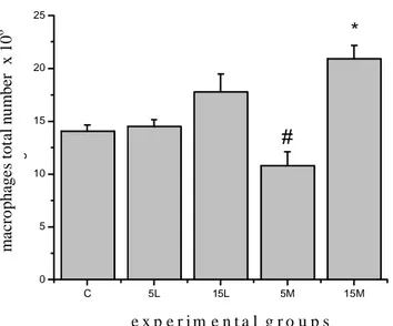
Total number and phagocytic capacity of macrophages A significant increase of 32.68% on total number of peritoneal macrophages was observed in the 15M group compared to the control (p= 0.0012). At the comparison between exercised groups, a statistically significant reduction of 34% of tissue macrophages was found only in the moderate intensity five minutes group in relation to the low intensity five minutes (p= 0.0021) (Figure 3). For phagocytic capacity, significant increases of 7% and 11.26% were observed respectively for the 5L and 15L groups (p= 0.0017 and p= 0.00001) compared to the control group. At groups comparison, a 6.4% lower phagocytic capacity was observed for the moderate five minutes group in relation to the group that exercised for the same duration at low intensity (p= 0.0032) (Figure 4).
DISCUSSION
Prescription of physical exercise has been used as an intervention tool by health professionals, especially Physical Therapists and Physical Educators. Exercise is often indicated with prophylactic or therapeutic objectives as well as to increase physical fitness. However, there is little evidence regarding physiological responses to short duration exercise, especially in relation to the immune response. Immune factors are directly linked to health in general.
Leukocytosis occurs as a response to acute physical stress as well as to intense short duration physical exercise. This response is well documented in the literature12. The effects of this kind of physical exercise on the increase of circulating leukocytes are mediated, at least partially, by the activation of the sympathetic nervous system13 and by an acute increase in the plasma levels of catecholamines during exercise14. Phagocytic capacity of peritoneal macrophages
Macrophages were incubated with 10 ml of PBS containing zymosan (zymozan: 50 µl, Cat. No. Z-7250, Sigma, St. Louis, MO USA) for 30 minutes at 37ºC. After incubation, a 100 µl aliquot was extracted and deposited in a plastic assay tube to which 100 µl of triplan blue were added. The Neubauer chamber was filled and 100 cells (macrophages) were identified. The number of cells among the 100 identified macrophages that had incorporated Zymozan was determined. Incorporation was only considered valid when at least 3 Zymozan particles had been incorporated. Therefore the counting defined the percentage of peritoneal macrophages that had incorporated at least 3 particles of zymozan according to methodological descriptions provided by Pithon-Curi et al.11.
Statistical analysis
All the data were described with means ± Standard Error of the Mean (SEM). Statistical analysis initially included the the Kolmorogorov-Smirnov normality test and homo-cedasticity test (Bartlett criterion). As all analyzed variables presented normal distributions and homocedasticity the two-way Anova was used (considering the independent variables intensity x duration). When significant differences were found, the Tukey’s post hoc test and F test were used for multiple comparisons. In all analysis the level of significance was fixed to p≤ 0.05. The software used in all statistical tests was the Statistica® 6.1.
RESULTS
Total leukocytes and monocytes number
When the groups that performed short duration exercise at low and moderate intensities were compared to the control group, a significant increase in the total leukocytes in all the exercise groups was observed. The increases were of 111%, 130.6%, 204.85% and 181.8% for the 5L, 15L, 5M and 15M groups (p= 0.0028, p= 0.0004, p= 0.00001 and p= 0.00002), respectively. In the comparison between exercised groups, a statistically significant leukocytosis was observed in the group that exercised for five minutes at low intensity. Leukocytosis in this group was 44.53% larger (p= 0.0244) (Figure 1).
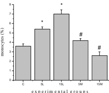
Regarding monocytes counting, increases of 68.42% and 84.21% were found in the 5L and 15L groups (p=0.0008 and p= 0.0001), respectively compared to the control group. At the comparison between experimental groups, a decrease of 52.38% was observed at the percentage of circulating monocytes in the 5M group compared to the 5L group (p= 0.0052), as well as the group that exercised for 15 minutes at moderate intensity. This last group presented a monocyte percentage 169.23% lower in relation to the low intensity group (p= 0.00008) (Figure 2).
C 5L 15L 5M 15M 0 10 20 30 40 50 60 70 80 90 B
#
*
*
ca pac ida de f ago ci tária de m
ac róf ago s (% )
grupo s exp erim entais
C 5L 15L 5M 15M
0 10 20 30 40 50 60 70 80 90 B
#
*
*
ca pac ida de f ago ci tária de m
ac róf ago s (% )
grupo s exp erim entais
e x p e r i m e n t a l g r o u p s
m a cr op h ag e s ph ag o c y ti c c ap ac it y ( % )
C 5L 15L 5M 15M
0 10 20 30 40 50 60 70 80 90 B
#
*
*
ca pac ida de f ago ci tária de m
ac róf ago s (% )
grupo s exp erim entais
C 5L 15L 5M 15M
0 10 20 30 40 50 60 70 80 90 B
#
*
*
ca pac ida de f ago ci tária de m
ac róf ago s (% )
grupo s exp erim entais
e x p e r i m e n t a l g r o u p s
m a cr op h ag e s ph ag o c y ti c c ap ac it y ( % )
Figure 3. Total peritoneal macrophages count. The results were expressed by average ± standard average error. * as compared to control group; # comparison between same duration, but different intensities groups.
Figure 4. Peritoneal macrophages phagocytic capacity. The results were expressed by average ± standard average error. * as compared to control group; # comparison between same duration, but different intensities groups.
Leukocytosis may increase linearly according to the increase in exercise intensity, which can also promote increased catecholamine responses12-14.
Such exercise-induced leukocytosis was found at the present study. Significant increases in the number of total circulating leukocytes were detected in all exercised groups compared to the non-exercised control group. In line with other authors12-14, the 5 and 15-minute groups of higher exercise intensity (moderate) demonstrated a higher percentage of leukocytosis compared to the low intensity groups.
Monocytes are precursors of macrophages in the circulation and can be found free or marginated15. In the case
of physical exercise, an increase in the number of monocytes has been observed as a result of the demargination mediated by catecholamines released during this type of activity6,16. These results were also observed at the present study. The percentage of monocytes increased only in the groups exercised at low intensity, possibly due to the mechanism discussed above.
Exhaustive exercise decreases the number of peritoneal macrophages in rats, and the magnitude of this effect is positively correlated to the concentration of plasmatic corticosterone6. In order to understand the physiological relevance of the glucocorticoids in the mediation of the effects of exercise on total number of peritoneal macrophages, the response with different concentrations of corticosterone was measured in rats17. Apparently there is a physiological concentration in which the glucocorticoids can stimulate macrophages, in accordance with the idea that low levels of glucocorticoids may enhance immunity instead of suppressing it13,17,18. For inhibition of monocyte and macrophage function to occur the mononuclear phagocyte must be exposed to a 50% or greater saturation of the available glucocorticoids receptors for at least 24 hours1.
In this study we found an increase in the amount of peritoneal macrophages in the group that performed the exercise for 15 minutes at low intensity compared to the control group, in accordance with the findings of Ortega1. Additionally, a greater phagocytic capacity was found in the groups that exercised for 5 and 15 minutes at low intensity, in line with what was found by other authors6,16,18. Exercise may modulate the number and function of the innate immune system cells, in this case, increasing the phagocytic capacity of peritoneal macrophages in the short duration exercise of low intensity. Thus, the present study demonstrates that 15 minutes of low intensity exercise may improve immune function through an increase in phagocytic capacity. This fact is of extreme importance for professionals involved with exercise prescription. This kind of activity produces benefits for the immune system and thus can be used as a safe and effective modality in rehabilitation. Increases in the concentration of glucocorticoids, catecholamines, prolactin, thyroid hormones, and β-endorphins may increase phagocytic capacity of the macrophages, and these substances may be stimulated by physical exercise1. In line with these findings, Ortega et al.19, evaluated cyclists during the competitive period and observed an increase in phagocytic capacity in relation to basal values. Additionally, increases of phagocytic capacity in cyclists have been shown to have a strong correlation with the concentration of catecholamines. Taken together, these two parameters are considered good indicators of immune function, not only in athletes, but as also in the general population. In line with these results, greater phagocytic capacity was found in basketball athletes compared to sedentary individuals20. This way, intensity of physical exercise appears to be a determining factor for immune function, and
C 5L 15L 5M 15M
0 5 10 15 20 25 A
#
*
no tot
al d e m ac róf ago s x 10 6 grupos exprimentais
C 5L 15L 5M 15M
0 5 10 15 20 25 A
#
*
no tot
al d e m ac róf ago s x 10 6 grupos exprimentais
e x p e r i m e n t a l g r o u p s
m a cr o p ha g e s to ta l nu mb er x 1 0 6
C 5L 15L 5M 15M
0 5 10 15 20 25 A
#
*
no tot
al d e m ac róf ago s x 10 6 grupos exprimentais
C 5L 15L 5M 15M
0 5 10 15 20 25 A
#
*
no tot
al d e m ac róf ago s x 10 6 grupos exprimentais
e x p e r i m e n t a l g r o u p s
m a cr o p ha g e s to ta l nu mb er x 1 0 6
C 5L 15L 5M 15M
0 5 10 15 20 25 A
#
*
no tot
al d e m ac róf ago s x 10 6 grupos exprimentais
C 5L 15L 5M 15M
0 5 10 15 20 25 A
#
*
no tot
al d e m ac róf ago s x 10 6 grupos exprimentais
e x p e r i m e n t a l g r o u p s
m a cr o p ha g e s to ta l nu mb er x 1 0 6
C 5L 15L 5M 15M
0 5 10 15 20 25 A
#
*
no tot
al d e m ac róf ago s x 10 6 grupos exprimentais
C 5L 15L 5M 15M
0 5 10 15 20 25 A
#
*
no tot
al d e m ac róf ago s x 10 6 grupos exprimentais
e x p e r i m e n t a l g r o u p s
the immune response differs according to the population involved.
At the present study, although total number of peritoneal macrophages did not increase, short duration physical exercise of low intensity increased phagocytic capacity of these cells. The effects of other short duration sessions may possibly favor an increase in immune protection, not as a chronic adaptation, but as a result of summed acute effects, since the phagocytic capacity of macrophages constitutes an important step at the first line of immune defense against infectious agents.
In this context, the present study demonstrated that low intensity exercise was more efficient in increasing phagocytic capacity of peritoneal macrophages as compared with moderate intensity, in line with what was verified by Ortega et al.21, and in contrast to what can be found in athletes19,20. This result is interesting and it can be used as a parameter in the prescription of physical exercise for sedentary individuals and/or patients with limiting pathologies, which is a fact of extreme importance for Physical Therapy.
CONCLUSIONS
Short length physical exercise performed at low intensity contributes mainly for maintenance in the number of tissue macrophages and an increase in their phagocytic capacity. These changes can enhance immune function. In this way, it is suggested that exercise of 5 to 15 minutes performed at low intensity may be used safely and effectively by health professionals involved with the prescription of exercise in clinical practice.
REFERENCES
1. Ortega E. Neuroendocrine mediators in the modulation of phagocytosis by exercise: physiological implications. Exerc Immunol Rev. 2003;9(1):70-93.
2. Asgeirsson G, Bellanti, J. Exercise, immunology and infection. Semin Adolesc Med. 1987;3(3):199-204.
3. Timmons BW, Tarnopolsky MA, Bar-Or O. Sex-based effects on the distribution of NK cell subsets in response to exercise and carbohydrate intake in adolescents. J Appl Physiol. 2006;100(5):1513-9.
4. Fitzgerald L. Exercise and the immune system. Immunol Today. 1988;9(1):337-9.
5. Friman G, Ilbäck NG. Acute infection: Metabolic responses, effects on performance, interaction with exercise, and myocarditis. Int J Sports Med. 1998;19 Suppl 3:S172-82.
6. Escribano BM, Castejon FM, Vivo R, Santisteban R, Aguera EI, Rubio MD. Effects of training on phagocytic and oxidative metabolism of peripheral neutrophils in horses exercised in the aerobic-anaerobic transition area. Vet Res Commun. 2005;29(2):149-58.
7. Saxton JM, Claxton D, Winter E, Pockley AG. Peripheral blood leucocyte functional responses to acute eccentric exercise in humans are influenced by systemic stress but not by exercise-induced muscle damage. Clin Sci. 2003;104(1):69-77.
8. Gobatto CA, Mello MAR, Sibuya CY, Azevedo JRM, Santos LAS, Kokubun E. Maximal lactate steady state in rats submitted to swimming exercise. Comp Biochem Physiol A Mol Integr Physiol. 2001;130(1):21-7.
9. Voltarelli FA, Gobatto CA, Mello MAR. Determination of anaerobic threshold in rats using the lactate minimum test. Braz J Med Biol Res. 2002;35(11):1389-94.
10. Dornfest BS, Lapin DM, Naughton BA, Adu S, Korn L, Gordon AS. Phenylhydrazine-induced leukocytosis in the rat. J Leuk Biol. 1986;39(1):37-48.
11. Pithon-Curi TC, Pires de Melo M, De Azevedo R, Zorn TMT, Curi R. Glutamine utilization by rat neutrophils. Presence of phosphate-dependent glutaminase. Am J Physiol Cell Physiol. 1997;273(42):C1124-9.
12. Barriga C, Pedrera MI, Maynar M, Maynar J, Ortega E. Effect of submaximal physical exercise performed by sedentary men and women on some parameters of the immune system. Rev Esp Fisiol. 1993;49(2):79-86.
13. Brenner I, Shek PN, Zamecnik J, Shephard RJ. Stress hormones and the immunological responses to heat and exercise. Int S Sports Med. 1998;19(2):130-43.
14. McCarthy DA, Dale MM. The leucocytosis of exercise. Sports Med. 1988;6(6):333-63.
15. Van Furth R, Sluiter W. Distribution of blood monocytes between a marginating and a circulating pool. J Exp Med. 1986;163(2): 474-9.
16. Woods JA. Exercise and neuroendocrine modulation of macrophage function. Int J Sports Med. 2000;21 Suppl 1: S24-30.
17. Forner MA, Barriga C, Rodriguez AB, Ortega E. A study of the role of corticosterone as a mediator in exercise-induced stimulation of murine macrophage phagocytosis. J Physiol. 1995;488(3):789-94.
18. Ortega E. Physiology and biochemistry: influence of exercise on phagocytosis. Int J Sports Med. 1994;15 Suppl 1:S172-8.
19. Ortega E, Marchena JM, Garcia JJ, Schmidt A, Schulz T, Malpica I, et al. Phagocytic function in cyclists: correlation with catecholamines and cortisol. J Appl Physiol. 2001;91(3): 1067-72.
20. Ortega E, Barriga C, De la Fuente M. Study of the phagocytic process in neutrophils from elite sportswomen. Eur J Appl Physiol. 1993;66(1):37-42.