DOI: 10.1590/0004-282X20160051
ARTICLE
A histomorphometric study of unmyelinated
fibers of the fibular nerve in Wistar rats
Histomorfometria das fibras amielínicas do nervo fibular em ratos
Wistar
Vânia Tognon-Miguel1, Adriana Helena do Nascimento-Elias1,2, Maria Cristina Lopes Schiavoni1, Amilton
Antunes Barreira1
In recent years, interest in the morphometry of the ibu -lar nerve has increased due to various experimental stud-ies, including its use as a model for nerve regeneration. he use of the ibular nerve in studies of nerve regeneration was boosted after the reintroduction of the end-to-side neuror -rhaphy1,2,3,4,5,6,7,8,9,10,11,23,13. We were not able to ind many
ref-erences to the unmyelinated axons of the ibular nerve in these and other recent studies. Few investigations have been devoted to gathering histomorphometric data about these axons. One research paper showed that unmyelinated ibers in the ibular nerve include both sympathetic (27%),
and sensory ibers (73%), and that the total number of un -myelinated axons ranged from 3,351 to 4,79214. Subsequent
investigators reported values ranging from a minimum of 2,960 unmyelinated axons15 to a maximum of 5,00016. Few
experiments using end-to-side neurorrhaphy with the previ -ously sectioned ibular nerve addressed the issue of regener-ation of unmyelinated ibers17-19. his study aims to evaluate
aspects of the histology and morphology of the unmyelin-ated ibers of the rat common ibular nerve, bringing new and signiicant baseline anatomical data to researchers in the ield of nerve regeneration and others.
1Universidade de São Paulo, Faculdade de Medicina de Ribeirão Preto, Departamento de Neurociências e Ciências do Comportamento, Ribeirão Preto SP, Brasil; 2Universidade de São Paulo, Faculdade de Medicina de Ribeirão Preto, Departamento de Biomecânica, Medicina e Reabilitação do Aparelho Locomotor,
Ribeirão Preto SP, Brasil.
Correspondence: Amilton Antunes Barreira; Faculdade de Medicina de Ribeirão Preto; Av. Bandeirantes 3900; 14049-900. Ribeirão Preto SP, Brasil. E-mail: [email protected]
Conflict of interest: There is no conlict of interest to declare.
Support: FAPESP (Processo: 2012/11012-7), CNPq (Processo: 141524/2015-4) and FAEPA (Fundação de Apoio ao Ensino, Pesquisa e à Assistência do Hospital das Clínicas da Universidade de São Paulo).
Received 08 December 2015; Accepted 16 March 2016.
ABSTRACT
There are few histomorphometric studies on the unmyelinated ibers of the ibular nerve in rats, and the number of experimental studies using this nerve has been increasing in the last years. Sixty-two percent of the endoneurial area from 10 ibular nerves of adult Wistar rats was scanned by electron microscopy, and digitized. The total number of unmyelinated axons (1.882 ± 271) was signiicantly lesser, and their axon diameters (0.2 µm to 2.8 µm) signiicantly higher than that determined in previous studies. The histogram peaked at 1 µm. The differences could be due to the nerve sampled area, the number and the age of the animals evaluated, and the laboratory techniques used. This study brings new andreferential data to be used in experimentalinvestigationsinvolvinghistomorphometric evaluation ofthe rat ibular nerve.
Keywords: unmyelinated ibers, unmyelinated; peroneal nerve; microscopy, electron, transmission; peripheral nerves.
RESUMO
Embora o nervo ibular de ratos venha sendo incluído progressivamente em maior número de estudos experimentais nos últimos anos, há poucos estudos a respeito das suas ibras amielínicas. Os nervos ibulares de 10 ratos Wistar adultos foram avaliados através de microscopia óptica e eletrônica. Varredura sistemática através de microscopia eletrônica de transmissão das áreas fasciculares da porção distal no nervo foi realizada. Em média, 62% da área endoneural foi digitalizada. O número total de axônios amielínicos encontrados (1.882 ± 271) foi signiicativamente menor e as medidas dos diâmetros axonais (0,2 µm a 2,8 µm) maiores do que o determinado em estudos prévios. O pico do histograma foi constituído por ibras de 1µm. As diferenças podem ser devidas à amostragem de maior área endoneural, ao número e à idade dos animais avaliados, e as técnicas laboratoriais utilizadas. Os dados obtidos podem ser considerados referenciais para o nervo ibular de ratos Wistar adultos.
METHOD
Animals
he institutional "Animal Experimentation Ethics Committee" at the Medical School of Ribeirão Preto ap-proved the study (024/2010). We performed analyzes of the ibular nerve using 10 adult female Wistar rats aged 130 days, and each weighing 250g–300g. hey were maintained with 12-hour light-dark cycle, a temperature of between 20–24°C, and free access to food and water.
Surgical procedures
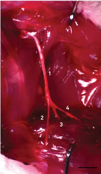
he animals were anesthetized using ketamine chlo-ridrate (75 mg/kg) and xylazine (15 mg/kg) administered intraperitoneally. After the sciatic nerve and its branches were exposed (Figure 1), the ibular nerve was dissected. he nerves were moistened in situ using 2% glutaraldehyde and removed. he distal portion of the left common ibular nerve was immersed in a 2% glutaraldehyde solution for 48h at 4°C. he nerve fragment was washed with sodium cacodylate bufer, postixed in 1% osmium tetroxide, and progressively
dehydrated and embedded in Epoxy resin (Epon 812®). After
removal of the nerve, the animals were euthanized using a double dose of the same anesthetics. he nerve was cut into 0.5 μm thick sections using a microtome (MT 6000XL-RMC), stained with toluidine blue, and mounted using Entellan® for
light microscopy analysis. Details of the methods used have been published in previous studies6,20,21.
Transmission electron microscopy procedures
he plastic embedded blocks were cut using an ultra -microtome (Carl Zeiss, model G/214711) into 80 nm-thick sections. he sections were put on oval grids covered with 5% Formvar ilm (Formvar Solution in Ethylene Dichloride, E.M.S. Inc) and stained with 5% uranyl acetate and 0.5% lead citrate. hey were analyzed using a transmission electron mi-croscope (JEM-100CXII, JEOL Ltda.) equipped with a digi -tal camera (Hamamatsu ORCA-HR, model C4742-51-12HR). he images were obtained sequentially to scan the complete cross-sectional area of each nerve fascicle. Even so, it was nec-essary to leave some gaps between two sequential samples to avoid overlapping of the scanned ields. Photomicrographs at 14,000X magniications were taken manually and sequential -ly while scanning (Figure 2). We scanned 62% of each fasci-cle area. Each digital image obtained from microscopic ields was 14.0 μm wide by 14.0 μm high and 1024x1024 pixels in tagged image ile format (TIFF), and all of them were ana -lyzed. Due to the variability of the fascicular area, the average number of images obtained was around 413 per nerve.
2
3
4
1
Figure 1. Wistar rat sciatic nerve and its branches. 1, sciatic nerve; 2, common ibular nerve; 3, tibial nerve; 4, sural nerve. Bar = 8 mm.
Unmyelinated axons assessment
The axonal area, total number of axons, axonal den -sity (axons/mm2), and the minimum axonal diameter
(μm) were measured using ImageJ software (ImageJ 1.47 National Institutes of Health (USA). The system was cal-ibrated to obtain measurements of the axonal area and minimum diameter of unmyelinated axons. At 14,000X, every 1024 pixels corresponded to 14 μm.This value was obtained using an electron microscopic image of a 1 μm mesh grid as reference. Each unmyelinated axon was sur -rounded using ImageJ’s “polygon tool” (Figure 3). The se -lected and surrounded axons measurements were stored using the “ROI Manager” tool. Unmyelinated axons were correctly identified using established criteria22: 1. The ax
-ons had a circular or oval profile; 2. They were surrounded by Schwann cell cytoplasm forming mesaxons; 3. The axo -plasm of unmyelinated axons was clearer than the cyto -plasm of the respective Schwann cell; 4. the axons were clustered into “units”, which had a direct relationship with Schwann cells; and 5. There was a basal lamina surround -ing each fiber unit externally (Schwann cells and axons). Only axons with a circular shape were measured, and the irregular and obliquely sectioned axons were only count -ed. The area of each fascicle was analyz-ed. The internal fascicular area measures (mm2) were similar to those
ob-tained in semithin sections, and so we measured fascicu -lar area in semithin sections (Figure 4). The methods that we used in this study are as reliable or more reliable than those used in our previous studies23-26. We represented the
frequency distribution of the unmyelinated axons as a his -togram, with axon diameters separated into class inter -vals increasing by 0.2 μm. All morphometric data were ex-pressed as mean ± standard deviation.
Statistical analysis
he obtained data were analyzed using SPSS v.17.0 (SPSS Inc., Chicago, IL, USA) statistical software. Student’s t-test al -lowed the comparison of the total number of unmyelinated
axons with other reports in the literature. Diferences were considered signiicant when p ≤ 0.05.
RESULTS
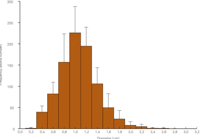
he morphometric data obtained from the ten ibular nerves is presented in Tables 1 and 2. he frequency dis -tribution obtained for axon diameter is presented as a his -togram (Figure 4). Typical ibular nerve samples exhibited a single fascicle surrounded by a well-deined perineurium (Figure 5). he perineurium consisted of a uniform cell lay-er that separates the epineurium from the endoneurium.
Figure 3. Example of the ImageJ tool use for measurement and counting of unmyelinated axons. An image with several unmyelinated ibers and myelinated ibers is showed at left. The red area highlights the enlarged portion of the frame at right. After manually outlining the axolemma (yellow line) the area and the axonal diameter are automatically measured.
A
B
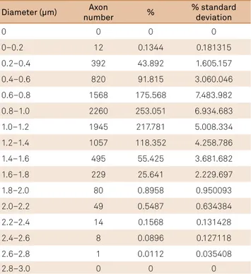
Table 2. Number of unmyelinated axons and their respective percentage regarding to the diameters.
Diameter (µm) Axon
number %
% standard deviation
0 0 0 0
0–0.2 12 0.1344 0.181315
0.2–0.4 392 43.892 1.605.157
0.4–0.6 820 91.815 3.060.046
0.6–0.8 1568 175.568 7.483.982
0.8–1.0 2260 253.051 6.934.683
1.0–1.2 1945 217.781 5.008.334
1.2–1.4 1057 118.352 4.258.786
1.4–1.6 495 55.425 3.681.682
1.6–1.8 229 25.641 2.229.697
1.8–2.0 80 0.8958 0.950093
2.0–2.2 49 0.5487 0.634384
2.2–2.4 14 0.1568 0.131428
2.4–2.6 8 0.0896 0.127118
2.6–2.8 1 0.0112 0.035408
2.8–3.0 0 0 0
Table 1. Histomorphometric parameters of the Wistar rat common ibular nerve and unmyelinated axons (n = 10).
Nerve and unmyelinated components Morphometric data
Cross-sectional data
Area (mm2) 0.13 ± 0.02
Diameter (mm) 0.42 ± 0.03
Unmyelinated Axons
Total number 1882 ± 270.9
Density (axons/mm2) 13935.0 ± 1875.8
Minimum diameter (µm) 0.162
Maximum diameter 2.651
Medium diameter 0.968 ± 0.10
Unmyelinated axons could be distinguished from myelinat-ed axons in photomicrographs. Unmyelinatmyelinat-ed axons could be characterized by the distinctive features of their mesax -ons, microtubules, neuroilaments and the mitochondria in the endoneurial space. heir diameters were highly vari -able as was the degree of circularity of their cross sections. We also identiied the following structures in endoneurial space: Schwann cells nuclei and cytoplasm, myelinated i -bers, endothelial cells of capillaries, mast cells, ibroblasts, and collagen ibers that had been crosswise or obliquely. Schwann cell nuclei were recognized by their closeness to myelinated and unmyelinated axons (Figure 6). We mea -sured 8,931 unmyelinated axons in the ten animals, and only counted the axons that at least partially appeared in the images. All studied nerves had a unimodal distribution of unmyelinated axon diameters. he peak of the histogram was at 1.0–1.2 μm (Figure 4).
DISCUSSION
We used previous data obtained in our laboratory to estimate that the ratio of myelinated ibers to unmyelin -ated axons in the common ibular nerve of adult Wistar
rats is approximately 1:120. Although the rat common
ibu-lar nerve is mainly motor, we expected to ind more unmy -elinated axons than we did. Our results showed less than half the number of unmyelinated axons in the typical com -mon ibular nerve than in a previous study, which estimated 4,171 ± 565 axons14. A further study by the same group in
-vestigated the regenerated ibular nerves of Wistar rats af -ter a nerve crush. hey estimated 2,960 unmyelinated axons in the distal portion of the nerve and gave no standard de-viations15. More than 4,600 unmyelinated axons were found
in a study aiming to evaluate the efect of laser beams on nerve ibers of the ibular nerve in rats16.
Differences in the sample size used for estimating the total number of fibers and other methodological differ -ences may at least partly explain the difference between our results and those of other authors. Our samples were fixed in 2% glutaraldehyde and embedded in Epoxy res -in (Epon 812®). The other authors perfused the hind paws
with 4.0%, 2.5%, and 5% glutaraldehyde respectively. The first two authors embedded their samples in Epon 812® as
we did14,15. One other author used Epon-Araldite®16. The
previous studies used conventional mesh grids to obtain their electron micrographs while we used oval grids with a single opening covered with a Formvar® film. Conventional
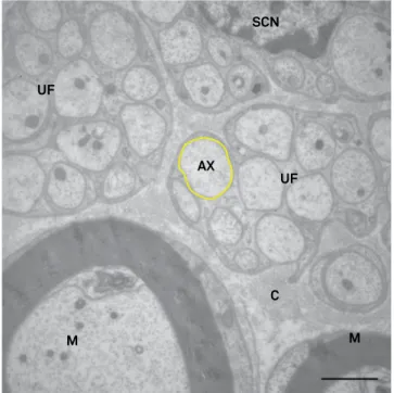
Figure 4. Transverse section through the distal level of a Wistar rat ibular nerve. Images of the following structures can be seen: unmyelinated ibers (UF) and axons (ax) of varied diameters, a Schwann cell nucleus (SCN), myelinated ibers (M), and collagen ibers (C). Yellow line surrounds an unmyelinated axon. Transmission electron microscopy – 14,000X. Bar = 2 µm.
Diameter (µm)
0,0 0,2 0,4 0,6 0,8 1,0 1,2 1,4 1,6 1,8 2,0 2,2 2,4 2,6 2,8 3,0 3,2
Frequency (axons number)
phase called the storage period goes from 48 to 160 days of age28. During the developmental period, the rat sciatic nerve
is known to have more axons than are present in adult rats. Profuse axonal branching is eliminated during the process of maturation29. Comparative experimental studies may difer
in their results due to the maturation of the rat’s body and the accompanying morphological changes30. One author studied
younger animals that were still growing, and we studied adult rats14. he axonal diameters found in the same study were
0.78 μm on average while ours were 0.96 μm. During the pro-cess of axonal maturation, the axonal diameter is less than in adult animals. Our sample size was larger than previous re-search groups. We evaluated ten animals while others stud -ies included four, one, and six animals, respectively14,15,16. It is
noteworthy that using Student’s t test, there are signiicant diferences between our results and those of two others au-thors (p = 0.004 and p < 0.001 respectively)14,16.
he methodology that we have employed and the larger size of the nerve area sampled, both argue for the reliability of the indings presented here. Our indings indicate that the number of unmyelinated axons of the common ibular nerve is smaller and their axon diameters larger than have been recognized hitherto.
Acknowledgments
The authors thank Mr. Antonio Renato Meirelles e Silva, Mrs. Aracy Edwirges Vieira da Silva Dias, Mrs. Maria Teresa Picinoto Maglia and Mr. José Augusto Maulin for technical assistance, as well as Mr. Geraldo Cássio dos Reis for statistical analysis.
grids are associated with a higher chance of error, due to the overlapping of thin sections on the grid meshes divi -sions, hiding part of the images and limiting the evalua -tion. Oval grids do not obstruct any portion of the full fas-cicle image, allowing its entire area to be scanned. This may be one of the reasons why the other authors present -ed data that was bas-ed on scanning a smaller area of the nerve fascicle than we did. In a study, data were obtained from 3 to 70% of the cross-sectional area of the nerves ex -amined and did not precisely indicate the fibular nerve area sampled14. Other investigation sampled 6 to 10% of
the sectional nerve area15. The total number of unmyelin
-ated axons was estim-ated based on the analysis of 15% of the nerve area in a third study16. Our quantification of the
number of unmyelinated axons in the typical fibular nerve was based, on average, on measurements conducted over 60% of the nerve’s cross-sectional area. The area sampled by the other authors was at least six times smaller. Their estimations were based on the proportion of both unmy-elinated axons and myunmy-elinated fibers (the specific ratio was not given). We extrapolated the total number of ax -ons from the scanned area to the complete fascicular area. Another reason for diferences between other authors’ data and ours could be the age of the animals studied. We ex -amined rats that were approximately 18 weeks old (130 days). Studies evaluated rats ive and half weeks old (39 days) but without giving any age information14-16. he observed difer
-ences may be related to difer-ences in the length of the devel -opmental period. he growth process of the skeleton is com-pleted between 120–140 days of age, and they are regarded as adults after that27. he process of growth and development in
rats involves three distinct phases. he initial phase is termed hyperplasia ( irst 17 days of life). he intermediate phase is re -ferred to as hyperplasia-hypertrophy (17 to 48 days). he last
Figure 5. Common ibular nerve histogram showing the distribution of unmyelinated axon diameters.
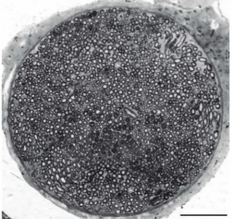
Figure 6. Transverse semithin section through the distal segment of the rat common ibular nerve. Toluidine blue. Bar = 100 µm.
M UF
AX
SCN
UF
C
References
1. Viterbo F, Trindade JC, Hoshino K, Mazzoni Neto A. Latero-terminal neurorrhaphy without removal of the epineural sheath. Experimental study in rats. Rev Paul Med.1992;110(6):267-75.
2. Viterbo F, Trindade JC, Hoshino K, Mazzoni Neto A. Two end-to-side neurorrhaphies and nerve graft with removal of the epineural sheath: experimental study in rats. Br J Plast Surg. 1994;47(2):75-80. doi:10.1016/0007-1226(94)90162-7
3. Viterbo F, Trindade JC, Hoshino K, Mazzoni Neto A. End-to-side neurorrhaphy with removal of the epineurial sheath: an
experimental study in rats. Plast Reconstr Surg. 1994;94(7):1038-47. doi:10.1097/00006534-199412000-00019
4. Zhang Z, Soucacos PN, Beris AE, Bo J, Ioachim E, Johnson EO. Long-term evaluation of rat peripheral nerve repair with end-to-side neurorrhaphy. J Reconstr Microsurg. 2000;16(4):303-11. doi:10.1055/s-2000-7338 5. Zhang Z, Soucacos PN, Bo J, Beris AE, Malizos KN, Ioachim E et al.
Reinnervation after end-to-side nerve coaptation in a rat model. Am J Orthop (Belle Mead NJ). 2001;30(5):400-6.
6. De Sá JM, Mazzer N, Barbieri CH, Barreira AA. The end-to-side peripheral nerve repair. Functional and morphometric study using the peroneal nerve of rats. J Neurosci Methods. 2004;136(1):45-53. doi:10.1016/j.jneumeth.2003.12.018
7. Brenner MJ, Dvali L, Hunter DA, Myckatyn TM, Mackinnon SE. Motor neuron regeneration through end-to-side repairs is a function of donor nerve axotomy. Plast Reconstr Surg. 2007;120(1):215-23. doi: 10.1097/01.prs.0000264094.06272.67
8. Silva DN, Silva AC, Aydos RD, Viterbo F, Pontes ER, Odashiro DN et al. Nerve growth factor with ibrin glue in end-to-side nerve repair in rats. Acta Cir Bras. 2012;27(4):325-32. doi:10.1590/S0102-86502012000400008
9. Maciel FO, Viterbo F, Chinaque LF, Souza BM. Effect of electrical stimulation of the cranial tibial muscle after end-to-side neurorrhaphy of the peroneal nerve in rats. Acta Cir Bras. 2013;28(1):39-47. doi:10.1590/S0102-86502013000100007 10. Yang LM, Wu YX, Zhang XP, Li XH. Experimental research on
end-to-side anastomosis of peripheral nerves and effect of FK506 on end-to-side anastomosis. Bratisl Lek Listy. 2014;115:625-31.
11. Liu HF, Chen ZG, Fang TL, Arnold P, Lineaweaver WC, Zhang J. Changes of the donor nerve in end-to-side neurorrhaphies with epineurial window and partial neurectomy: a long-term evaluation in the rat model. Microsurgery. 2014;34(2):136-44. doi:10.1002/micr.22167
12. Liu HF, Chen ZG, Shen HM, Zhang H, Zhang J, Lineaweaver WC et al. Eficacy of the end-to-side neurorrhaphies with epineural window and partial donor neurectomy in peripheral nerve repair: an experimental study in rats. J Reconstr Microsurg. 2015;31(1):31-8. doi:10.1055/s-0034-1382263
13. Fagotti de Almeida CE, Farina Junior JA, Colli BO. Morphometric and functional analysis of axonal regeneration after end-to-end and end-to-side neurorrhaphy in rats. Plast Reconstr Surg Glob Open. 2015;3(3):e326. doi:10.1097/GOX.0000000000000280
14. Schmalbruch H. Fiber composition of the rat sciatic nerve. Anat Rec. 1986;215(1):71-81. doi:10.1002/ar.1092150111
15. Toft PB, Fugleholm K, Schmalbruch H. Axonal branching following crush lesions of peripheral nerves of rat. Muscle Nerve. 1988;11(8):880-9. doi:10.1002/mus.880110813
16. Wesselmann U, Kerns JM, Rymer WZ. Laser effects on myelinated and nonmyelinated ibers in the rat peroneal nerve: a quantitative ultrastructural analysis. Exp Neurol. 1994;129(2):257-65. doi:10.1006/exnr.1994.1168
17. Kovacic U, Tomsic M, Sketelj J, Bajrović FF. Collateral sprouting of sensory axons after end-to-side nerve coaptation: a longitudinal study in the rat. Exp Neurol. 2007;203(2):358-69. doi:10.1016/j.expneurol.2006.08.018
18. Kovacic U, Sketelj J, Bajrović FF. Sex-related differences in recovery of cutaneous nociception after end-to-side nerve repair in the rat. J Plast Reconstr Aesthet Surg. 2009;62(6):806-13. doi:10.1016/j.bjps.2007.09.046
19. Kovacic U, Sketelj J, Bajrović FF. Effect of aging on recovery of cutaneous nociception after end-to-side nerve repair in the rat. Ann Plast Surg. 2009;62(4):439-45. doi:10.1097/SAP.0b013e318180c8cb 20. Nascimento-Elias AH. Veículos, indutores e tubulização
látero-terminal na regeneração do nervo periférico [thesis]. Ribeirão Preto: Universidade de São Paulo; 2013.
21. Santos AP, Suaid CA, Fazan VP, Barreira AA. Microscopic anatomy of brachial plexus branches in Wistar rats. Anat Rec (Hoboken). 2007;290(5):477-85. doi:10.1002/ar.20519
22. Morris JH, Hudson AR, Weddell G. A study of degeneration and regeneration in the divided rat sciatic nerve based on electron microscopy II. The development of the “regenerating unit”. Z Zellforsch Mikrosk Anat. 1972;124(1):103-30. doi:10.1007/BF00335457
23. Fazan VP, Salgado HC, Barreira AA. Aortic depressor nerve unmyelinated ibers in spontaneously hypertensive rats. Am J Physiol Heart Circ Physiol. 2001;280(4):H1560-4.
24. Fazan VP, Ma X, Chapleau MW, Barreira AA. Qualitative and quantitative morphology of renal nerves in C57BL/6J mice. Anat Rec. 2002;268(4):399-404. doi:10.1002/ar.10174
25. Marques VD, Barreira AA, Davis MB, Abou-Sleiman PM, Silva WA Jr, Zago MA et al. Expanding the phenotypes of the Pro56Ser VAPB mutation: proximal SMA with dysautonomia. Muscle Nerve. 2006;34(6):731-9. doi:10.1002/mus.20657
26. Oliveira AL, Fazan VP, Marques Junior W, Barreira AA. Dorsal cutaneous branch of the ulnar nerve: a light and electron microscopy histometric study. J Peripher Nerv Syst. 2011;16(2):98-101. doi:10.1111/j.1529-8027.2011.00326.x
27. Hughes PCR, Tanner JM. The assessment of skeletal maturity in the growing rat. J. Anat. 1970;106(2):371-402.
28. Enesco M, Leblond P. Increase in cell number as factor in the growth of the organs and tissues of the young male rat. J Embryol Exp Morphol. 1962;10:530-62.
29. Jenq CB, Chung K, Coggeshall RE. Postnatal loss of axons in normal rat sciatic nerve. J Comp Neurol. 1986;244(4):445-50. doi:10.1002/cne.902440404
ERRATUM DOI: 10.1590/0004-282X20160051erratum
Erratum
Arquivos de Neuropsiquiatria. 2016;74(5):367-72. doi:10.1590/0004-282X20160051
he correct legends for the Figures 4, 5 and 6 are:
Figure 4. Common fibular nerve histogram showing the distribution of unmyelinated axon diameters.
Diameter (µm)
0,0 0,2 0,4 0,6 0,8 1,0 1,2 1,4 1,6 1,8 2,0 2,2 2,4 2,6 2,8 3,0 3,2
Frequency (axons number)
0 50 100 150 200 250 300
Figure 5. Transverse semithin section through the distal segment of the rat common fibular nerve. Toluidine blue. Bar = 100 µm.
Figure 6. Transverse section through the distal level of a Wistar rat fibular nerve. Images of the following structures can be seen: unmyelinated fibers (UF) and axons (ax) of varied diameters, a Schwann cell nucleus (SCN), myelinated fibers (M), and collagen fibers (C). Yellow line surrounds an unmyelinated axon. Transmission electron microscopy – 14,000X. Bar = 2 µm.
M UF
AX SCN
UF
C