Late Clinical and Functional Assessment of Arrhythmias in Children
after Repair of Tetralogy of Fallot
Maria Eulália Thebit Pfeiffer
1, Eduardo Machado Andrea
1, Salvador Manoel Serra
1, Claudio Roberto Assumpção
1,
Gesmar Volga Haddad Herdy
2Instituto Estadual de Cardiologia Aloysio de Castro (IECAC)1, Rio de Janeiro, RJ, Universidade Federal Fluminense (UFF)2, Niterói, RJ - Brazil
Mailing address: Maria Eulalia Thebit Pfeiffer •
Av. Gilberto Amado, 970/ CBO - 301 - Barra da Tijuca - 22621-232 - Rio de Janeiro, RJ - Brazil
E-mail: [email protected], [email protected]
Manucript received July 30, 2009; revised mansucript received December 24, 2009; accepted March 18, 2010.
Abstract
Background: Cardiac arrhythmias are the major cause of late sudden death in patients undergoing repair of Tetralogy of Fallot (TF).
Objective: To evaluate the occurrence of cardiac arrhythmias in children and adolescents undergoing repair of TF, and to associate them with clinical aspects and laboratory tests.
Methods: Cross-sectional study of 37 patients undergoing repair of TF at Instituto Estadual de Cardiologia Aloysio de Castro (Rio de Janeiro). After review of the medical records and clinical assessment, the patients underwent electrocardiography (ECG), echocardiography (Echo), 24-h Holter monitoring and exercise test (ET), whose results were subjected to statistical analysis.
Results: A total of 37 patients of whom 54% were males with a mean age of 9.7 ± 3.5 years and mean follow-up period of 4.7 ± 1.9 years were studied. The abnormalities most frequently found were: on ECG: right bundle branch block (89%); Echo: severe pulmonary regurgitation (43%), mild pulmonary stenosis (73%), moderate right ventricular hypertrophy (RVH, 57%); on ET: low exercise capacity (90%), impaired chronotropic response (40%), arrhythmias (20%); on Holter monitoring: arrhythmias (59%, of which 44% were ventricular, 38% supraventricular, and 24% both ventricular and supraventricular, with predominance of infrequent and benign ventricular premature beats). Five patients (15%) presented with multiform ventricular premature beats. There was an association of ventricular arrhythmia with moderate and severe RVH (p=0.026), as well as with right ventricle-to-pulmonary artery gradient (RV/PA) > 45 mmHg (p=0.004). The logistic regression analysis showed that increased RV/PA gradient was an independent predictor of ventricular arrhythmia (p=0.017).
Conclusion: Cardiac arrhythmia was a common finding in a large proportion of children and adolescents after surgical repair of TF; however, it was infrequent and benign in most of the cases. The RV/PA gradient was considered a strong predictor of ventricular arrhythmia. (Arq Bras Cardiol 2010; 95(3): 295-302)
Key words: Tetralogy of Fallot/surgery; heart defects, congenital; arrhythmias, cardiac; child; health services evaluation.
serious events such as sustained ventricular tachycardia and ventricular fibrillation1,5,6.
Studies show that the most important factors influencing a poorer patient outcome are: age at surgery above five years; longer postoperative follow-up; a more aggressive operative technique with utilization of patches for more extensive transannular pulmonary enlargement, thus leading to more severe pulmonary regurgitation and progressive RV dilation or, also, residual obstructive pulmonary injury with increased RV systolic pressure; surgical scars; and increased duration of the QRS complex longer than 180ms7,8.
Patients older at repair have a less satisfactory response with a higher predisposition to arrhythmias, since their right ventricle (RV) shows more marked hypertrophy, more fibrous tissue, and requires more extensive surgery9,10. Zeltser et
al11, in an experimental study, observed that increased right
ventricular systolic pressure (RVSP) was considered a predictive factor for atrial and ventricular arrhythmias11. According to
Introduction
Tetralogy of Fallot (TF) is the most common cyanotic congenital heart disease, with a favorable outcome after total surgical repair1. In addition to a longer survival, surgically
treated patients have a better quality of life, they can work, and practice physical exercises2,3. However, in the mid and
long terms, they may present with hemodynamic changes due to residual lesions, which lead to the onset of several arrhythmias, thus resulting in higher morbidity and mortality4.
Daliento et al12, the increased right ventricular volume resulting
from hypertrophy, fibrosis and different anatomical changes due to residual lesions may lead to severe arrhythmias12.
Chandar et al13 conducted a multicenter study of 359 patients
with a mean age of five years at surgery and mean follow-up of seven years, and found an incidence of 48% of spontaneous ventricular premature beats and 17% of laboratory-induced ventricular tachycardia by electrophysiologic stimulation, both related to older patients, longer follow-up, symptoms of syncope or presyncope, and RVSP > 60 mmHg13.
Several studies have been conducted in an attempt to improve the knowledge on predictors of arrhythmias; however their pathophysiological mechanism has not yet been fully explained4,8,14.
The objective of this study was to specially investigate the occurrence of cardiac arrhythmias in children and adolescents after surgical repair of TF, associating them with clinical and laboratory findings.
Methods
Cross-sectional study conducted in 37 children and adolescents undergoing surgical repair of TF in the period from 2000 to 2007 at Instituto Estadual de Cardiologia Aloysio de Castro (IECAC-RJ). In order to be included in the study, the patients should have undergone total repair and given written informed consent.
The study was approved by the IECAC Ethics Committee on Human Research, protocol 2006/05, and was conducted between March 2006 and March 2008.
The medical records of 51 consecutive patients with TF referred for surgery were reviewed. Of these, four underwent the Blalock procedure alone, four died in the immediate postoperative period, and six missed the medical visit and the study protocol tests; thus, a total of 37 patients were included in the study. After clinical assessment, collection of surgical data and of previous test results, these patients were referred for new tests, namely: ECG, Echo, Holter monitoring, and ET. All tests were performed in IECAC.
A baseline 12-lead ECG was obtained from all patients (Dixtal model EP-3, and Ecafix), with analysis of rhythm, heart rate (HR), presence or absence of arrhythmias and duration of the QRS complex (QRSd).
Echo was also performed in all patients (Toshiba Nemio 30, 3.75 and 5.0-MHz transducers) and analyzed by two observers in the M-mode and two-dimensional mode, and the values of cardiac chamber measurements were considered in relation to the body surface15. Left ventricular (LV) function was assessed
by means of the ejection fraction in the M-mode, using the Teichholz method15. Qualitative RV function was subjectively
assessed using the two-dimensional mode, and was classified as normal or abnormal16-18. Tricuspid regurgitation was
quantified as severe if the regurgitant jet reached the posterior wall of the right atrium, and the gradient was quantified by continuous Doppler for the assessment of the pulmonary pressure18. Pulmonary regurgitation was quantified according
to the beginning of the regurgitant jet: as mild when it was close to the pulmonary valve; as moderate, when originated
in the pulmonary trunk; and as severe, if originated in the branch bifurcation. The RV outflow tract obstruction was estimated by continuous Doppler, and was classified by the author, for the purpose of statistical analysis, as: <10 (normal),
≥10 up to <45 (mild), ≥45 up to <75 (moderate), and >75
(severe) mmHg15-18.
All patients underwent 24-h Holter monitoring (Dynamis-CARDIOS, São Paulo). The tracings were analyzed both by the researcher and by a specialist from the department of arrhythmia. HR, the presence or absence of arrhythmias and their description were analyzed. The description of the arrhythmias was based on Lown’s classification for ventricular arrhythmias, as follows: grade zero (no arrhythmia); grade I (ventricular premature beats - VPB - isolated or < 30/min); grade II (frequent VPB, > 30/ min); grade III (multiform VPB); grade IV A (couplets) and IV B (non-sustained ventricular tachycardia); and grade V (sustained ventricular tachycardia - SVT)19.
ET was performed in the patients clinically fit for the test by means of the Bruce protocol, which was administered in 21 patients, and the ramp protocol in nine patients, according to the guidelines of the department of ergonomics. The exercise capacity was evaluated using percentiles (P), according to Cumming: P10 (very weak), P25 (weak), P50 (reasonable), P75 (good) and P90 (excellent)20. The following parameters
were also evaluated: characteristics of the arrhythmias detected; exercise time (in minutes); chronotropic reserve by means of the chronotropic index; autonomic response, by means of HR reduction in the 1st minute of recovery; and functional class, according to the NYHA, where class I (7 METs or more), class II (5 and 6 METs), class III (3 and 4 METs) or class IV (less than 3 METs)21,22.
For the statistical analysis, numeric variables were expressed as mean, standard deviation, median, minimum and maximum values. Categorical variables were expressed as frequencies and percentages. The Mann-Whitney test was used for the comparison of quantitative data; the chi-square test and Fisher’s exact test, for qualitative data. Logistic regression analysis was used to identify independent variables. The significance level was set at 5% (p<0.05).
The statistical analysis was processed by the SAS software 6.04 (SAS Institute, Inc. Cary, North Carolina).
Results
Background
other due to severe pulmonary regurgitation (PR). One patient had a permanent pacemaker due to complete atrioventricular block in the immediate postoperative period.
Evaluation of ECG, Echo and Holter monitoring
Baseline ECG was normal in two patients (5.4%); right bundle branch block (RBBB) was found in 33 (89.0%); RBBB associated with left anterior hemiblock (LAHB) in two (5.4%); 1st-degree atrioventricular block (AVB) in two (5.4%); mean QRSd was 108.6 ms, and > 120 ms in six patients (16.2%). Three patients (8.1%) showed isolated SVPB and VPB.
Echo showed diameters and LV function, as well as RV function, which were normal in 100% of the patients. However, LV diameters were increased in 94.6%, as shown in Graph 1.
Residual ISD of varying diameters was observed in 10 patients (27%). RV outflow tract obstruction was detected in 34 patients (91.9%); its classification is shown in Table 1. PR was found in 34 patients (91.9%); it was severe in 16 (43.2%) and moderate in nine (24.3%). Tricuspid regurgitation was moderate in four patients (10.8%) and severe in one (2.7%). Holter monitoring was performed in 34 patients, because in three patients the tracing could not be recorded due to technical problems. Twenty patients (59%) presented with arrhythmia: SVPB (38%), VPB (44%), and both (24%), which were infrequent or occasional in most of them. Frequent SVPB were observed in four patients (>500/day). VPB were monomorphic (Lown I) in most of the patients, but were multimorphic (Lown III) with varying frequency in five patients (15%). The mean maximum HR was 137 ± 18 bpm; the mean HR, 86 ± 14 bpm; and mean minimum HR, 56 ± 12 bpm.
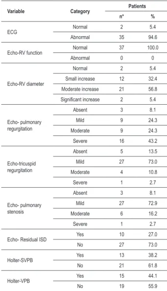
The overall descriptive analysis of the qualitative variables of ECG, Echo, and Holter monitoring is shown in Table 2.
Functional assessment at exercise
Exercise test (ET) was performed in 30 patients, because the other seven could not perform the exercise due to lack of medical conditions or of ability. The mean exercise time was 10.3 ± 2.2 min; mean baseline HR 85 ± 17 bpm, and peak HR 170 ± 24 bpm. The mean baseline systolic blood pressure (BP) was 98 ± 13 mmHg, and mean peak systolic BP was 119 ± 25 mmHg. The mean peak double product (HR x systolic BP) was 21.173 ± 6.442 mmHg.bpm. The chronotropic index was lower than 80% in 12 patients (40%) and the autonomic response was inadequate in two patients (7%). VPB during exercise were observed in six patients: they were rare in five and frequent in one. Exercise capacity was classified as weak or very weak in 90% of the patients (Graph 2), although 97% were in functional class I (NYHA).
Statistical analysis
The following variables were used: age at surgery and at assessment; follow-up period; RVH (RV hypertrophy); PR (pulmonary regurgitation); RV-PA gradient; and QRSd
Table 1 -Classiication of the gradients found in the right ventricular
outlow tract
Gradient Patients
n %
<10 mmHg 3 8.1
≥10 - <45 mmHg 27 73.0
≥ 45 - <75 mmHg 6 16.2
≥75 mmHg 1 2.7
Total 37 100
Table 2 -Overall descriptive analysis of the ECG, Echo and Holter
monitoring variables
Variable Category Patients
n* %
ECG Normal 2 5.4
Abnormal 35 94.6
Echo-RV function Normal 37 100.0
Abnormal 0 0
Echo-RV diameter
Normal 2 5.4
Small increase 12 32.4
Moderate increase 21 56.8
Signiicant increase 2 5.4
Echo- pulmonary regurgitation
Absent 3 8.1
Mild 9 24.3
Moderate 9 24.3
Severe 16 43.2
Echo-tricuspid regurgitation
Absent 5 13.5
Mild 27 73.0
Moderate 4 10.8
Severe 1 2.7
Echo- pulmonary stenosis
Absent 3 8.1
Mild 27 72.9
Moderate 6 16.2
Severe 1 2.7
Echo- Residual ISD Yes 10 27.0
No 27 73.0
Holter-SVPB Yes 13 38.2
No 21 61.8
Holter-VPB Yes 15 44.1
No 19 55.9
*ECG and Echo: n = 37; Holter: n = 34.
regression showed that in the sample studied the numerical RV-PA gradient (in mmHg) was a significant predictor of VA, in a direct relation: the higher the gradient the greater the tendency to ventricular arrhythmia (coefficient of 0.0761, standard error of 0.0320, p value = 0.017).
Discussion
In the present study, the mean age at surgery was five years. Most of theresearchers consider that the optimal age for repair of Tetralogy of Fallot is between three and 11 months, although the procedure of each institution depends on its expertise and appropriate conditions23. Many patients are diagnosed
late, and therefore the repair is made at an older age. Older patients are more prone to develop arrhythmias, because their RV is exposed to the harmful effects of pulmonary obstruction for a longer time, and they also require more extensive surgeries7,9,10. The mean age at assessment was 9.8 years, and
the mean follow-up period was 4.8 years; arrhythmias and SD were more prevalent among patients with longer follow-up, because the RV tends to undergo progressive dilation due to residual lesions and surgical scars4,8,9,24.
Most of the patients (95%) were asymptomatic. Studies show that symptoms occur more frequently in cases with longer follow-up18,24.
In our case series, ventriculoseptoplasty was performed via the transatrial approach, and half of the patients required a pulmonary patch. A higher incidence of arrhythmias is usually correlated in the literature with the transventricular approach and placement of larger patches8,14,23.
Resting ECG showed a high prevalence of RBBB (89%),
and QRSd was > 120 ms in 16.2%, although QRSd ≥ 180
ms, which is considered an important predictor for the occurrence of severe arrhythmias and sudden death8,12,25,
was not observed in any of the patients. Progressive QRS prolongation suggests RV dilation and severe PR for a longer time, although when occurring in the immediate postoperative period it reflects surgical myocardial injury or direct His bundle injury, in this case, with a more benign character25,26.
Only one patient had a baseline ECG showing complete AVB; two had AVB (1st degree), and other three, sparse SVPB and VPB. According to some studies, and as corroborated by our case series, the prevalence of complete AVB is low - of approximately 3%, and few patients require a permanent pacemaker4,27. Complete RBBB occurs in approximately 80%
of the patients, and in approximately 10% it is associated with LAHB, the latter being a more serious condition due to the possibility of progressing to late complete AVB, severe arrhythmias and SD25,26.
On the echocardiographic study, we observed that all patients had normal overall cardiac function, although the RV diameters were increased in 95%, with the presence of moderate to severe RVH in most of them (62%). Overload of the right chambers is a direct consequence of pulmonary regurgitation, as well as of RV outflow tract obstruction. However, PR is better tolerated during childhood and in the first postoperative years, with a lower incidence of arrhythmias7,14.
PR was moderate and severe in 68% of the cases, and RV (QRS duration); based on data from Holter monitoring.
Statistical tests were applied to verify the association between ventricular arrhythmias (VA) or ventricular plus supraventricular arrhythmias (VA + SVA). In relation to the qualitative variables, the tests showed that moderate and severe RVH were significantly more frequent in patients with VA than in the group without VA (80% and 42%, respectively,
p = 0.026), same as for the RV-PA gradients ≥ 45 mmHg
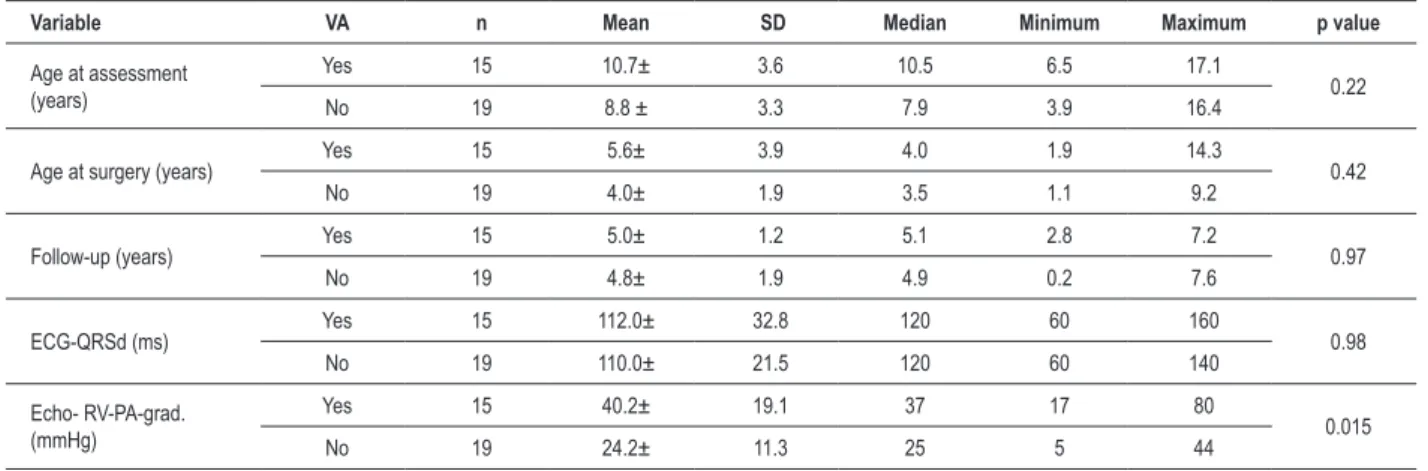
(40% and 0%, respectively, p = 0.004) (Table 3, Graphs 3 and 4). This correlation also occurred in patients with VA+SVA in comparison to those without arrhythmia (p = 0.028). In relation to the numerical variables, in patients with arrhythmia (Table 4), the mean RV-PA gradient was also significantly higher among those with VA (p = 0.015); likewise, it was higher among patients with VA+SVA (p = 0.049) than in the groups without VA or without VA+SVA.
Graph 2 - Patient distribution according to their exercise capacity20.
Graph 3 - Association between right ventricular hypertrophy and ventricular arrhythmia (VA).
outflow tract obstruction showed RV-PA gradients above 45 mmHg in 19% of the patients. Just as in our cases, studies show the presence of some degree of residual pulmonary flow obstruction in most of the patients, and a 60% to 90% prevalence of PR resulting from commisurotomy and transannular enlargement7,14.Some authors believe that a more
restrictive right ventricular physiology, with less pulmonary regurgitation, is more beneficial in the long term, preventing severe RV dilation and causing less QRSd prolongation28.
In our case series, there were two reoperations, one for heart
valve replacement, and both patients had arrhythmias, although they were stable. Pulmonary valve replacement has been indicated to reduce regurgitation, although much discussion still exists in relation to the optimal time for theprocedure17.
Patients undergoing pulmonary valve replacement, whether for severe regurgitation or stenosis, show clinical improvement, with less arrhythmias and QRSd stabilization17,29.
Table 3 -Analysis of qualitative (categorized) variables according to the presence of ventricular arrhythmias
Variable Category
Ventricular arrhythmias (n = 34)
Present Absent
p value
n %* n %*
Follow-up ≥5 years 8 53.3 9 47.4 0.73
<5 years 7 46.7 10 52.6
QRS duration ≥120 ms 9 60.0 13 68.4 0.61
<120 ms 6 40.0 6 31.6
RVH Mod/severe 12 80.0 8 42.1 0.026
Norm/mild 3 20.0 11 57.9
Pulmonary regurgitation
Severe 9 60.0 6 31.6
0.097
Not severe 6 40.0 13 68.4
RV-PA gradient ≥45 mmHg 6 40.0 0 0 0.004
<45 mmHg 9 60.0 19 100
Graph 4 - Association of the RV-PA gradient with ventricular arrhythmia (VA).
that the risk of developing arrhythmias is proportional to the age at repair24. A patient 30 years old at the time of repair has
a 17 times greater risk of developing arrhythmic symptoms than another one who was five years old at repair, and a five-year-old patient has a 1.4 times greater risk than a patient two years old at repair24. Matina et al30 conducted a study in
59 patients with a mean follow-up of 7.5 years and observed the occurrence of arrhythmias six years after repair, and none of the patients less than two years old at repair presented with ventricular arrhythmic events30. Our patients underwent
surgery at a mean age of 5.0 years, and the mean follow-up period was also 5.0 years, and they had an incidence of arrhythmias similar to that found in the literature24,30-32.
Exercise test detected arrhythmias in 20%, characterized by
infrequent VPB. Among all patients, the highest frequency of VPB was recorded in the patient who had the highest RV-PA gradient. Exercise capacity was classified as very weak in most of the patients, and 40% of them had a chronotropic index considered inadequate. Some authors correlate a significantly reduced exercise capacity with the use of patches and severe pulmonary regurgitation24. We also believe that the very weak
exercise capacity observed in most of the cases studied may be associated with irregular exercise practice induced by family protection and by the habit of limitation established by the underlying disease22.
Table 4 -Statistical data of numerical variables according to the presence of ventricular arrhythmias
Variable VA n Mean SD Median Minimum Maximum p value
Age at assessment (years)
Yes 15 10.7± 3.6 10.5 6.5 17.1
0.22
No 19 8.8 ± 3.3 7.9 3.9 16.4
Age at surgery (years) Yes 15 5.6± 3.9 4.0 1.9 14.3 0.42
No 19 4.0± 1.9 3.5 1.1 9.2
Follow-up (years) Yes 15 5.0± 1.2 5.1 2.8 7.2 0.97
No 19 4.8± 1.9 4.9 0.2 7.6
ECG-QRSd (ms) Yes 15 112.0± 32.8 120 60 160 0.98
No 19 110.0± 21.5 120 60 140
Echo- RV-PA-grad. (mmHg)
Yes 15 40.2± 19.1 37 17 80
0.015
No 19 24.2± 11.3 25 5 44
n = 34. SD - standard deviation.
the cut-off point between children with heart diseases and normal children21.
In our case series, we did not observe any association between the different variables studied and the presence of cardiac arrhythmias, unlike in other studies in the literature. However, there was an association between RV-PA gradients
≥ 45 mmHg and the presence of VA(p=0.004) and VA+SVA (p=0.028)4,8,25. These results corroborate the findings from
other studies which consider a high RV systolic blood pressure to be a predictor of arrhythmias and sudden death11,13,24,29.
Garson et al2 observed that a poor surgical outcome,
with residual RV pressure elevation and development of dysfunction, was associated with the occurrence of arrhythmias and sudden death2.
We also observed an association between moderate and severe RVH, which is also considered in the literature as a predictor of arrhythmias, and the presence of VA (p = 0.026)4,9,14.
The most relevant result of the present study, obtained by means of simultaneous analysis of variables using the logistic regression analysis, was the observation that the RV systolic gradient was able to predict VA in a direct relation, i.e., the higher the gradient, the greater the tendency to VA in this sample (p = 0.017).
Conclusion
Although a common finding, cardiac arrhythmias in children and adolescents undergoing TF repair are infrequent and benign in most of the cases. However, in patients with greater overload and higher right ventricular systolic pressure, the arrhythmic events are more frequent or potentially more
severe, the increased RV pressure gradient being considered an independent risk factor for ventricular arrhythmias.
Recommendations
A regular follow-up of these patients after surgical repair, by means of clinical and laboratory tests, is important for the early detection of complications subsequent to residual lesions, since it can probably reduce the risk of late cardiac arrhythmias in these children.
Acknowledgements
We extend special thanks to the colleagues from the Department of Pediatric Cardiology, IECAC, for their support and help with the performance of the echocardiographic studies, and also those from the Center of Exercise Cardiology, for helping with the exercise tests. To Professor Rosangela Noé (UFRJ) for the statistical analysis, and Professor Maria do Carmo L. Gomes, for proofreading this manuscript.
Potential Conflict of Interest
No potential conflict of interest relevant to this article was reported.
Sources of Funding
There were no external funding sources for this study.
Study Association
References
1. Folino AF, Daliento L. Arrhythmias after Tetralogy of Fallot repair. Indian Pacing Electrophysiol J. 2005; 5 (4): 312-24.
2. Garson Jr A, Nihill MR, McNamara DG, Cooley DA. Status of the adult and adolescent after repair of Tetralogy of Fallot. Circulation. 1979; 59 (6):1232-40.
3. Atik FA, Atik E, Cunha CR, Caneo LF, Assad RS, Jatene MB, et al. Long term results of correction of Tetralogy of Fallot in adulthood. Eur J Cardiothorac Surg. 2004; (25): 250-5.
4. Freedom RM, Yoo S. Tetralogy of Fallot. In: Freedom RM, Yoo S, Mikailian H, Williams WG, editors. The natural and modified history of congenital heart disease. New York: Futura; 2004. p. 186-211.
5. Bricker JT. Sudden death and Tetralogy of Fallot: risks, markers, and causes. Circulation. 1995; 92 (2): 158-9.
6. Günal N, Tokel K, Kahramanyol O, Ozer S, Celiker A, Ekici E, et al. Incidence and severity of arrhythmias and conduction disturbance after repair of Tetralogy of Fallot. Turk J Pediatr. 1997; 39 (4): 491-8.
7. Bouzas B, Kilner PJ, Gatzoulis MA. Pulmonary regurgitation: not a benign lesion. Eur Heart J. 2005; (26): 433-9.
8. Gatzoulis MA, Balaji S, Webber SA, Siu SC, Hokanson JS, Poile C, et al. Risk factors for arrhythmia and sudden cardiac death late after repair of Tetralogy of Fallot: a multicentre study. Lancet. 2000; 356 (9234): 975-81.
9. Murphy JG, Gersh BJ, Mair DD, Fuster V, McGoon MD, Ilstrup DM, et al. Long term outcome in patients undergoing surgical repair of Tetralogy of Fallot. N Engl J Med. 1993; 329 (9): 593-9.
10. Joffe H, Georgakopoulos D, Celermajer DS, Sullivan ID, Deanfield JE. Late ventricular arrhythmia is rare after early repair of Tetralogy of Fallot. J Am Coll Cardiol. 1994; 23 (5): 1146-50.
11. Zeltser I, Gaynor JW, Petko M, Myung RJ, Birbach M, Waibel R, et al. The roles of chronic pressure and volume overload states in induction of arrhythmias: an animal model of physiologic sequelae after repair of Tetralogy of Fallot. Thorac Cardiovasc Surg. 2005; 130 (6): 1542-8.
12. Daliento L, Rizzoli G, Menti L, Baratella MC, Turrini P, Nava A, et al. Accuracy of electrocardiographic and echocardiographic indices in predicting life threatening ventricular arrhythmias in patients operated for Tetralogy of Fallot. Heart. 1999; 81 (6): 650-5.
13. Chandar JS, Wolff GS, Garson A Jr, Bell TJ, Beder SD, Bink-Boelkens M, et al. Ventricular arrhythmias in postoperative Tetralogy of Fallot. Am J Cardiol. 1990; 65 (9): 655-61.
14. Amorim S, Cruz C, Macedo F, Bastos PT, Gonçalves FR. Tetralogy of Fallot: prognostic factors after surgical repair. Rev Port Cardiol. 2005; 24 (6): 845-55.
15. Silverman NH. Tetralogy of Fallot and related lesions. In: Pediatric Echocardiography. Baltimore: Ed. Williams and Wilkins; 1993. p. 195-214.
16. Mesquita SF, Snitcowsky R, Lopes AA. Right ventricular structure and function as possible determinants of surgical outcome 30 years after repair of Tetralogy of Fallot. Arq Bras Cardiol. 2003; 81 (5): 458-61.
17. Lim C, Lee JL, Kim WH, Kim SC, Song JY, Kim SJ, et al. Early replacement
of pulmonary valve after repair of tetralogy: is really beneficial? Eur J Cardiothorac Surg. 2004; 25: 728-34.
18. Hesselink JR, Perlroth MG, McGhie J, Spitaels S. Atrial arrhythmias in adults after repair of Tetralogy of Fallot: correlations with clinical, exercise and echocardiographic findings. Circulation. 1995; 91 (8): 2214-9.
19. Vlay SC. Manual of cardiac arrhythmias: a practical guide to clinical management. Boston: Litle Brown and Company; 1988.
20. Cumming GR. Maximal exercise capacity of children with heart defects. Am J Cardiol. 1978; 42 (4): 613-9.
21. Bozza A, Loos L. O teste de esforço em crianças e adolescentes: experiência com brasileiros normais. Rev SOCERJ. 1995; 7: 19-25.
22. Silva OB, Saraiva LCR, Sobral Filho DC. Teste ergométrico em crianças e adolescentes: maior tolerância ao esforço com o protocolo em rampa. Arq Bras Cardiol. 2007; 89 (6): 391-7.
23. van Arsdell GS, Maharaj GS, Julie Tom RN, Rao VK, Coles JG, Freedom RM, et al. What is the optimal age for repair of Tetralogy of Fallot? Circulation. 2000; 102 (19 Suppl 3): 123-34.
24. Katz NM, Blackstone EH, Kirklin JW, Pacifico AD, Bargeron Jr LM. Late survival and symptoms after repair of Tetralogy of Fallot. Circulation. 1982; 65 (2): 403-10.
25. Gatzoulis MA, Somerville J, Redington AN. Mechanoelectrical interaction in Tetralogy of Fallot: QRS prolongation relates to right ventricular size and predicts malignant ventricular arrhythmias and sudden death. Circulation. 1995; 92 (2): 231-7.
26. Neches WH, Park SC, Ettedgui JA. Tetralogy of Fallot and Tetralogy of Fallot with pulmonary atresia. In: Garson A Jr, Bricker JT, Fisher DJ, Neish SR, editors. The science and practice of pediatric cardiology. Baltimore: Williams and Wilkins; 1990. p. 1383-411.
27. Nakazava M, Shinohara T, Sasaki A, Echigo S, Kado H, Niwa K, et al. Arrhythmias late after repair of Tetralogy of Fallot: a Japanese multicenter study. Circulation. 2004;68:126-30.
28. Cardoso SM, Miyague NI. Right ventricular diastolic dysfunction in the postoperative period of Tetralogy of Fallot. Arq Bras Cardiol. 2003; 80 (2): 198-201.
29. Frigiola A, Redington AN, Cullen S, Vogel M. Pulmonary regurgitation is an important determinant of right ventricular contractile dysfunction in patients with surgically repaired Tetralogy of Fallot. Circulation. 2004; 110 (11 Suppl 1): 153-7.
30. Matina D, Mouly A, Massol J, Gatau-Pelanchou J, Blin D, Langlet F, et al. Ventricular arrhythmia following repair of Fallot’s tetralogy: a propos of 59 cases. Arch Mal Coeur Vaiss. 1985; 78 (1):103-10.
31. Kuzevska-Maneva K, Kacarska R, Gurkova B. Arrhythmias and conduction abnormalities in children after repair of Tetralogy of Fallot. Vojnosanit Pregl. 2005; 62 (2): 97-102.