DOI: 10.5935/2359-4802.20170054
Mailing Address: Nicolle Lopes Guenther
Av. Ari Parreiras, 174, apto. 101. Postal Code: 24230-322, Icaraí, Niterói, RJ – Brazil E-mail: [email protected]
Use of Thrombolysis in PE Said to be of no High-Risk, which Presents Acute Cor Pulmonale
Nicolle Lopes Guenther,1 Nágela Simão Vinhosa Nunes,2 Valdênia Pereira de Souza,2 Ronaldo Vegni e Souza2 Universidade Federal Fluminense,1 Complexo Hospitalar de Niterói (CHN),2 Niterói, Rio de Janeiro, RJ – BrazilManuscript received July 24, 2016; revised manuscript January 23, 2017; accepted March 20, 2017. Pulmonary Embolism; Thromboembolism; Ventricular
Dysfunction,Right; Hypertension, Pulmonary; Indicators of Morbidity and Mortality.
Keywords
Introduction
PE is a condition that is often dificult to recognize due to its varied clinical presentation, ranging from unclear symptoms to severe conditions that quickly leading the
patient to shock and death.1 It is the third most common
cardiovascular disease worldwide, of high mortality and morbidity, with an estimated incidence of 100-200 per 100,000 inhabitants, according to the Guidelines of the
European Society of Cardiology.2
There is a variety of related risk factors, including major orthopedic surgeries, traumas, prolonged immobilization, malignant neoplasia, spastic and laccid paralysis, hormonal contraception, congestive
heart failure, thrombophilia, obesity and pregnancy.3,4
However, PE can occur even in the absence of known risk factors.2
About 10% of all PE patients die within three months after the diagnosis, which is established by right ventricular failure caused by massive occlusion of
pulmonary circulation.2 Acute cor pulmonale in PE is an
important determinant of the severity and early clinical outcome. Therefore, rapid recognition of serious medical conditions is of the essence.
Diagnosis should be suspected in patients with suggestive clinical symptoms in the presence of risk factors. The most common symptoms are non-speciic,
such as dyspnea, pleuritic pain and hemoptysis.1-3
Syncope is not a common symptom and may occur
even in the absence of hemodynamic instability, where brief and self-limited hypotension may occur due to vasovagal relection.5,6
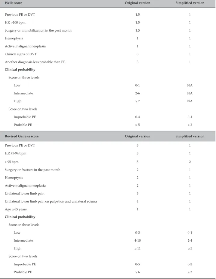
The Wells and Geneva probability scores include
clinical criteria for the diagnosis of PE,7 simply obtained
and validated on three levels (high, intermediate and low probability) or two levels (probable or improbable
PE), as shown in Table 1.2 The investigation continues
with complementary tests to conirm or rule out the diagnosis. For patients with improbable PE, a D-dimer test is run. D-dimer is a ibrin degradation product with high negative predictive value for the diagnosis of PE. A low D-dimer value in this context can virtually rule out
such diagnosis.2,3 The method of choice for diagnosing
patients with probable PE and D-dimer greater than 1. 600 is the computed tomography angiography of the chest,
which shows thrombi in the pulmonary tree.8
Despite the applicability of probability scores, diagnosis only considers clinical parameters, leaving out an essential tool for the evaluation of most cardiac disorders: ECG. PE may present abnormalities on electrocardiogram, such as T-wave inversion on leads V1 to V4, QR complex on V1, S1Q3T3 pattern and right bundle branch block, suggesting RV overload.1,2,9
These signs point to a more serious condition associated with pulmonary hypertension and acute cor pulmonale in cases of suspected PE, conirmed with relative ease by
transthoracic echocardiogram (TTE).2,3
Table 1 - Wells and revised Geneva score
Wells score Original version Simplified version
Previous PE or DVT 1.5 1
HR >100 bpm 1.5 1
Surgery or immobilization in the past month 1.5 1
Hemoptysis 1 1
Active malignant neoplasia 1 1
Clinical signs of DVT 3 1
Another diagnosis less probable than PE 3 1
Clinical probability
Score on three levels
Low 0-1 NA
Intermediate 2-6 NA
High ≥ 7 NA
Score on two levels
Improbable PE 0-4 0-1
Probable PE ≥ 5 ≥ 2
Revised Geneva score Original version Simplified version
Previous PE or DVT 3 1
HR 75-94 bpm
≥ 95 bpm
3
5
1
2
Surgery or fracture in the past month 2 1
Hemoptysis 2 1
Active malignant neoplasia 2 1
Unilateral lower limb pain 3 1
Unilateral lower limb pain on palpation and unilateral edema 4 1
Age ≥ 65 years 1 1
Clinical probability
Score on three levels
Low 0-3 0-1
Intermediate 4-10 2-4
High ≥ 11 ≥ 5
Score on two levels
Improbable PE 0-5 0-2
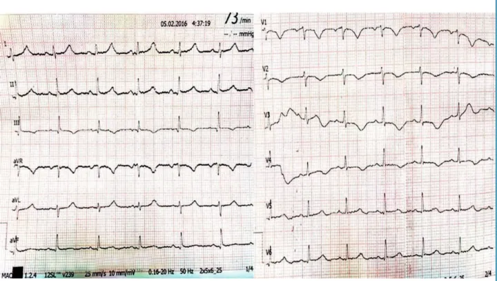
Figure 1 – ECG showing S1Q3T3 pattern and RV strain.
Case report
Female, 35-year-old patient, black ethnicity, overweight, sedentary and hypertensive, under regular use of clonidine, losartan and amlodipine. She was an assistant nurse and reported palpitation preceding fatigue and dyspnea on great exertion for three months, progressively worsening in the last month, which made it impossible for her to work.
The patient was admitted to the emergency service after a syncope episode preceded by tachycardia and dyspnea on moderate exertion. That episode was the third similar episode in the last four days. The patient had a questionable history of thrombophlebitis a year ago and miscarriage 10 years ago in the fourth month of pregnancy. Patient is currently not using contraception.
On admission, she reported dyspnea and fatigue on exertion and 90% saturation in ambient air, blood pressure (BP): 140x100 mmHg; heart rate (HR): 98 bpm and respiratory rate (RR): 22 irpm. On physical examination, the patient was bruised on her left eyebrow and in the parietal and occipital region due to traumatic brain injury (TBI) in the irst two syncope episodes. There were no signs of deep vein thrombosis.
Laboratory tests found 0.035 troponin (VR<0.015) CK-mass 5.3, D-dimer 6,400 and BNP 729. On admission, ECG showed S1Q3T3 pattern and right ventricular (RV) strain, and negative and symmetric T-waves in THE right precordial leads, from V1 to V4 (Figure 1). Chest X-ray was normal and computed tomography of the head revealed subgaleal hematomas in the parietal, occipital and frontal left regions.
Full anticoagulation with enoxaparin 1 mg/kg 12/12h, O2 by nasal catheter and medication reconciliation
were initiated.
Figure 2 – TCA angiography with bilateral thrombosis of pulmonary arteries.
The therapy chosen was chemical thrombolysis with intravenous rTPA 100 mg as the patient was young, at major negligible risk of bleeding and computed tomography scans of the chest showing only subgaleal hematomas, no intracranial bleeding or subarachnoid hemorrhage. After thrombolysis, there was signiicant increase of subgaleal hematomas, reports of pain, but without major bleeding. Thrombolysis was successful, regular analgesia was established for pain control, as well as ice packs on hematomas.
The day after thrombolysis, further TTE was done, showing increased right cavities with signiicant drop in PAP from 86 to 35 mmHg and normal RV systolic function. Fatigue and shortness of breath disappeared
completely after the 2nd day of hospitalization. TTE and
lower limb venous Doppler were repeated on the 5th
day of hospitalization with evolutionary improvement in volumetric RV overload, maintaining normal function, and no more thrombi in the deep lower limb veins. Computed tomography of the head CT was also
repeated on the 6th day of hospitalization and showed
persisting yet not increased left parietal, occipital and frontal subgaleal hematomas. There was no bleeding at other sites.
The patient was discharged from the Intensive Care
Unit (ICU) to the room on the 4th day of admission under
coumarin, full anticoagulation and enoxaparin to stay in hospital until the target International Normalized Ratio (INR) was reached — between 2 and 3 — and was
discharged on the 9th day of hospitalization.
Discussion
The case described above is about a young woman with few comorbidities, with submassive PE, as she was not in shock, but with signs, symptoms and laboratory tests
compatible with the presence of acute cor pulmonale.2,3
Although controversial and open to interpretation, the case is undoubtedly rich in terms of lessons learned.
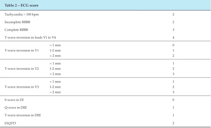
The irst lesson was the need to give greater value to easier and cheaper cardiology tests, which can give valuable information about the cardiac impact of PE. The patient was not in shock, but ECG showed inverted T-waves deeper than 2 mm, rS pattern in D1, qR in D3 and T-wave inverted in D3. In 2001, Kurt et al.9 published a paper studying
Table 2 – ECG score
Tachycardia > 100 bpm 2
Incomplete RBBB 2
Complete RBBB 3
T-wave inversion in leads V1 to V4 4
T-wave inversion in V1
< 1 mm 1-2 mm > 2 mm
0 1 2
T-wave inversion in V2
< 1 mm 1-2 mm > 2 mm
1 2 3
T-wave inversion in V3
< 1 mm 1-2 mm > 2 mm
1 2 3
S-wave in DI 0
Q-wave in DIII 1
T-wave inversion in DIII 1
S1Q3T3 2
The patient had slow onset of clinical symptoms, which started three months ago, but with signiicant worsening in the last month and three episodes of syncope in the past four days, which could be signs of chronic PE with new acute episodes in past days which eventually led the patient to present acute cor pulmonale with very high PAP, but with no circulatory shock. Another interesting fact is the absence of tachycardia that accompanies severe cases as the one reported above, which may be due to the chronic use of centrally acting alpha 2 agonist medication (clonidine) which, therefore, had a lowering effect on adrenergic low, which may have contributed to the absence of tachycardia in the context of acute cor pulmonale. Therefore, we assumed that the absence of tachycardia in this patient was not synonymous with benignity, so we proceeded with the treatment.
The main treatment for PE is systemic anticoagulation allowing the dissolution of the thrombus by the ibrinolytic system of the individual itself. For PE with circulatory shock and persistent hypotension, the use of thrombolytic agents is well established to speed up this process and reduce mortality, but in the absence of hemodynamic impairment, its use is controversial. About 10% of patients die three months after diagnosis,
and acute cor pulmonale in PE is an important determinant of the severity and early clinical outcome of this disease.1-3
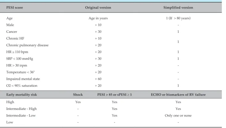
According to the Guidelines of the European Society of Cardiology, the best-validated prognostic score
is the PESI (Pulmonary Embolism Severity Index).2
Analyzing it in Table 3, we found that the patient had an intermediate to high risk of death, with an original PESI=105 or simpliied PESI=2, echocardiographic image and laboratory markers showing RV failure.
The multicenter, double-blind, randomized, placebo-controlled PEITHO (Pulmonary Embolism
Thrombolysis)10 study has recently revealed that rapid
thrombolysis reduces the combination of death from any cause and hemodynamic instability in seven days in patients with RV failure and myocardial injury without circulatory shock and this is mainly due to decreased risk of hemodynamic decompensation in
these patients.10 Therefore, these patients, stratiied as
Table 3 – PESI score
PESI score Original version Simplified version
Age Age in years 1 (If > 80 years)
Male + 10
-Cancer + 30 1
Chronic HF + 10
1
Chronic pulmonary disease + 20
HR ≥ 110 bpm + 20 1
SBP < 100 mmHg + 30 1
HR > 30 irpm + 20
-Temperature < 36º + 20
-Impaired mental state + 60
-O2 < 90% saturation + 20 1
Early mortality risk Shock PESI > 85 or sPESI ≥ 1 ECHO or biomarkers of RV failure
High Yes Yes Yes
Intermediate - High - Yes Yes
Intermediate - Low - Yes Only one or none
Low - -
-population without many comorbidities.10 Our case,
a young patient with intermediate risk PE, RV failure and myocardial injury, underwent thrombolysis successfully and without major bleeding, despite mild TCE prior to thrombolysis, which we believe to be not enough to contraindicate the thrombolytic drugs. Subsequent head scans and clinical outcome without signiicant increase in subgaleal hematomas or emergence of further intracranial hematomas corroborate our initial thoughts. However, the PEITHO study found potential bleeding risk, stressing the importance of proper selection of patients, as well as the need of an experienced medical team and the support of a tertiary institution prepared to address potential
bleeding complications.10
Thrombolysis does not change the hospital mortality rate in these cases of intermediate risk but ensures clinical improvement and decreases the chance of hemodynamic decompensation and progression to chronic cor pulmonale, which may lead to late mortality, at the expense of greater bleeding risk, though.10
Further studies comparing different doses of thrombolytics and methods for local thrombolysis with
lower risk of bleeding can bring great beneits to such
patients, with a smaller risk of bleeding.
Author contributions
Conception and design of the research: Guenther NL, Nunes NSV. Acquisition of data: Guenther NL, Nunes NSV, Souza RV. Analysis and interpretation of the data: Guenther NL, Nunes NSV, Souza VP. Writing of the manuscript: Guenther NL. Critical revision of the manuscript for intellectual content: Nunes NSV, Souza VP, Souza RV.
Potential Conflict of Interest
No potential conlict of interest relevant to this article was reported.
Sources of Funding
There were no external funding sources for this study.
Study Association
1. Álvares F, Ignácio de Pádua A, Terra Filho J. Tromboembolismo pulmonar: diagnóstico e tratamento. Medicina (Ribeirão Preto). 2003;36:214-40.
2. Konstantinides SV, Torbicki A, Agnelli G, Danchin N, Fitzmaurice D,
Galiè N, et al; Task Force for the Diagnosis and Management of Acute Pulmonary Embolism of the European Society of Cardiology (ESC). 2014 ESC guidelines on the diagnosis and management of acute pulmonary embolism. Eur Heart J. 2014;35(43):3033-69.
3. Caramelli B, Gottschall CA, Blacher C, Casagrande EL, Lucio EA,
Manente ER, et al; Sociedade Brasileira de Cardiologia. Diretriz de
embolia pulmonar. Arq Bras Cardiol. 2004;83(supl 11)1-9.
4. Anderson FA Jr, Spencer FA. Risk factors for venous thromboembolism. Circulation. 2003;107(23 Suppl 1):I9-16.
5. Thames MD, Alpert JS, Dalen JE. Syncope in patients with pulmonary embolism. JAMA. 1977;238(23):2509-11.
6. Castelli R, Tarsia P, Tantardini C, Pantaleo G, Guariglia A, Porro F. Syncope in patients with pulmonary embolism: comparison between patients with syncope as the presenting symptom of pulmonary embolism and patients with pulmonary embolism without syncope. Vasc Med. 2003;8(4):257-61.
7. Wells PS, Ginsberg JS, Anderson DR, Kearon C, Gent M, Turpie AG, et al. Use of a clinical model for safe management of patients with suspected pulmonary embolism. Ann Intern Med. 1998;129(12):997-1005.
8. Silva CI, Muller NL. Imaging of acute pulmonary thromboembolism. J Bras Pneumol. 2004;30(5) 474-9.
9. Daniel KR, Courtney DM, Kline JA. Assessment of cardiac stress from massive pulmonary embolism with 12-lead ECG. Chest. 2001;120(2):474-81.
10. Meyer G, Vicaut E, Danays T, Agnelli G, Becattini C, Beyer-Westendorf J, et al; PEITHO Investigators. Fibrinolysis for patients with intermediate risk pulmonary embolism. N Engl J Med. 2014;370(15):1402-11.