442
Maia et al
Brugada Syndrome
Arq Bras Cardiol volume 74, (nº 5), 2000
Hospital Pró-Cardíaco - Hospital de Cardiologia de Laranjeiras Mailing address: Ivan G. Maia Rua Raul Kennedy, 81 22631200 -Rio de Janeiro, RJ, Brazil
Ivan G. Maia, Magda Wanderley Soares, Silvia H. Boghossian, Roberto Sá
Rio de Janeiro, RJ - Brazil
The Brugada Syndrome. Outcome of one Case
Case Report
The Brugada syndrome is a rare condition, and due to its mutating manner of presentation it may be difficult to diagnose. We report one case and discuss the diagnostic aspects and the clinical outcome of one patient with charac-teristic findings of this syndrome. These findings are espe-cially defined by J-ST elevation in the right leads of serial electrocardiographic records, wide oscillations of J points and ST segments during 24-hour Holter monitoring, and nocturnal sudden death. We stress the importance of the Holter monitor findings for diagnostic complementation. Through this method it is possible to establish a correlation between vigil activities and sleep and the variability of the degree of impairment in ventricular repolarization.
In the last decade, interest in the study of the primary electrical diseases of the myocardium has progressively risen. The conjunct effort of specialists in arrhythmias, electrophysiologists, and geneticists has allowed clarifica-tion of several pathophysiological and clinical characte-ristics of those diseases with a favorable influence on their prognosis. Among those that have been drawing more at-tention, we can cite the congenital long QT syndrome and,
more recently, the Brugada syndrome1,2. Both have as a
common characteristic, when evolving in their natural form, the presence of a high potential for developing severe ven-tricular arrhythmias producing syncopal findings, sudden death, or both. Therefore, taking this prognosis and an often times dubious diagnosis into consideration, publi-shing data related to the evolution of a patient with the Bru-gada syndrome seemed worthwhile.
Case Report
The patient was a 40-year-old white male, from the Sta-te of Rio de Janeiro, where he had always lived. He had a his-tory of hypertension for 5 years and use of captopril (25 mg/ day) and hydrochlorothiazide (50 mg/day).
The patient reported that in July 1998 he had hematu-ria and lumbar pain. Due to the presence of renal calculi, surgery (left pyelolithotomy) was indicated.
In the preoperative assessment, the patient was asymp-tomatic, smoked 40 cigarettes per day and did not consume alcohol. Prior personal and familial morbidity was not notable. On clinical examination, the patient was eupneic, in re-gular condition, without any alteration on inspection. His radial pulse was regular and 66 bpm; his blood pressure was 140/80mm Hg.
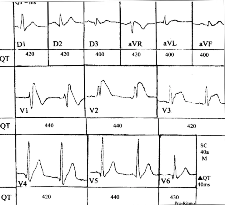
Complementary tests, all of which were normal, were as follows: coagulation tests, hemogram, urea, glucose, elec-trolytes, and urinary sediment. Transthoracic echocar-diogram showed that the myocardial and valvular structu-res and functions were normal. Chest x-rays showed no abnormalities. Electrocardiogram showed a pattern of com-plete right bundle-branch block, normal QT intervals (mean of 430ms), and a degree of dispersion of ventricular repola-rization of 40 ms, similar to that observed in healthy indivi-duals (fig. 1).
The patient sought our hospital for assessment of the surgical risk. After a new electrocardiogram, he was advised to postpone the surgery until a complete clarification of the diagnosis could be made. The electrocardiogram record was similar to the previous one (fig. 1). During this period, the patient’s preoperative assessment was completed in the hospital of origin and the patient underwent surgery to re-move the renal calculus under general anesthesia. The postoperative period was uneventful, and the patient was discharged 72 hours after the procedure.
As the patient had not returned to our hospital, we cal-led him. At the beginning of November 98, he returned and a new electrocardiogram was performed, the results of which were similar to those of the previous ones. Twenty-four-hour Holter monitoring was also performed. Forty-eight hours after the recording, at dawn, while the patient was lying down, he had a sudden stertorous breath, a convulsive crisis, and ended up dying without medical assistance. His mother and sister were with him at the time of sudden death.
The result of the Holter monitoring was as follows: sinus rhythm, atrioventricular conduction 1:1, and normal QT intervals. Ventricular repolarization showed wide oscillations of the J points and ST segments during the 24 hours, and also in the superior channel of the recording (V1). The incidence of active arrhythmias was negligible (11 ventricular extrasys-toles and 2 atrial extrasysextrasys-toles during the 24 hours). The pati-ent remained asymptomatic during the recording.
Arq Bras Cardiol volume 74, (nº 5), 2000
Maia et al Brugada Syndrome
443
elevation of the J point and ST segment can be seen, as well as its disappearance in the recordings in D (rapid walk).
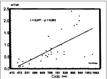
We correlated the degree of elevation of the J point (in mVolt), starting at the base line and in the V1 lead, with dura-tion of the previous cardiac cycle (in ms). We determined a total of 35 cycles ranging from 460 to 1040 ms (the 2 extre-mes of frequency observed in the recordings). We found an excellent linear correlation between the 2 variables with an r=0.697 (p=0.001). Longer cardiac cycles produced a greater degree of elevation of the J point and vice-versa (fig. 3).
As the syndrome is genetically transmitted, we carried out a familial screening of the patient. Data were obtained through electrocardiographic findings with no therapeutical test. No episode of sudden death had occurred in the pa-tient’s family. One of his brothers had Wolff-Parkinson-White syndrome, a condition that is also described as of possible genetic transmission. A nephew had a mild delay
in the right branch conduction, which may represent a nor-mal variation of the electrocardiogram.
Discussion
In 1992, Brugada and Brugada 1 reported a new clinical
syndrome characterized by the presence of an electrocar-diographic pattern similar to a right bundle-branch block as-sociated with an elevation of the J point and ST segment (superior concavity), in the V1, V2, and V3 leads. It occurs mainly in men, who have a structurally normal heart but who may experience frequent episodes of syncope, or sud-den death, or both. The condition is genetically transmitted with a dominant autosomal pattern. Elevation of the J point and ST segment is related to alterations in the dynamics of the transient current of potassium (Ito), which occurs at the end of the phase 0 and beginning of the phase 2 (phase 1)
444
Maia et al
Brugada Syndrome
Arq Bras Cardiol volume 74, (nº 5), 2000
Fig. 2 – Holter monitoring recordings where the great oscillations in the degree of J-ST elevation, such as during sleep, occur (C). By walking rapidly, these alterations practically disappeared (D). In the superior channel, the V1 lead, and in the medium and inferior channels, the V5 lead.
BEGINNING OF THE TRACING
ON THE BUS
Arq Bras Cardiol volume 74, (nº 5), 2000
Maia et al Brugada Syndrome
445 Fig. 3 – Curve of linear correlation between the degree of the J point elevation
(mVolt) and duration of the previous RR intervals (ms).
of the transmembrane action potential 2. The syndrome has a
high incidence in some regions of the Asian continent, such as Laos and Thailand, where it accounts for most of the sud-den deaths in individuals under the age of 30 years (it is es-timated to cause 10 sudden deaths per 10,000 inhabitants per
year) 3. Even though the syndrome has already been
des-cribed in North 3 andSouth 4 America and Europe 1,2,5, its
ac-tual incidence in those regions is unknown.
The initial diagnostic suspicion occurs because of an electrocardiographic recording with the pattern shown in fi-gure 1, with gross alterations of ventricular repolarization in right precordial leads. This abnormal pattern, even though possible, is extremely rare in classical right bundle-branch block 6, and has never been described as a form of
manifes-tation of an electrolytic disorder (the patient used diuretics), raising the possibility of a Brugada syndrome. As the J-ST alterations observed in this syndrome are mutant, where occult forms may occur with an apparently normal electro-cardiogram (the diagnosis may only be established through a pharmacological test using ajmaline or procainamide) 2,3,
Holter monitoring has become fundamental for diagnostic confirmation. The relations between the cycle and the J-ST elevation that we observed confirm what has already been described, i. e., the exercise test tends to normalize the
repo-larization alterations in this syndrome3. Considering that the
presence of the J-ST elevation represents a signal of increa-sed myocardial vulnerability, our findings explain, at least partially, the frequent occurrence of nocturnal sudden death in this syndrome, suggesting a vagal modulation for it. The nocturnal death is so frequent in certain regions of Thailand that the natives attribute it to ghosts of widows that take young males during the night. In an attempt to avoid it, young males sleep dressed in female clothes 3.
This seems to be the second case of the Brugada syn-drome entirely published in Brazil 4. Its diagnosis was based
on electrocardiographic findings, with a characteristic J-ST elevation in right precordial leads and a so-called pattern of right bundle-branch block associated with data obtained through Holter monitoring and the final clinical outcome (sudden death). From our viewpoint, these findings were enough to confirm the diagnosis of the Brugada syndrome. Unfortunately, we were not able to deepen the studies with electrophysiologic evaluations and attempt to induce ven-tricular tachyarrhythmias through programmed venven-tricular electrical stimulation.
The case is reported here to highlight the obscure prognosis of the condition. At the beginning we did not give the appropriate importance to the syndrome, and we have learned that a precocious intervention is fundamen-tal. Even among asymptomatic patients, it is estimated that at least 1/3 of them will develop ventricular tachycardia or ventricular fibrillation within the 24 months following the initial diagnosis3. This initial diagnosis is based on
electro-cardiographic findings, data from Holter monitoring, and on the patient’s report about previous syncopes or a familial history of sudden death. Those cases of occult manifestation are of difficult diagnosis. In case of sus-picion, the provocative pharmacological test with
ajma-line, considered a specific drug 3, is very useful. Other
drugs, such as procainamide, may induce a similar
res-ponse 7. The control of ventricular tachyarrhythmias with
the use of antiarrhythmic drugs has not been a feasible op-tion. Implantation of a cardioverter/defibrillator seems to be the most appropriate management, because it modifies in a very favorable way the natural history of this severe
and curious syndrome 8.
1. Brugada P, Brugada J. Right bundle branch block, persistent ST segment eleva-tion and sudden cardiac death: a distinct clinical and electrocardiographic syn-drome. J Am Coll Cardiol 1992; 20: 1391-6.
2. Gussak I, Antzelevitch C, Bjerregaard P, et al. The Brugada syndrome: clinical, electrophysiologic and genetic aspects. J Am Coll Cardiol 1999; 33: 5-11. 3. Antzelevitch C, Brugada P, Brugada J, et al. The Brugada syndrome. In: Camm AJ.
Ed. Clinical Approaches to Tachyarrhythmias (vol 10). Armonk: Futura, 1999. 4. Villacorta H, Torres RAF, Castro IRS, et al. Morte súbita em paciente com blo-queio de ramo direito e elevação persistente de segmento ST. Arq Bras Cardiol 1996; 66: 229-31.
5. Lorga Filho A, Brugada P. Bloqueio de ramo direito, elevação de segmento ST de
References
V1 a V3 e morte súbita. O que sabemos sobre esta peculiar síndrome clínico-ele-trocardiográfica. Arq Bras Cardiol 1997; 68: 205-8.
6. Tohyou JA, Nakazawa K, Ozawa A, et al. A survey in the incidence of right bun-dle branch block with ST segment elevation among normal population. Jpn J Electrocardiol 1995; 15: 223-6.
7. Fujuki A, Usui M, Nagasawa H, et al. ST segment elevation in the right precor-dial leads induced with class IC antiarrhythmic drugs: insight into the mecha-nisms of Brugada syndrome. J Cardiovasc Electrophysiol 1999; 10: 214-8. 8. Brugada J, Brugada R. Right bundle-branch block and ST-segment elevation in