Haptoglobin study in MyastHenia gravis
Leonardo H. Mendonça Oliveira
1a, Marcondes C. França Jr
1b, Anamarli Nucci
1c,
Denise Madureira de Oliveira
2a, Elza Myiuki Kimura
2a, Maria de Fátima Sonati
2bAbstract – Objective: A cross-sectional study of haptoglobin (Hp) in myasthenia gravis (MG) was designed, with the objective to identify its values and correlate them with different disease status. Method: 46 patients were enrolled in the study, all having disease severity established according to the quantitative myasthenia gravis strength scores (QMGSS). Based on the functional scale determined by Myasthenia Gravis Foundation of America (MGFA) recommendations, patients were classified as having: complete stable remission (CSR; n=10); minimal manifestations–0 (MM0; n=6), minimal manifestations–1 (MM1; n=4); pharmacological remission (PR; n=6). Two other groups participated: thymomatous patients (T; n=10) and patients without imunosuppression or thymectomy, until the assessment for Hp (WIT; n=10). Hp dosage was done by immunonephelometry, blindly to clinical data. Student´s t-test, Anova test and linear regression were employed for statistical analyses. Results:
Statistically significant differences occurred between CSR+MM0xWIT groups (86.62x157.57, p<0.001) and PR+MM1xWIT groups (73.93x157.57, p<0.001). Linear regression showed correlation between Hp levels and QMGSS (r=0.759, p<0.001). Conclusion: Our results suggest that Hp may be useful in clinical practice as a disease severity marker in MG.
KEY WORDS: haptoglobin, acute phase response, myasthenia gravis, immune disorders.
estudo sobre a haptoglobina na miastenia grave
Resumo – Objectivo: Desenhou-se estudo transversal sobre a haptoglobina (Hp) na miastenia grave (MG) com o objetivo de identificar seus valores e correlacioná-los a diferentes condições na doença. Método: 46 pacientes foram incluídos, todos tendo a gravidade da doença estabelecida segundo escores internacionais (QMGSS). Os pacientes tiveram seu estado funcional determinado de acordo com a Myasthenia Gravis Foundation of América (MGFA) e classificados em: remissão completa estável (CSR; n=10); mínima manifestação–0 (MM0; n=6), mínima manifestação–1 (MM1; n=4); remissão farmacológica (PR; n=6). Dois outros grupos participaram: pacientes timomatosos (T; n=10) e pacientes sem imunossupressão ou timectomia, até o momento da inclusão no estudo (WIT; n=10). A dosagem de Hp foi realizada por imunonefelometria, de modo cego quanto à clínica. As análises estatísticas incluíram o teste de Student, Anova e regressão linear. Resultados: Observou-se diferença significativa entre os grupos CSR+MM0xWIT (86,62x157,57, p<0,001) e entre PR+MM1xWIT (73,93x157,57, p<0,001). A regressão linear mostrou correlação positiva entre os valores de Hp e os escores QMGSS (r=0,759, p<0,001). Conclusão: O estudo sugere que valores altos de Hp se correlacionaram a maior gravidade da MG. PALAVRAS-CHAVE: haptoglobina, resposta de fase aguda, miastenia grave, doença autoimune.
Departments of Neurology1 and Clinical Pathology2, Faculty of Medical Sciences, Campinas State University (UNICAMP), São Paulo SP, Brazil: 1aMedical student; 1bNeurologist, PhD student; 1cAssociated Professor of Neurology; 2aBiologist; 2bAssociated Professor of Clinical Pathology. Financial Support: FAPESP (Fundação de Amparo à Pesquisa do Estado de São Paulo). Grant 05/58716-5.
Received 18 September 2007, received in inal form 13 December 2007. Accepted 12 February 2008.
Dra. Anamarli Nucci – Departamento de Neurologia / Faculdade de Ciências Médicas (UNICAMP) - Cx. Postal 6111 - 13083-970 Campinas SP - Brasil. E-mail: [email protected].
Haptoglobin (Hp) is an acute phase inlammatory a 2-glicoprotein synthesized in the liver1,2. It binds free hemo-globin in the plasma and thereby inhibits its oxidative ac-tivity in other tissues1-3. In the immune system, Hp mod-ulates the function of T helper lymphocytes and inhibits the CD22 binding to activatedTNF-a3. Due to polymor-phisms in the gene encoding Hp on chromosome 16q22, there are three distinct human Hp phenotypes: Hp 1-1, Hp 2-1 and Hp 2-21,3,4. Hp has been used as a diagnostic and
Myasthenia gravis (MG) is the most frequent disor-der of the neuromuscular junction and is characterized by luctuating weakness either restricted to extrinsic oc-ular muscles or expressed in a more generalized distribu-tion10,11. It is an autoimmune disease caused by antibodies against acetylcholine receptors (Ab-AChR) in about 95% of cases, and may be associated with other autoimmune diseases10. T lymphocytes and proinlammatory cytokines play key roles in the pathogenesis of MG10,12. Thymus is in-volved in the disease process since thymic hyperplasia oc-curs in 60% of MG cases and thymoma in 15%10,11.
As MG shares some pathogenic features with auto-immune diseases, such as GBS8,10, in which Hp was proven useful, we investigated Hp values in MG patients looking for its possible utility as a marker of disease activity.
MetHod
This study was approved by the Ethics Committee of Faculty of Medical Sciences, Clinical Hospital, Campinas State Universi-ty. A written informed consent was obtained from all patients.
Study design
In a cohort of prospectively selected MG patients, we per-formed a cross-sectional study of Hp values.
Subjects selection
Adult patients fulilling clinical and laboratorial criteria for
MGand attending the Neuromuscular outpatient clinic were
enrolled in the study. Quantitative myasthenia gravis strength
score (QMGSS)13 was employed to assess disease severity.
Pa-tient’s functional status was determined in accordance to
Myas-thenia Gravis Foundation of America (MGFA) recommendations13
and divided into four functional groups: 1) Complete stable
re-mission (CSR): no symptoms or signs of MG (clinical score=0) for
at least one year and without immunosuppressive therapy during
that time; 2) Minimal manifestations – 0 (MM0): no symptoms
or functional limitations due to MG, but patient had weakness of some muscles upon examination (clinical score=1) without
immunosuppressive therapy for at least one year; 3)
Pharma-cologic remission (PR), the same criteria as for CSR, except that
patient continued to take immunosuppressive therapy; 4)
Mini-mal manifestations – 1 (MM1), the same criteria as for MM-0,
ex-cept that patient continued to take immunosuppressive thera-py. Two other groups of patients were included, also evaluated
by QMGSS: 5) Thymomatous (T) patients with histopathological
diagnosis, regardless of symptoms or treatment in the moment
of the study; 6) MG symptomatic patients (QMGSS>1) without
immunosuppression or thymectomy (WIT). Patients in the WIT
group were referred to our institution, eventually using anticho-linesterasic drugs, and they were submitted to immunosuppres-sive treatment after venous puncture for Hp study.
Haptoglobin dosage
Blood samples from each patient were collected and the Hp
concentration was determined by immunonephelometry (Dade Behring, Marbung, Germany), blindly to the classiication of pa-tients in the study.
Statistical analyses
Differences between two groups were compared by Stu-dent’s t-test. Comparison of Hp levels in multiple groups was accomplished by the Anova test and Tukey´s post-hoc analysis. We employed linear regression to analyze correlations between Hp levels and QMGSS. MG patients with other active immune-mediated co-morbidity were excluded from inal analysis. Anal-yses were performed on SYSTAT 10.2 software and the level of signiicance was assumed at p<0.05.
results
Forty-six patients participated in the study, 27 women and 19 men. Mean age was 41.7 years (range 18 to 76 years) and the mean duration of disease was 7.1 years. There were 10 patients in the CSR group, 6 in MM-0, 6 in PR, 4 in MM-1, 10 in T and 10 in WIT group. For statistical analyses, groups CSR and MM0 were fused into inal fCSR group. Similarly, groups PR and MM1 were fused into inal fPR group. Clinical and laboratorial data of patients on group fCSR, fPR, T and WIT are displayed on Tables 1 to 4.
In Tables 1 and 2 we can observe three patients with active autoimmune co-morbidities, indicated by clinical history through chart revision, previous specialized clin-ical and laboratory follow-up and speciic treatment in course. They were cases 7 and 13 in the group CSR and case 22 in the group PR; their Hp values were the highest
in each respective group so these patients were excluded from inal analysis to avoid a statistical bias.
Figure 1 shows Hp levels in the different groups of MG patients. The mean Hp value in fCSR, fPR, WIT and T groups were 86.62, 73.93, 157.57 and 126.39 mg/dL, respec-tively. Comparing Hp values in these different groups (Fig
1), we found statistically signiicant differences only be-tween groups fCSRxWIT (86.62x157.57, p<0.001) and be-tween groups fPRxWIT (73.93x157.57, p<0.001).
We hypothesized that Hp levels correlated with dis-ease severity in MG patients and performed two addi-tional analyses. First, we compared Hp in symptomatic (QMGSS>1) versus asymptomatic (QMGSS≤1) patients (re-gardless of medication status) and found signiicantly low-er results in the lattlow-er group (154.1x95.5, p<0.001 – Fig 2). Second, we used linear regression analyses that showed a signiicant correlation between Hp levels and QMGSS (r=0.759, p<0.001 – Fig 3).
Fig 2. Box-and-whiskers plot showing Hp levels in asymptomatic (QMGSS≤1) and symptomatic (QMGSS>1) MG patients. The box ex-tends from the 25th percentile to the 75th percentile, with a horizon-tal line at the median (50th percentile). Whiskers extend down to the smallest value and up to the largest.
Fig 3. Linear regression analysis of Hp values and QMGSS in MG patients.
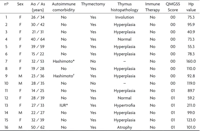
Table 1. Hp levels on fCSR (CSR+MM1) group.
nº Sex Ao / As
(years)
Autoimmune comorbidity
Thymectomy Thymus
histopathology
Immune Therapy
QMGSS Score
Hp value
1 F 26 / 34 No Yes Involution No 00 75.3
2 F 30 / 42 No Yes Hyperplasia No 00 95.9
3 F 21 / 31 No Yes Hyperplasia No 00 40.9
4 F 40 / 64 No Yes Normal No 00 73.3
5 F 39 / 59 No Yes Hyperplasia No 00 55.3
6 F 15 / 22 No Yes Hyperplasia No 00 78.3
7 F 32 / 53 Hashimoto* No – No 00 160.0
8 F 19 / 28 No Yes Hyperplasia No 00 110.0
9 M 23 / 36 Hashimoto# Yes Hyperplasia No 00 92.8
10 M 28 / 35 No No – No 00 119.0
11 F 14 / 25 No Yes Hyperplasia No 01 89.7
12 F 28 / 39 No Yes Normal No 01 59.2
13 F 27 / 33 IUR* Yes Hypertroia No 01 211.0
14 M 22 / 27 No Yes Hyperplasia No 01 99.0
15 F 32 / 39 No Yes Hyperplasia No 01 123.0
16 M 50 / 62 No Yes Atrophy No 01 101.0
Table 2. Hp levels on fPR (PR+MM1) group.
nº Sex Ao / As
(years)
Autoimmune comorbidity
Thymectomy Thymus
histopathology
Immune therapy
QMGSS score
Hp value
17 M 20 / 23 No No – Aza / Pred 00 139.0
18 F 61 / 66 No No – Pred 00 83.8
19 M 62 / 68 No No – Aza 00 72.5
20 M 64 / 72 No No – Aza 00 63.7
21 F 18 / 22 No No – Aza 00 26.7
22 M 27 / 35 Pemphigus Yes Hyperplasia Aza / Pred 00 187.0
23 M 33 / 44 IUR# Yes Atrophy Pred 01 63.4
24 F 48 / 51 No No – Aza 01 49.2
25 F 32 / 34 No No – Aza / Pred 01 96.0
26 M 24 / 26 No No – Aza 01 71.1
F, female; M, male; Ao, age of disease onset; As, age at study;*active illness; #illness in remission; IUR, inlammatory ulcerative rectocolitis; Aza, azatioprine; Pred, prednisone.
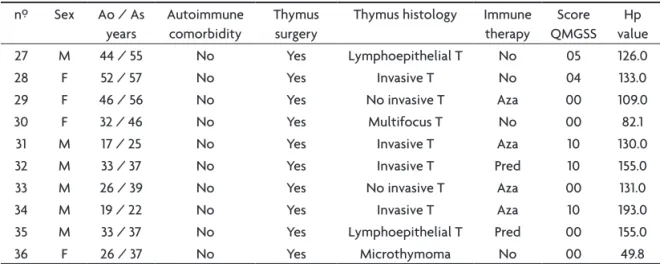
Table 3. Hp levels on T group.
nº Sex Ao / As
years
Autoimmune comorbidity
Thymus surgery
Thymus histology Immune
therapy
Score QMGSS
Hp value
27 M 44 / 55 No Yes Lymphoepithelial T No 05 126.0
28 F 52 / 57 No Yes Invasive T No 04 133.0
29 F 46 / 56 No Yes No invasive T Aza 00 109.0
30 F 32 / 46 No Yes Multifocus T No 00 82.1
31 M 17 / 25 No Yes Invasive T Aza 10 130.0
32 M 33 / 37 No Yes Invasive T Pred 10 155.0
33 M 26 / 39 No Yes No invasive T Aza 00 131.0
34 M 19 / 22 No Yes Invasive T Aza 10 193.0
35 M 33 / 37 No Yes Lymphoepithelial T Pred 00 155.0
36 F 26 / 37 No Yes Microthymoma No 00 49.8
F, female; M, male; Ao, age of disease onset; As, age at study; Aza, azatioprine; Pred, prednisone; T, thymoma.
discussion
To our knowledge, this is the first study of Hp val-ues in patients with MG. We found that groups fCSR and fPR presented lower Hp levels when compared to WIT group, composed of MG patients in their natural disease state, eventually taking symptomatic drugs, but without effective treatment. Similarly, symptomatic patients had higher Hp levels in comparison to asymptomatic patients. Group T did not present signiicantly different Hp levels in comparison to other groups. This is possibly explained by the clinical heterogeneity of the group, which includes ive patients in remission and ive patients with active dis-ease. Further, there was a positive correlation between Hp levels and QMGSS among symptomatic MG individ-uals. Overall, these indings indicate that active disease (QMGSS>1) is associated with raised Hp levels, regardless of thymus status or medication in use.
The production of Ab-AChR is mediated by cytokines produced by CD4+ and CD8+ T helper (Th) cells12. IL-4, IL-6
and IL-10, all Th2 cytokines, are an eficient growth pro-moter for B-cell proliferation and differentiation, acting mainly in disease progression and persistence12.
Interferon-γ, a Th1 cytokine, is important in inducing B-cell maturation, acting more agile at the onset of MG, probably being one of the initiating factors in the induction of the disease12. In contrast, transforming growth factor- β (TGF-β) and
IFN-a exerts immunosuppressive effects, which include the down regulation of both Th1 and Th2 cytokines in MG12.
bind-ing to glycoprotein3. When Hp binds to CD22 it inhibits the TNF-a-activated action3. TNF-a is a proinlammatory cytokine implicated in MG pathogenesis14,15. The soluble recombinant TNF receptor Fc protein, etanercept, has been tested in MG patients showing beneit in those with low plasma IL-6 levels14.
The high serum Hp levels observed in the study are consistent with an active inlammatory state in MG pa-tients1,3. Hp is closely related to IL-6, which is the main stimulatory cytokine responsible for its production in the liver1,3. Interestingly, IL-6 acts in MG as an important growth and differentiation factor of B lymphocytes, which are directly related to the damage in the mioneural plate12. Moreover, lower IL-6 levels have been associated with bet-ter therapeutic results in MG patients treated with etan-ercept14. It is thus expected that in cases of active disease, there is aggression to the mioneural plate and therefore, high levels of IL-6. So, indirectly there would be raised Hp values. This reasoning could also explain the relation between the clinical score and Hp levels, since more in-tense inlammatory process may be associated with more prominent symptoms11. Similarly, there are elevated serum Hp levels in GBS patients, which have been explained by the concomitant increase of IL-6 levels8.
Hp values are not disease-speciic, explaining that pa-tients in clinical remission of MG, but presenting another active autoimmune illness had high values of Hp than MG patients without active autoimmune comorbidity. In these cases, Hp would relect the activity of disease comorbid-ity, not that of MG. According to Christensen et al.16, 14% of MG patients presented concomitant autoimmune dis-ease, therefore clinicians must be alert to evaluate each autoimmune illness independently, and consider Hp val-ues in a contextual setting.
In conclusion, we suggest that Hp can be useful as a marker of clinical activity in MG without another associated illness.
ACknOwledgeMents – The authors wish to express special thanks
to Prof. Dr. Konradin Metze for his assistance with thymus histopa-thology; to the staff of the Thoracic Surgery Department (Profs. Drs. José Cláudio Seabra, José Geraldo dos Santos, Ivan C Toro and Ricardo M Kalaf).
references
1 Sadrzadeh SMH, Bozorgmehr J. Haptoglobin phenotypes in health and disorders. Am J Clin Pathol 2004;121:97-104.
2. Melamed-Frank M, Lache O, Enav BI, et al. Structure-function analysis of the antioxidant properties of haptoglobin. Blood 2001;98:3693-3698.
3. Langlois MR, Delangue JR. Biological and clinical signiicance of hap
-toglobin polymorphism in humans. Clin Chem 1996;42:1589-1600. 4. Wobeto VPA, Rosim ET, Melo MB, Calliari LEP, Sonati MF.
Haptoglo-bin polymorphism and diabetic retinopathy in Brazilian patients. Diab Res Clin Pract 2007;doi:10.1016/j.diabres.2006.12.018.
5. Panter SS, Sadrzadeh SMH, Hallaway PE, Haines JL, Anderson VE, Ea-ton JW. Hypohaptoglobinemia associated with familial epilepsy. J Exp Med 1985;161:748-754.
6. Saccucci P, Verdecchia M, Piciullo A, et al. Convulsive disorder and genetic polymorphism. Association of idiopathic generalized epilepsy with haptoglobin polymorphism. Neurogen 2004;5:245-248. 7. Borsody M, Burke A, Coplin W, Miller-Lotan R, Levy A. Haptoglobin
and development of cerebral artery vasoespasm after subaracnoid hem-orrhage. Neurology 2006;66: 634-640.
8. Gutowski NJ, Pinkham JM, Akanmu D, Chirico S, Murphy RP. Free
radicals in inlamatory neurological disease: increased lipid peroxida
-tion and haptoglobin levels in Guillain Barré syndrome. Ir J Med Sci 1998;167:43-46.
9. Lehmensiek V, Süssmuth SD, Brettschneider J, et al. Proteome
analy-sis of cerebrospinal luid in Guillain-Barré syndrome. J Neuroimmu
-nol 2007;185:190-194.
10. Vicent A. Immunology of disorders of neuromuscular transmission. Acta Neurol Scand 2006;113:1-7.
11. Romi F, Gilhus NE, Aarli JA. Myasthenia gravis: clinical, immunolog-ical and therapeutic advances. Acta Neurol Scand 2005;111:134-141. 12. Zhang G, Navikas V, Link H. Cytokines and pathogenesis of
myasthe-nia gravis. Muscle Nerve 1997;20:534-551.
13. Jaretki A, Barohn RJ, Ernstoff RM, et al. Myasthenia Gravis: recommen-dations for clinical research standards. Neurology 2000;55:16-23. 14. Tüzün E, Meriggioli MN, Rowin J, Yang H, Christadoss P. Myasthenia
gravis patients with low plasma IL-6 and IFN-γ beneit from etanercept
treatment. J Autoimmunity 2005;24:261-268.
15. Rowin J, Meriggioli MN, Tüzun E, Leurgans S, Christadoss P. Etaner-cept treatment in corticosteroid-dependent myasthenia gravis. Neurol-ogy 2004;63: 2390-2392.
16. Christensen PB, Jensen TS, Tsiropoulos I et al. Mortality and survival in myasthenia gravis: a Danish population based study. J Neurol Neu-rosurg Psychiatry 1998;64:78-83.
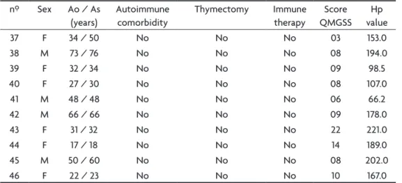
Table 4. Hp levels on WIT group.
nº Sex Ao / As
(years)
Autoimmune comorbidity
Thymectomy Immune
therapy
Score QMGSS
Hp value
37 F 34 / 50 No No No 03 153.0
38 M 73 / 76 No No No 08 194.0
39 F 32 / 34 No No No 09 98.5
40 F 27 / 30 No No No 08 107.0
41 M 48 / 48 No No No 06 66.2
42 M 66 / 66 No No No 09 178.0
43 F 31 / 32 No No No 22 221.0
44 F 17 / 18 No No No 14 189.0
45 M 50 / 60 No No No 08 202.0
46 F 22 / 23 No No No 10 167.0