Presentation and treatment of
Carotid Cavernous aneurysms
Lucas Perez de Vasconcellos
1, Juan Antônio Castro Flores
2,
José Carlos Esteves Veiga
3, Mário Luiz Marques Conti
4, Pedro Shiozawa
5Abstract – We analyzed a group of patients with the diagnosis of internal carotid aneurysms in its intracavernous segment, with emphasis in prevalence, clinical features, treatments, evolution and neurological prognosis. Neurological signs and symptoms at initial presentation were registered and compared with final outcome. Patients were divided into two stratified groups, one with 19 patients which underwent interventionist treatment, and another with 21 patients who were conservatively treated. The present study demonstrated that intervention is significantly correlated with a better prognosis considering evolution of pain symptoms secondary to neurovascular compression (p=0,002). Regarding neurological deficits, an interventionist approach was also significantly correlated with better outcome in comparison with initial presentation (p=0,008). These results indicate that interventionist treatment determines improvement or resolution of pain symptoms in comparison with patients conservatively treated, as well as stabilization or partial improvement of neuro-ophthalmological deficits.
Key WoRds: carotid cavernous aneurysm, clinical treatment, interventionist treatment, prognosis.
apresentação e tratamento dos aneurismas intracavernosos
Resumo – Analisamos um grupo de pacientes com diagnóstico de aneurismas da artéria carótida interna, em sua porção intracavernosa, estudando-se: prevalência, apresentação clínica, formas de tratamento, evolução e prognóstico neurológico. os sintomas e sinais neurológicos da apresentação foram registrados e comparados ao término do acompanhamento, com um grupo de 21 aneurismas submetidos a tratamento conservador e outro com 19 a tratamento intervencionista. o estudo demonstrou que a intervenção está relacionada a um melhor prognóstico, quanto à evolução do quadro álgico secundário à compressão neurovascular (p=0,002). em relação ao déficit neurológico, a abordagem intervencionista pôde ser associada com uma melhora do quadro inicial (p=0,008). estes resultados indicam que o tratamento intervencionista proporcionou melhora ou resolução do sintoma dor em comparação ao grupo de pacientes com tratamento conservador, além de levar a uma estabilização ou melhora parcial dos déficits neuro-oftalmológicos.
PAlAvRAs-chAve: aneurisma intracavernoso, tratamento clínico, tratamento intervencionista, prognóstico.
santa casa Medical school, discipline of Neurosurgery, são Paulo sP, Brazil: 1Resident of Neurosurgery; 2Assistant Physician; 3Adjunct Professor and
head of Neurosurgery; 4Assistant Physician and head of Interventionist Neuroradiology service; 5Medical student.
Received 26 september 2007, received in inal form 6 February 2008. Accepted 23 February 2008.
Dr. Lucas Perez de Vasconcellos – Rua Desembargador Joaquim Barbosa de Almeida 368 - 05463-010 São Paulo SP - Brasil. E-mail: lucasvasconcellos@ hotmail.com
Internal carotid aneurysms in its intracavernous seg-ment represent approximately 3-5% of all intracranial an-eurysms1 and 15% of those originated in the internal
ca-rotid2. carotid cavernous aneurysms (ccA) can arise from
any segment of cavernous carotid artery (Fig 1), but most commonly are originated in the horizontal segment, being projected forwardly and laterally, with the superior orbi-tary issure and below the anterior clinoid process³. This preferential site is related with the three most common branches of this segment (Mcconnell´s capsular artery, in-ferolateral trunk and meningohypoisary trunk)4. This
sug-gests that the hemodynamic stress veriied in these bifur-cations can contribute to the aneurysms´ genesis3,5.
oth-er aneurysmatic sites within the intracavoth-ernous segment are also common, what can interrogate the existence of other pathogenic mechanisms as atherosclerosis and dis-section, spontaneous or traumatic6.
ccA morbidity and mortality indices are low7-10,
how-ever, pain and neuro-ophthalmologic deicits due to neu-rovascular compression are frequent, what highlights the possibility of surgical treatment6,11. The vast majority of
treated, commonly through aneurysmatic isolation with-out vascular occlusion, while ccA, when operated, fre-quently are through occlusion of ipsilateral internal ca-rotid artery (IcA), with cerebral ischemia and amaurosis risks12-14. IcA endovascular occlusion has apparently a
bet-ter outcome than IcA ligature13,15,16, although there is still
much controversy around this matter, with authors in fa-vor of surgical treatment of ccA patients with or with-out symptoms15,17, and others which are contrary to
surgi-cal treatment in both groups18. The reason for this
contro-versy is in the lack of data on the natural history and long term outcome of ccA surgical patients16,19,20.
The following study has the objective to determine the long-term neurological outcome of the patients di-agnosed with carotid cavernous aneurysms in our cen-ter, treated conservatively or surgically, with emphasis in prevalence, clinical presentation, therapeutical strategies, outcome and neurological prognostic.
method
After approval from the Institutional Review commission, the discipline of Neurosurgery of santa casa Medical school of são Paulo studied patients with the diagnostic of cerebral aneu-rysms in the period between January 1989 and April 2007.
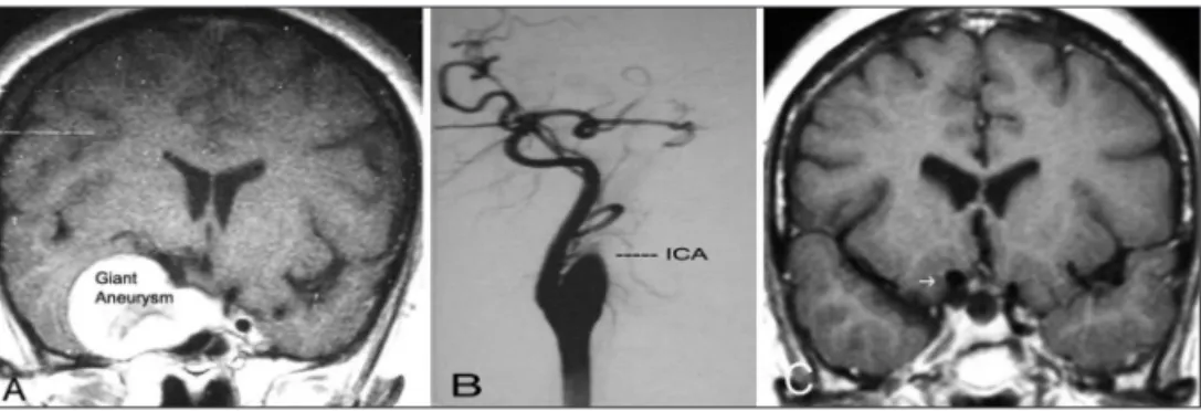
These patients were analyzed regarding genre, age, site and number of aneurysms, being selected for a second phase of the study those with ccA between (c3) lacerus segment and (c5) cli-noid segment of IcA4. There were excluded from the study pa-tients that presented aneurysms with partial or total intradural or subarachnoid colon, displasic aneurysms (beyond segment c4 of IcA) and traumatic or infectious aneurysms. All the selected patients were submitted to a full neurological exam and under-went radiological study with contrasted cranial computed to-mography scan (cT), complete cerebral angiography (cAG) with and without subtraction and magnetic resonance imaging scan (MRI) with slices from the paraselar region after 90’s (Fig 2).
The data from patients´ iles were completed afterwards during medical appointments. The following items were veri-ied: age of diagnostic, age during treatment, genre, ethnic, mor-bid antecedents, site and size of aneurysm, presence of other aneurysms, neurological and visual signs and symptoms, thera-peutic options and complications after treatment. The patients were divided into two groups, one which underwent conserva-tive treatment and other, interventionist treatment. It was con-sidered to be interventionist treatment endovascular approach with coils, stent and IcA occlusion with ballon as well as IcA lig-ature with or without external carotid by-pass to media cerebral artery or IcA trapping.
As to measure, according to liskey et al6, pain symptoms and neurological deicits, the pain was graduated in severe, moder-ate, weak or absent, while neurological deicits were classiied as severe, in the presence of cavernous sinus syndrome includ-ing trigeminal neuropathies; moderated, if there were complete
involvement of III, Iv and vI cranial nerves; weak, if there were deicits in one or two cranial nerves; and absent.
each patient was classiied taking into account his initial and inal presentation during overcome: 0, absence of symptoms; 1, weak pain or neurological deicit; 2, moderate pain or neurolog-ical deicit; 3, severe pain or neurologneurolog-ical deicit.
statistical analyses were performed using the [chi]2 test with analysis of covariance and multinomial logistic regression. We demonstrated with statistical signiicance the impact of treat-ment regarding pain and neurological deicits, considering val-ues of p<0,05 for bicaudal tests.
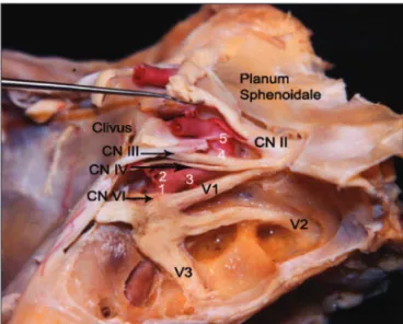
Fig 1. Cavernous sinus dissection, demonstrating cranial nerves: II-III-IV-V1-V2-V3-VI; 1, the posterior vertical segment; 2, the posterior bend; 3, the horizontal segment; 4, the anterior bend; 5, the anterior vertical segment (By Castro JAF, MD).
results
There were identiied 711 patients, with average age of 41 years (41±11.3), 73% females and 27% males, with a total of 802 aneurysms which underwent either conser-vative or interventionist treatment by the Neurosurgery discipline. of those patients, 35 presented with ccA with a total of 40 aneurysms, 5% of all intracranial aneurysms. Thirty-two patients (91.4%) were alive until the end of the study, while 3 (8.6%) were not. None of the obits was re-lated to the ccA or its treatment, two were due to sur-gical complications of other intracranial aneurysms and one was due to undetermined factors.
Thirty-two patients (91.4%) were females and three, male (8.6%). Average age by the time of diagnostic was 48.4 years (48.4±57.1 months), with a median of 47 years (total variation between 19 and 67 years) and an average time of follow of 60.5 months. There was a higher preva-lence of white middle-age females, being arterial hyper-tension the only consistent systemic alteration found, presented in 48,5% of the patients studied. Thirty from the 35 patients had unilateral ccA (85.7%), from a total of 40 aneurisms, 50% of the aneurysms were from the right side. Fifty percent were classiied as giant aneurysms (>2.5 cm) as shown in Figures 2 and 3 and thirteen were classi-ied as large (1.0–2.5 cm). seven patients (21.2%) had an-eurysms in other sites.
Thirty-three patients (94.2%) had symptoms due to ccAs (Table 1). The most frequent was headache, present-ed in 33 patients (94%), followpresent-ed by diplopic vision due to compromised vI cranial nerve (82.8%). sequentially, there
was retro bulbar pain in 18 patients (54.5%), deicit in visu-al accuracy in 9 patients (17.8%), photophobia in two cas-es and facial pain in one case (Table 1). All cascas-es of IcA an-eurysms with incidental diagnostic were clinically treated and observed without surgical intervention.
The clinical sign of highest prevalence was ophthal-mic paresis, observed in 33 patients (94.2%), being multi-ple in 23 cases (69.6%) and isolated in 10 patients (30.3%). The most affected cranial nerve was the abducent (90%). other signs were sensorial trigeminal neuropathy in 11 pa-tients (31.4%), followed by three cases of ischemic cerebral vascular accident (IcvA) and two of epistaxis.
Nineteen aneurysms (47.5%) underwent surgical treat-ment (Table 2). The most frequent surgery was IcA occlu-sion (11 cases). Ten of these patients were submitted to ca-rotid occlusion test with balloon (Fig 2) and one with sil-verstone clamp. eight patients (23.5%) underwent surgery with IcA ligature after cervicotomy (Fig 2). Three patients were treated with detachable balloon allocated by endo-vascular technique after collateral circulation test. Four Fig 3. (A) MRI scan presenting a coronal slice in which can be seen giant aneurysm in right ICA in a 57
year old patient with headache and right III, IV and VI. (B) After months, presented complete remis-sion of headache with angiography showing Right ICA spontaneous thrombosis. (C) Axial slice from a MRI scan showing remission of aneurysmatic dilatation of right ICA after a 10 year follow up.
Table 1. Incidence of symptoms in the presentation of patients with intracavernous aneurysms.
symptom surgery (n=19)% conservative (n=21)%
diplopya 16 (84.2%) 18 (85.7%)
Painb 19 (100%) 9 (42.8%)
Incidental 0 2 (9.5%)
visual deicit 5 (26.3%) 4 (19%)
ap=0.008; bp=0.002.
Table 2. Distribution of terapeutical strategies of patients with intracavernous aneurysms.
N aneurisms clinical surgical IcA ligature IcA balloon embolization By pass Trapping
RIcA 20 10 10 6 1 1 1 1
lIcA 20 11 9 2 2 4 1
Total 40 21 19 8 3 5 1 2
patients were treated with simple aneurysmatic embo-lization and one with emboembo-lization associated with IcA stent. Two cases were submitted to IcA trapping and one patient was treated with high low by-pass (safen vein in external carotid artery – media cerebral artery).
Twenty-one aneurysms (52.5%) had conservative treat-ment (Table 2), 10 were giant, there were five cases of spontaneous thrombosis: one of the aneurysm and four (20%) of IcA (Fig 3). In these ive cases there was impor-tant remission of the pain symptoms.
Among the 19 patients that underwent surgical treat-ment, seven (36.8%) had surgical complications. Two pa-tients had transitory motor deicit, manifested as a tran-sitory ischemic attack. considering permanent complica-tions, one patient presented ophthalmic paresis associat-ed with amaurosis and another patient had a cerebrovas-cular accident. As late complications there was a case of carotid-cavernous istula and two cases of collateral cav-ernous aneurysms.
In the group of patients surgically treated, 85% had improvement of pain and 15% did not present alterations in initial pain symptoms. Regarding neurological deicits, there was improvement of symptoms in 70% of patients while 30% were stable.
Patients treated conservatively presented in 16.7% of the cases improvement of pain while 58.3% were stable and 25% had intensiication of the initial pain symptoms. Regarding neurological deicits, patients treated clinically did not have improvement of symptoms, 66.7% were sta-ble and 33.3% presented intensiication of deicits.
Analyzing all patients without therapeutical strategy distinction, there was improvement of pain symptoms in 56.7% of the cases, stability in 33.3% and increase in pain in 10%. considering neurological deicits, there was im-provement of symptoms in 36.7% of the patients, stabili-ty in 50% and decrease of the deicit in 13.3%.
statistical analysis was performed through [chi2] test
with analysis of covariance and multinomial logistic re-gression, which demonstrated that surgical intervention is related with better neurological prognostic in comparison with the group treated conservatively; i.e., resolution of pain scenario independently of initial intensity (p=0.002) and stabilization or resolution of neurological deicits, in-dependently of initial intensity of clinical features used for classiication (p=0.008).
Regarding aneurysms´ size (large or giant) there was no statistically signiicant relation with neurological deicit outcome (p=0.836) or initial pain symptoms (p=0.909).
With the purpose of verifying the impact of age on the prognostic of both, pain and neurological symptoms, we divided the population studied into two groups using as limit value between the groups the mediana of ages.
Group A was composed of individuals with less then 55 years of age and group B of individuals with 55 years old or more. comparing both groups, we observed that age could not be statistically related to neither pain nor neu-rological symptoms (respectively, p=0.714 and p=0.916).
disCussion
Using Pubmed data base there were identiied only three studies with large series of ccAs, including one with natural history of 79 aneurysms with a follow up of 10 years14 and other two studies which emphasized patients´
neuro-ophtalmological prognostic: one with 40 ccAs for 30 years19 and other, a multi-centric study with 206
aneu-rysm with a follow up of 16 years20. In the present study, we
observed the clinical outcome for both conservative and interventionist therapeutical strategies, with emphasis on neurological prognosis, including statistical analysis of all patients with intracranial aneurysms in a 19-year period of segment, all treated and followed in our institution. ccAs are more frequent in white females, with the beginning of symptoms around 5th and 6th decades6. similarly, systemic
arterial hypertension is a risk factor for the development of intracranial aneurysms, this disease was present in 17 of the 35 patients when the aneurysms were irst detect-ed, independently of previous history of hypertension. The presence of bilateral cavernous aneurysms in 5 patients, was common and in accordance with data from previous studies4,6,14,19. seven patients had intracranial
an-eurysms in other sites, suggesting that a degenerative process secondary to a genetic factor of fragility in the vascular wall should be present.
Rarely do ccAs suffer rupture and subarachnoid hem-orrhage by the time of diagnostic, due to the fact that cavernous sinus are composed by dural slices, which lay over the body of sphenoid and are, infrequently, project-ed towards the subarachnoid space4,11 .
Two patients were asymptomatic when the diagnostic was made, and the presence of ccAs was only veriied after angiography for investigation of other brain aneu-rysms. In the presence of rupture, commonly, there are carotid-cavernous istulas or epistaxis8,19,21 with low
mor-bidity and mortality rates if surgical treatment is adopt-ed. In our study two patients had epistaxis when ccAs were diagnosed and two who underwent interventionist treatment presented later carotid-cavernous istulas, with satisfactory evolution: both patients with epistaxis were submitted to IcA ligature and one patient with istula was submitted to endovascular treatment.
cranial nerve lesions due to its position within cavernous sinus, as demonstrated in Image 14,10. The association with
other nerves (III, Iv, v1 and v2), located in the lateral wall of cavernous sinus characterizes the complete cavernous sinus syndrome9,10,19,20.
considering the group of patients submitted to con-servative treatment, it could be veriied a relapsing and luctuating course of pain symptoms and a progressive im-pairment of cranial nerves´ function, determining a poor neurological outcome.
Patients submitted to interventionist treatment, i.e., endovascular treatment with coils, stent and IcA occlusion with balloon, as well as simple IcA ligature, IcA ligature associated with external carotid to media cerebral artery by-pass and IcA trapping, could be compared and included in a same group for having as inal result of surgical strat-egy, aneurysmatic or carotid exclusion from circulation. The interventionist therapy altered signiicantly the outcome of neurological deicits: the surgically treated group presented improvement of symptoms in 70% of the cases (p=0.008), avoiding in all cases the progression of installed neurological deicits.
The pain scenario, determined by headache, hemi fa-cial pain and retro bulbar pain, was improved or extin-guished in 100% of the patients submitted to interven-tionist treatment (p=0.002).
Brain infarcts occurred in three of nineteen patients that underwent interventionist treatment, two were tran-sitory in consequence of trantran-sitory ischemic attach and one after ischemic cerebral vascular accident11.
complica-tion rates in our study are comparable to those veriied in literature13,19,21
In conclusion, cavernous carotid artery aneurysms present a satisfactory long-term neurological outcome with low complication rates if submitted to interven-tionist treatment, what determines improvement or res-olution of pain symptoms in comparison with patients conservatively treated, as well as stabilization or partial improvement of neuro-ophthalmological deicits.
referenCes
1. Bars HW, Blackwood W, Meadows SP. Intracavernous carotid aneu-rysms. A clinical-pathological report. Brain 1971;94:607-622. 2. Bavinzski G, Killer M, Ferraz-Leite H, et al. Endovascular therapy
of idiopathic cavernous aneurysms over 11 years. Am J Neuroradiol 1998;19:559-565.
3. Derdeyn CP, Cross DT, Moran CJ, et al. Postprocedure ischemic events after treatment of intracranial aneurysms with Guglielmi detachable coils. J Neurosurg 2002;96:837-843.
4. German WJ, Black SP. Cervical ligation for internal carotid aneurysms: an extended follow-up. J Neurosurg 1965;23:572-577.
5. Goddard AJ, Annesley-Williams D, Gholkar A. Endovascular manage-ment of unruptured intracranial aneurysms: does outcome justify treat-ment? J Neurol Neurosurg Psychiatry 2002;72:485-490.
6. Goldenberg-Cohen N, Curry C, Miller NR, et al. Long term visual and neurological prognosis in patients with treated and untreated cavern-ous sinus aneurysms. J Neurosurg 2004;75:863-867.
7. Higashida RT, Halbach VV, Dowd C, et al. Endovascular detachable balloon embolization therapy of cavernous carotid artery aneurysms: results in 87 cases. J Neurosurg 1990;72:857-863.
8. Inagawa T. Follow-up study of unrupted aneurysms arising from C3 and C4 segments of the internal carotid artery. Surg Neurol 1991;36:99-105. 9. Inoue T, Rhoton AL Jr, Theele D, et al. Surgical approaches to
cavern-ous sinus: a microsurgical study. Neurosurgery 1990;26:903-932. 10. Johnston SC, Wilson CB, Halbach VV, et al. Endovascular and surgical
treatment of unruptured cerebral aneurysms: comparison of risks. Ann Neurol 2000;48:11-19.
11. Kupersmith MJ, Berenstein A, Choi IS, et al. Percutaneous
transvascu-lar treatment of giant carotid aneurysms: neuro-ophthalmologic ind
-ings. Neurology 1984;34:328-335.
12. Kupersmith MJ, Hurst R, Berenstein A, et al. The benign course of cav-ernous carotid artery aneurysms. J Neurosurg 1992;77:690-693. 13. Kupersmith MJ, Stiebel-Kalish H, Huna-Baron R, et al. Cavernous
ca-rotid aneurysms rarely cause subarachnoid hemorrhage or major neu-rologic morbidity. J Stroke Cerberovasc Dis 2002;11:9-14.
14. Kupersmith MJ. Aneurysms involving the motor and sensory visu-al pathways. In: Neuro-Vascular Neuro-Ophthvisu-almology. Heidelberg: Springer-Verlag, 1993:254-261.
15. Linskey ME, Sekhar LN, Hirsch WL Jr, et al: Aneurysms of the intra-cavernous carotid artery: natural history and indications for treatment. Neurosurgery 1990;26:933-937.
16. Locksley HB. Natural history of subarachnoid hemorrhage, intracranial an-eurysms and arteriovenous malformations. J Neurosurg 1966;25:321-368.
17. Miller NR. Carotid-cavernous sinus istulas. In: Miller NR, Newman
NJ (Eds). Welsh & Hoyt’s clinical neuro-ophthalmology. Baltimore: Wil-liams and Wilkins, 1997:3263-3322.
18. Newman SA. Aneurysms. In Miller NR, Newman NJ (Eds). Walsh & Hoyt’s clinical neuro-ophthalmology. Baltimore: Williams and Wilkins, 1997:2975-3261.
19. Stiebel-Kalish H, Kalish Y, et al. Presentation, natural history, and man-agement of carotid cavernous aneurysms. Neurosurgery 2005;57:850-857. 20. van der Schaaf EH, Buskens E, Rinkel GJ. Endovascular treatment of
aneurysms in the cavernous sinus: a systematic review on balloon oc-clusion of the parent vessel and embolization with coils. Stroke 2002;33: 313-318.