ANTERIOR SURGICAL MANAGEMENT OF THE
CERVICOTHORACIC JUNCTION LESIONS AT
T1 AND T2 VERTEBRAL BODIES
Asdrubal Falavigna
1, Orlando Righesso
2, Darcy Ribeiro Pinto-Filho
3, Alisson Roberto Teles
4Abstract – Lesions of the cervicothoracic junction have a high propensity for causing instability and present unique challenges in the surgical treatment. Several surgical approaches to this region have been described in the literature. We report our experience in the surgical treatment of six patients with unstable lesions involving the cervicothoracic junction at T1 and T2 vertebral bodies. The patients underwent an anterior left Smith-Robinson approach and manubriotomy. Mesh and cervical plate system were used for stabilization and reconstruction of the region. No complication related to the surgical procedure was observed. In our experience, in injuries involving the T1 and T2 vertebral bodies, the transmanubrial approach offers good working room to remove the lesions and anterior reconstruction.
KEY WORDS: spine, cervicothoracic junction, instability, surgical treatment.
Manejo cirúrgico via anterior das lesões da junção cérvico-torácica nos corpos vertebrais de T1 e T2 Resumo – Lesões da junção cérvico-torácica têm alta tendência em causar instabilidade e apresentam grandes desafios ao tratamento cirúrgico. Diversas abordagens cirúrgicas a esta região foram descritas na literatura. Relatamos nossa experiência no tratamento cirúrgico de seis pacientes com lesões instáveis envolvendo a junção cérvico-torácica em corpos vertebrais de T1 e T2. Os pacientes foram submetidos a uma abordagem anterior de Smith-Robinson pela esquerda e manubriotomia. Mesh e placa cervical foram utilizados para estabilização e reconstrução da região. Nenhuma complicação relacionada ao procedimento cirúrgico foi observada. Em nossa experiência, em lesões que envolvem os corpos vertebrais de T1 e T2, a abordagem transmanubrial oferece bom campo de trabalho para remoção das lesões e estabilização anterior.
PALAVRAS-CHAVE: coluna, junção cervico-torácica, instabilidade, tratamento cirúrgico.
1Department of Neurosurgery, Universidade de Caxias do Sul, Hospital Saúde, Caxias do Sul RS, Brazil; 2Department of Orthopedics, Hospital Saúde, Caxias do Sul RS, Brazil; 3Department of Thoracic Surgery, Universidade de Caxias do Sul, Hospital Geral, Caxias do Sul RS, Brazil; 4Medical student, Universidade de Caxias do Sul, Caxias do Sul RS, Brazil
Received 5 December 2007. Accepted 23 February 2008.
Dr. Asdrubal Falavigna – Rua General Arcy da Rocha Nóbrega 401/602 - 95040-290 Caxias do Sul RS - Brasil. E-mail: [email protected]
The cervicothoracic junction is deined as the area ex-tending from vertebral segments C7 to T4, and includes the lower brachial plexus, the thoracic outlet and the par-enchymatous, vascular and nervous structures of the up-per mediastinum. This region is a transition area from a mobile, lordotic cervical spine to a rigid, kyphotic thorac-ic spine1. It is susceptible to injury because of the weight
transfer from the anterior column to the posterior col-umn2 and the vertebral index decreases from C6 to T1
ver-tebrae, causing added stress to be applied to the more narrow and thinner vertebrae3. Several studies
demon-strate that surgical procedures in the cervicothoracic junc-tion can destabilize this region, mainly laminectomies at C7–T14, 5 and spinal fusions ending at this junction5,6.
Pathological processes such as tumors, trauma,
degen-eration and infection, which usually occur in the anterior segment of the vertebrae, frequently determine instabil-ity of this segment7. Progressive instability of this area
ul-timately leads to kyphosis and spinal cord compression, neurological involvement being a common complication as high as 80%7,8. This predisposition may be due to the
small spinal canal and the tenuous blood supply7,8. The
surgical treatment goals of cervicothoracic pathologies are neural decompression, immediate stabilization, res-toration of anatomical spinal alignment and early reha-bilitation. Different surgical approaches to the cervico-thoracic junction have been described1,4,7-29. As most
Table. Summary of patients. Case Age/ Sex Level Clinical manifestations Pathology Recon-struction Surgical time Bleeding Hospital stay (days) Outcome
1 65/F T1 Severe local pain Left Cervicobrachialgia Strength grade III at left C7 and T1
Lung metastasis mesh and plate 2.8 hours
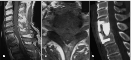
200 mL 7 No complications, marked neu-rological improvement with pain relief and strength improvement (grade V), died 18 months later of progressive systemic disease. 2 53/F T1 Severe local pain
Bilateral cervicobrachialgia Thyroid metastasis mesh and plate 3.2 hours
350 mL 8 No complications, pain relief, 9 months of follow-up.
3 68/M T1–T2 Severe local pain Crural paraparesis grade III
Lung metastasis mesh and plate 4.2 hours
400 mL 6 No complications, marked neu-rological improvement with pain relief and strength improvement (grade V), died 6 months after surgery of the progressive sys-temic disease.
4 30/M T1 Crural paraparesis grade II Fracture and luxation at C7–T1 mesh and posterior ixation 5.2 hours
520 mL 7 No complications, strength grade IV with 24 months of fol-low-up.
5 62/F T1–T2 Severe local pain Bilateral cervicobrachialgia Melanoma metastasis mesh and plate 3.7 hours
180 mL 6 No complications, pain relief, died 6 months after surgery of the progressive systemic disease. 6 72/M T1 Severe local pain
Bilateral cervicobrachialgia Strength grade III at T1
Lung metastasis mesh and plate 3.3 hours
220 mL 9 Lung infection in the immediate pos-op, pain relief and strength improvement, follow-up of 13 months.
and potentially dangerous because of bony obstructions such as the manubrium, clavicle and ribs and because of nearby vital structures such as great blood vessels, esoph-agus, trachea, recurrent laryngeal nerve, thoracic duct and sympathetic ganglions.
We report our experience with the anterior approach to the cervicothoracic junction at T1 and T2 vertebral bodies and its results.
METHOD
We reviewed the records of six patients with unstable le-sion of the cervicothoracic junction at T1 and T2 vertebral bod-ies surgically treated by anterior surgical approach. All patients had metastatic or traumatic vertebral body lesions diagnosed by computed tomography and magnetic resonance image. Clin-ical data of patients are summarized in Table.
All patients underwent an anterior approach to the cervi-cothoracic junction by a left Smith-Robinson approach and ma-nubriotmy. The patient was placed in the supine position on the operating table under general endotracheal anaesthesia. The neck was extended slightly using a folded sheet. Both wrists have traction bands applied to pull the arms down for lateral radio-graphic imaging during the procedure. Left vertical incision was performed along the medial aspect of the sternocleidomastoid extending along the midline of the sternum down two centime-ters from the Louis angle to permit manubriotomy.
Subplatys-mal laps were elevated and retained by sutures. The sternomas-toid, sternohyoid and sternothyroid muscles were sectioned, allowing connection of the region of the lower cervical and the upper thoracic spine. The sternum was split partially (manubri-otomy) and the level of dissection was conirmed by luoros-copy. The lesion was removed using a microscope and the re-construction was done using mesh and a cervical plate system. A suction drain was used in the substernal space. The manubri-otomy was closed with steel wires number three. The patients kept 48 hours in the intensive care unit and then discharged to a hospital room.
RESULTS
was submitted to posterior ixation of the spine because of posterior ligament and bone instability. The patients stayed in hospital for 7 days in average (range, 6–9 days). Figure illustrates the case 2.
DISCUSSION
Lesions of the cervicothoracic junction have a high propensity for causing instability and present unique challenges in surgical treatment. The pathologies involv-ing this region often produce spinal cord compression, which was noted in three of seven cases presented in this series. Several surgical approaches to the cervicothoracic junction have been described in the literature. Posterior approaches are disadvantageous because of a destabiliza-tion effect, inadequate visualizadestabiliza-tion of the vertebral body pathology, and the need for a long posterior construct to restore stability with a higher complication rate than anterior or lateral approaches7,15,28,30.
The limitations of a posterior exposure have resulted in the development of various posterolateral and anterior approaches7-26,29. The irst description of a
posterolater-al approach to the cervicothoracic area was the costo-transversectomy described in 1894 by Ménard22. Capener10
described the lateral thoracotomy approach, which pro-vided a more extensive posterolateral exposure afforded by a resection of a longer rib segment. A modification of Capener’s technique was described by Larson into the lateral extracavitary approach18, which improved exposure
and reduced morbidity. Fessler and colleagues13 proposed
the parascapular extrapleural lateral approach, which im-proved exposure of all the upper thoracic vertebrae. The disadvantages of this procedure are prolonged surgery (10–12 hours), excessive blood loss and ineficiency for the pathologies extending into the C7 vertebrae15.
Fur-thermore, pulmonary-related complications are common following this approach31.
Many authors suggest a simple anterior supramanu-brial cervicotomy to be used to reach the anterior por-tion of the proximal dorsal vertebrae26,32,33. Although this
approach is not very invasive, it does not permit good visualization and anterior reconstruction below T1, which results from the patient’s anatomical characteristics, such as marked junctional kyphosis, congenital high sternum, short neck or large shoulders7,15,24. Cauchoix and Binet11
proposed an anterior approach combining the supracla-vicular approach with a median sternotomy. This sternal splitting approach enables the exposure of the whole cervicothoracic junction up to T416,24. Hodgson et al.14
re-ported a surgical mortality rate of 40% with the sternal splitting approach and recommended the anterolateral thoracotomy approach to the cervicothoracic junction, which had only 4% mortality. However, the anterolat-eral thoracotomy approach provides limited access to the lower cervical spine because of obstruction by the scapula and upper ribs. Louis20 improved the sternal
split-ting approach combining this procedure with the anterior Smith-Robinson approach27, permitting access from C2 to
T516. In our experience, performing a partial sternotomy
(manubriotomy) we are able to reach until T4, but are un-able to achieve additional caudal exposure despite using a complete sternotomy because of the limited retraction of the aortic arc15 and this procedure can minimize the risks
of sub-sternal dissection21. Upper lateral transthoracic or
extrapleural approach is mandatory for total removal and reconstruction of the lesions of T4 and below15. Despite
the high mortality reported by Hodgson et al.14
follow-ing an anterior surgical procedure of the cervicothoracic junction, many authors8,12,15,16,21,23,24,26,29,32,33 believe that the
direct anterior approach to the region is safe and effec-tive, as demonstrated in our cases.
Some authors reported the need for clavicle resec-tion for both more extended lateral exposure and bone fusion9,17,19,29. This technique was described by Sunderasan
et al.29 in a series of seven patients with tumors
involv-ing the cervicothoracic junction until T2. They proposed removing of a rectangular block of sternum instead of complete splitting and adding a resection of the medial third of the clavicle for better exposure and to use it as a strut graft. For tumors with signiicant intrathoracic exten-sion, a trapdoor technique has been described to achieve gross-total resection25. Although this approach improves
anterior visualization and reduces the risk of spinal cord manipulation, it carries the potential risks associated with thoracotomy. In our experience, using microscopic visual-ization and the new systems for spinal reconstruction we can achieve good results in terms of tumor removal and stabilization using cervical exposure and manubriotomy.
The anterior approach to the cervicothoracic junction require accurate knowledge of the numerous anatomical structures that hinder this region. The right brachiocephalic vein passes vertically, joining the left brachiocephalic or in-nominate vein and coursing obliquely in front of the aortic arch behind the manubrium to form the superior vena cava at the level of the right second rib. The aortic arch limits this surgical approach to the area along T4-T5. It is orient-ed behind the lower aspect of the manubrium and left bra-chiocephalic vein. The brabra-chiocephalic arterial trunk pass-es behind the manubrium. The recurrent laryngeal nerve arises from the vagus and courses around the subclavian artery on the right and around the aortic arch on the left. It crosses the operative ield obliquely at a higher level on the right side; contrary on the left side, it reaches the tracheoesophageal groove more caudally having a vertical trajectory less liable to injury with left-sided exposure24.
Besides, the right nerve may not be recurrent from the va-gus in one percent of the patients and rise at a higher level in the neck34. A left approach is desirable to decrease the
risk of recurrent laryngeal nerve injury, knowing that, on this side, the thoracic duct is presented ascending on the left side of the thoracic inlet and the esophagus, behind the left subclavian, lowing into the internal jugular vein.
Steinmetz et al.5 studied the factors associated with
treatment failure in cervicothoracic junction surgery. In their large series of 593 patients, they observed treat-ment failure in 14 patients. Uninstrutreat-mented laminecto-my and ventral multilevel corpectomies (two or three levels) across the cervicothoracic junction were associ-ated with fusion failure in 38% and 16.7%, respectively. A trend toward treatment failure was noted in cases in
which dorsal constructs ended at the C7 vertebrae, al-though this was not statistically signiicant. The authors recommended supplemental dorsal instrumentation in cases of multilevel corpectomies, posterior instability or dorsal cervicothoracic laminectomies with extension of the dorsal hardware to T1 or T2. Besides, treatment fail-ure was also associated with histories of prior cervical surgery, deformity correction and tobacco use. In case 4, we performed an anterior decompression followed by a posterior stabilization because of posterior ligament and bone instability.
In our experience, a left Smith-Robinson approach com-bined with manubriotomy offers good exposure and work-ing room for the cervicothoracic lesions involvwork-ing T1 and T2 vertebral bodies. This approach requires accurate knowl-edge of surgical anatomy, as many vital structures are pres-ent in this area. We used mesh and a cervical plate system with good results for stabilize and reconstruct this region.
REFERENCES
1. An HS, Wise JJ, Xu R. Anatomy of the cervicothoracic junction: a study of cadaveric dissection, cryomicrotomy, and magnetic resonance imag-ing. J Spinal Disord 1999;12:519-525.
2. Pal GP, Routal RV. A study of weight transmission through the cervi-cal and upper thoracic regions of the vertebral column in man. J Anat 1986;148:245-261.
3. Boyle JJ, Singer KP, Milne N. Morphological survey of the cervicotho-racic junctional region. Spine 1996;21:544-548.
4. An HS, Vaccaro A, Cotler JM, Lin S. Spinal disorders at the cervicotho-racic junction. Spine 1994;19:2557-2564.
5. Steinmetz MP, Miller J, Warbel A, Krishnaney AA, Bingaman W, Ben-zel EC. Regional instability following cervicothoracic junction surgery. J Neurosurg Spine 2006;4:278-284.
6. Drennan JC, King EW. Cervical dislocation following fusion of the up-per thoracic spine for scoliosis. A case report. J Bone Joint Surg Am 1978;60:1003-1005.
7. Le H, Balabhadra R, Park J, Kim D. Surgical treatment of tumors in-volving the cervicothoracic junction. Neurosurg Focus 2003;15:E3. 8. Sapkas G, Papadakis S, Katonis P, Roidis N, Kontakis G. Operative
treatment of unstable injuries of the cervicothoracic junction. Eur Spine J 1999;8:279-283.
9. Birch R, Bonney G, Marshall RW. A surgical approach to the cervico-thoracic spine. J Bone Joint Surg Br 1990;72:904-907.
10. Capener N. The evolution of lateral rhachotomy. J Bone Joint Surg Br 1954;36-B:173-179.
11. Cauchoix J, Binet JP. Anterior surgical approaches to the spine. Ann R Coll Surg Engl 1957;21:234-243.
12. Charles R, Govender S. Anterior approach to the upper thoracic verte-brae. J Bone Joint Surg Br 1989;71:81-84.
13. Fessler RG, Dietze DD Jr., Millan MM, Peace D. Lateral parascapu-lar extrapleural approach to the upper thoracic spine. J Neurosurg 1991;75:349-355.
14. Hodgson AR, Stock FE, Fang HS, Ong GB. Anterior spinal fusion: the
operative approach and pathological indings in 412 patients with Pott’s
disease of the spine. Br J Surg 1960;48:172-178.
15. Kaya RA, Turkmenoglu ON, Koc ON, et al. A perspective for the selec-tion of surgical approaches in patients with upper thoracic and cervi-cothoracic junction instabilities. Surg Neurol 2006;65:454-463. 16. Knoller SM, Brethner L. Surgical treatment of the spine at the
cervico-thoracic junction: an illustrated review of a modiied sternotomy ap
-proach with the description of tricks and pitfalls. Arch Orthop Trauma Surg 2002;122:365-368.
17. Kurz LT, Pursel SE, Herkowitz HN. Modiied anterior approach to the
cervicothoracic junction. Spine 1991;16(Suppl 10):S542-S547. 18. Larson SJ, Holst RA, Hemmy DC, Sances A, Jr. Lateral extracavitary
19. Lesoin F, Thomas CE 3rd, Autricque A, Villette L, Jomin M. A transste-rnal biclavicular approach to the upper anterior thoracic spine. Surg Neurol 1986;26:253-256.
20. Louis R. Die Chirurgie der Wirbelsäule. Chirurgische Anatomie und operative Zugangswege. Berlin: Springer-Verlag, 1985.
21. Luk KD, Cheung KM, Leong JC. Anterior approach to the
cervicotho-racic junction by unilateral or bilateral manubriotomy: a report of ive
cases. J Bone Joint Surg Am 2002;84-A:1013-1017.
22. Ménard V. Causes de la paraplégie dans le mal de Pott. Son traitement
chirurgical par l’ouverture directe du foyer tuberculeux des vertebres.
Rev Orthop 1894;5:47-64.
23. Micheli LJ, Hood RW. Anterior exposure of the cervicothoracic spine using a combined cervical and thoracic approach. J Bone Joint Surg Am 1983;65:992-997.
24. Miscusi M, Bellitti A, Polli FM. Surgical approaches to the cervico-tho-racic junction. J Neurosurg Sci 2005;49:49-57.
25. Nazzaro JM, Arbit E, Burt M. “Trap door” exposure of the cervicotho-racic junction: technical note. J Neurosurg 1994;80:338-341.
26. Resnick DK. Anterior cervicothoracic junction corpectomy and plate
ixation without sternotomy. Neurosurg Focus 2002;12:E7.
27. Robinson RA, Smith GW. Anterolateral disc removal and interbody fusion for cervical disc syndrome. Johns Hopkins Hosp Bull 1953;96:223-224. 28. Sundaresan N, DiGiacinto GV, Krol G, Hughes JE. Spondylectomy for
malignant tumors of the spine. J Clin Oncol 1989;7:1485-1491. 29. Sundaresan N, Shah J, Foley KM, Rosen G. An anterior surgical
ap-proach to the upper thoracic vertebrae. J Neurosurg 1984;61:686-690. 30. Siegal T, Siegal T. Surgical decompression of anterior and posterior
malignant epidural tumors compressing the spinal cord: a prospective study. Neurosurgery 1985;17:424-432.
31. Resnick DK, Benzel EC. Lateral extracavitary approach for thoracic and thoracolumbar spine trauma: operative complications. Neurosurgery 1998;43:796-802.
32. Gieger M, Roth PA, Wu JK. The anterior cervical approach to the cervi-cothoracic junction. Neurosurgery 1995;37:704-709.
33. Comey CH, McLaughlin MR, Moossy J. Anterior thoracic corpectomy without sternotomy: a strategy for malignant disease of the upper tho-racic spine. Acta Neurochir (Wien) 1997;139:712-718.