IMPROVEMENT IN HIPPOCAMPAL KINDLING ANALYSIS
THROUGH COMPUTATIONAL PROCESSING DATA
Joacir Graciolli Cordeiro
1,2, Alberto Capurro
3,4, Ad Aertsen
3,4,
Karina Kohn Cordeiro
1,2, João Cândido Araújo
2, Andreas Schulze-Bonhage
1,3Abstract – The kindling phenomenon is classically investigated in epileptology research. The present study aims to provide further information about hippocampal kindling through computational processing data. Adult Wistar rats were implanted with dorsal hippocampal and frontal neocortical electrodes to perform the experiment. The processing data was obtained using the Spike2 and Matlab softwares. An inverse relationship between the number of “wet dog shakes” and the Racine’s motor stages development was found. Moreover it was observed a significant increase in the afterdischarge (AD) duration and its frequency content. The highest frequencies were, however, only reached at the beginning of behavioral seizures. During the primary AD, fast transients (ripples) were registered in both hippocampi superimposed to slower waves. This experiment highlights the usefulness of computational processing applied to animal models of temporal lobe epilepsy and supports a relevant role of the high frequency discharges in temporal epileptogenesis.
Key WoRDS: hippocampal kindling, temporal epilepsy, experimental model.
Aprimorando a análise do modelo de kindling hipocampal com o auxílio de processamento computacional Resumo – o fenômeno de kindling é classicamente utilizado no campo da epileptologia experimental. este trabalho objetiva aprofundar a análise do modelo kindling hipocampal através de processamento computacional. Ratos wistar adultos receberam eletrodos hipocampais dorsais e neocorticais frontais para a realização do experimento. o processamento dos dados encontrados foi realizado pelos softwares Matlab e Spike2. encontrou-se uma relação inversa entre wet dog shakes e o desenvolvimento dos estágios motores de Racine. A duração e o conteúdo de freqüência das pós-descargas hipocampais aumentaram durante o processo, sendo observadas descargas de alta freqüência (ripples) em ambos os hipocampos durante as pós-descargas primárias, superimpostas a ondas lentas. As mais altas freqüências, entretanto, foram apenas atingidas com o início das crises epilépticas. A utilização de sistemas computacionais para a confecção e análise do modelo de epilepsia temporal é ressaltada e reforça-se a relevância do papel das altas freqüências na epileptogênese temporal.
PAlAvRAS-chAve: kindling hipocampal, epilepsia temporal, modelo animal.
Albert-ludwigs-University, Freiburg, Germany: 1center for epilepsy, Dept. Neurosurgery, University Medical center Freiburg, Albert-ludwigs-University, Freiburg, Germany; 2Department of Neurosurgery, hospital de clínicas, Federal University of Paraná, curitiba PR, Brazil; 3Bernstein center for com-putational Neuroscience Freiburg, Albert-ludwigs-University, Freiburg, Germany; 4Neurobiology and Biophysics, Faculty of Biology, Albert-ludwigs-University, Freiburg, Germany. Supported by BMBF- Germany (01GQ0420, c3).
Received 11 December 2008, received in inal form 14 May 2009. Accepted 17 June 2009.
Dr. Joacir Graciolli Cordeiro – Breisacherstr. 64 - PLZ 79106 Freiburg im Breisgau - Germany. E-mail: [email protected]
Temporal lobe epilepsy (Tle) is the most frequent ep-ilepsy form in adults1 and its major cause is mesial
tem-poral sclerosis (MTS), which comprises around 60–70% of Tle cases2. MTS presents with a high clinical
refractori-ness rate, medical treatment being able to provide seizure control in only 20–40% of the cases1,3,4.In refractory
pa-tients surgical treatment may be indicated, with the cur-rent rate of favorable outcome with microsurgery
reach-ing around 67 to 85% of the patients4,5. Despite the
high-cate local ield potentials (lFP) signal analysis. An exam-ple is the coupling with the Matlab software (The Math-Works, Natick, USA), which integrates the signal process-ing form to the construction of spectrograms, which fa-cilitates the signal evaluation. The objective of this study is to evaluate the use of computational analytical tools applied to the lFP signal recorded from rats submitted to the hippocampal kindling model.
METHOD
In order to perform the experiment, 12 adult Wistar rats of both sexes (non-pregnant), with weight between 250 and 350 grams were used. The animals were obtained through the Uniklinik Freiburg (Germany) and their use was approved by its ethical commission in compliance with the regulation for exper-imental animal models. The animals were individually housed during all the experiment period in transparent cages alternat-ing, 12 hours in daylight and 12 hours in darkness. All experiments were held at the same time during the day light cycle. Food and
water intake was ad libitum. Three rats were excluded due to
technical problems with the electrodes. Anesthesia for cere-bral electrode implantation was performed with intraperitone-al injection of ketamine10% (100 mg/kg), xylosine 2% (3 mg/kg) and atropine 0.5 mg/ml (0.1 mg/kg). For post-operative analge-sia, subcutaneous buprenorine (0.05 mg/kg) was used. The ani-mals were placed in the stereotactic frame and burr holes were drilled in the skull according to the coordinates for the dorsal
hippocampus8. A bipolar twisted platinum electrode was
im-planted (125 μm diameter and mean impedance of 413±220 kΩ
at 10 hz) on the right dorsal hippocampus (y= –3.6; X= –2.4; Z= –3.5). A tungstenium monopolar electrode was also implanted
(60 μm diameter and mean impedance of 1220±544 kΩ at 10hz)
on both dorsal hippocampi anteriorly and posteriorly to the platinum electrode (y= –2.6; X=±1.5; Z= –3.2 and y= –4.6; X=±3.5; Z= –3.4). Additionally, two monopolar tungstenium electrodes were implanted in both frontal neocortices aiming at the prima-ry motor area (1 mm anterior to the coronal suture), one monop-olar reference electrode above the left cerebellum and one oc-cipital screw for grounding. Fixation to the skull was performed with dental cement and cranial screws and the electrodes were plugged into the multiple channel connector.
A multiple channel connector was plugged into the MPA8I
tained behavioral response was classiied according to the Ra-cine and Pinel and Rover seizure scales9,10 (Table 1).
Sessions were individually repeated until each rat reached ten consecutive behavioral seizures with a minimum Racine score of ive (i.e. fully kindled state). After process completion, the animals underwent intracardiac perfusion under deep anes-thesia for histological veriication of electrode position. Record-ed lFP were analyzRecord-ed in order to determinate the number of wet dog shakes (WDS) and the primary after discharge (1AD) dura-tion. WDS are a stereotyped motor patter (mimicking the shake of a wet dog), which are associated to a characteristic electro-encephalographic discharge. WDS quantiication was done by
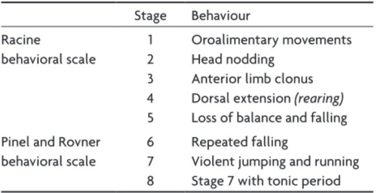
Table 1. Behavioral seizure classiication.
Stage Behaviour
Racine behavioral scale
1 2 3 4 5
oroalimentary movements head nodding
Anterior limb clonus Dorsal extension (rearing)
loss of balance and falling Pinel and Rovner
behavioral scale
6 7 8
Repeated falling
violent jumping and running Stage 7 with tonic period
Source: References 9 and 10. Pinel and Rovner behavioral scale adds three posterior stages to the Racine scale.
Table 2. Kindling process subdivision.
D0 irst session
D1 irst quarter
D2 second quarter
D3 third quarter
D4 fourth quarter (irst generalized behavioral seizure)
D5 ifth generalized behavioral seizure
D6 fully kindled state
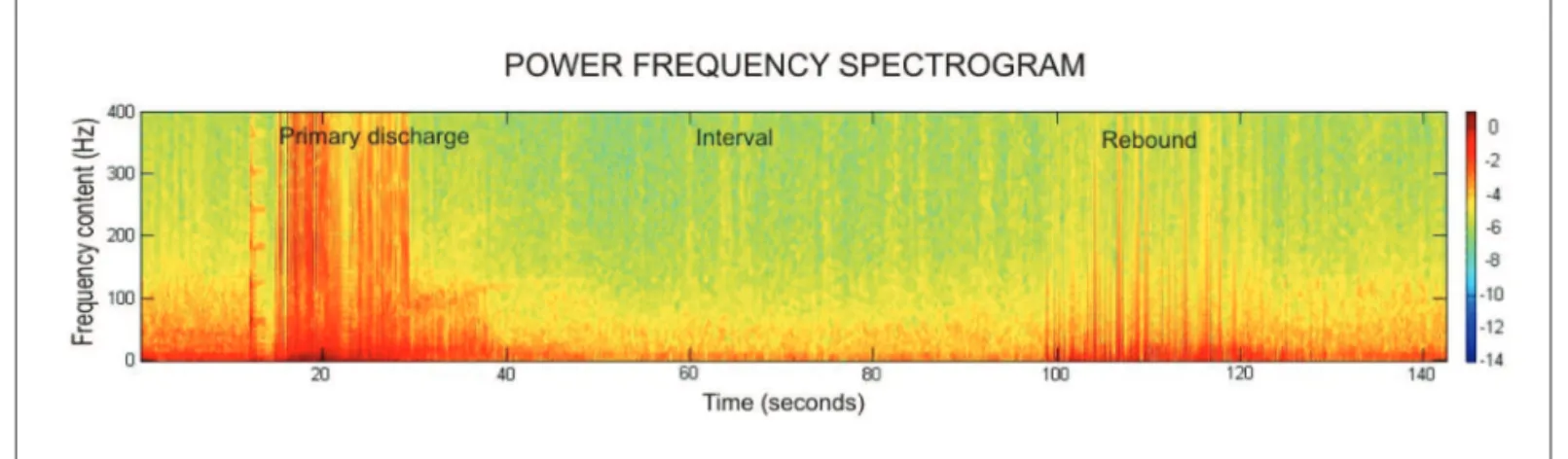
direct observation of the characteristic behavioral and electro-physiological patterns. For the 1AD determination, amplitude and frequency analyses were utilized. Amplitude analysis was performed by means of the Spike2 software. For frequency anal-ysis the lFP signal was processed into power frequency spectro-grams by the Matlab6.5 software (Fig 1)11-12.
Due to the number of animals and non-normal distribution of data, non-parametric Mann-Whitney-U-test was used at signif-icance levels of 95% and 99%. For statistical analysis the JMS soft-ware (SAS Softsoft-ware corporation, cambridge, england) was utilized.
RESULTS
The kindling process was divided into seven phases in order to be studied in details (Table 2). The irst kindling session of each rat was named D0 (irst phase). The indi-vidual number of kindling sessions needed to the appear-ance of the irst generalized behavioral seizure was di-vided into four quarters. The session referred to the irst quarter was named D1, D2 to the second quarter, D3 to the third quarter, D4 for the fourth quarter (the session when the irst generalized behavioral seizure appeared). D5 cor-respons to the ifth generalized behavioral seizure and D6 to the end of the kindling process (fully kindled state).
Behavioral response
During the hippocampal kindling process, each rat presented the irst generalized behavioral seizure after a
mean of 22.6 sessions (SD=9.2). eight animals completed the kindling process after a mean of 42.4 sessions (SD=8.8). one rat showed a connector problem after D5. The du-ration of the behavioral response of each seizure (Racine score of three or more) was analyzed on D4, D5 and D6 phases. A statistically signiicant difference was observed between D4 and D6 (p<0.05), presenting D6 a longer du-ration (Table 3).
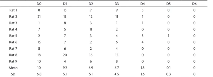
The WDS amount of each session was quantiied and a progressive decrease along the kindling process was ob-served. As the behavioral manifestation reached a high-er Racine score, the numbhigh-er of WDS rate decreased, es-tablishing an inverse correlation between the WDS oc-currence and the development of the Racine motor stag-es (Table 4). A statistically signiicant fall on the number of WDS after the irst generalized behavioral seizure (D4) was noticed (p<0.05).
Amplitude and frequency content
Through the visual lFP inspection before (control peri-od) and after the triggering stimulation, it was noted that 1AD consisted of waves of higher frequency and ampli-tude. After 1AD a relatively silent period was visualized, in which frequency and amplitude were inferior to the control period. The 2AD period (rebound) was marked by slow and high amplitude waves, which ceased simultane-ously in all the eight recording channels, inalizing the
kin-Fig 1. AD power frequency spectrogram. Representative power frequency spectrogram processed from the LFP signal recorded from a kindling session. In the frequency spectrogram a clearer differentiation and analysis of the three typical periods of a hippocampal AD (primary AD, in-terval and rebound) was observed. Scale bar in Log10[mV2]. Source: Experiment data.
Table 3. Behavioral seizure duration.
Phase Rat1 Rat2 Rat3 Rat4 Rat5 Rat6 Rat7 Rat8 Rat9 Mean SD
D4 25.8 30 25 32 6.4 11.7 14.2 8.8 31.4 20.6 10.3
D5 29.3 31.8 38.4 33.2 14.7 9.1 14 26.5 23 24.4 10
D6 36 37.2 34.9 28.9 – 18.9 28.3 34.1 37.2 32.2 5.9
dling sessions. After D4 (generalized behavioral seizure) an increase on 1AD complexity was observed and the visu-al differentiation between 1AD, intervvisu-al and 2AD became dificult. Nevertheless, the after discharge end remained clear on all channels.
The lFP analysis coupled to the power frequency spec-trograms showed high frequency discharges above 100 hz (including ripples). Ripples (high frequency oscillations be-tween 100 and 250 hz) were visualized superimposed to slower waves and were present on both hippocampi even before the behavioral seizures appearance (D4) (Fig 2).
1AD duration analysis
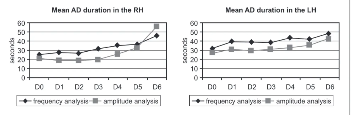
The lFP frequency and amplitude increase were used to determine 1AD duration on both hippocampi. 1AD start was marked when the frequency after the stimu-lus overcame the control period frequency and the end was marked when it returned to the control period val-ue. The same procedure was used for amplitude analy-sis. Duration measurements were performed on signal re-corded from posterior electrodes situated on both dor-sal hippocampi.
A signiicant duration increase after the irst behav-ioral seizure was observed in the right hippocampus (Rh) through the frequency analysis, being D0 shorter than D4, D5 and D6 (p<0.05, p<0.01 and p<0.05 respectively). D1 was shorter than D5 and D6 (p<0.05 and p<0.01), as well as D2 was shorter than D4 and D5 (p<0.05). The same record-ings submitted to frequency analysis were used for am-plitude analysis. D0, D1, D2 and D3 1AD duration were in-dividually shorter than D5 and D6 (p<0.05 and p<0.01). In summary, a statistically signiicant 1AD duration increase was observed in the right hippocampus (Rh) after behav-ioral seizures appearance.
Regarding to frequency analysis performed on the
Rat 9 10 4 6 8 0 0 0
Mean 10 9.2 6.9 6.7 1.3 0.1 0
SD 6.8 5.1 5.1 4.5 1.6 0.3 0
An inverse relationship between WDS and epileptic behavioral was observed. There was an accentuated WDS fall with the appearance of the irst generalized behavioral seizure (after D4, p<0.01).
Fig 2. LFP signal evidencing high frequency oscillations (ripples) in both hippocampi. A period between the 50th and the 51th recording
seconds was ampliied to better demonstrate the ripples, which were found superimposed to slower waves. RH1 to RH4 channels recorded from the right hippocampus, LH1 and LH2 from the left hippocam-pus, RC from the right frontal neocortex frontal and LC from the left frontal neocortex. Source: Experiment data.
was observed that D0 was shorter than D4, D5 and D6 (p<0.05, p<0.05 and p<0.01). D1, D2 and D3 were shorter than p<0.01. Thus, a statistically signiicant and progres-sive increase on 1AD duration was also observed in the left hippocampus during the kindling process.
For an evolutional view of the duration increase, the means of each session are exposed (Fig 3).
By comparing both analytical tools, no signiicant dif-ference in the 1AD duration between amplitude and fre-quency analysis in the lh was detected. Nevertheless, in the Rh D1, D2 and D3 duration was shorter by amplitude analysis. (p<0.01, p<0.05 and p<0.01).
DISCUSSION
Kindling phenomenon was irst described in 1967 and since then has been extensively studied in animal mod-els of temporal lobe epilepsy6. The phenomenon is
char-acterized by a progressive installation through electrical stimulation repetition. Stimulation is delivered through deep brain electrodes and the obtained response is an afterdischarge (AD). The typical hippocampal AD seen in the initial kindling sessions is characterized by a low fre-quency basal pattern and has few behavioral correspon-dents. Ictal and behavioral events increase in complexity during the process, culminating with the appearance of high frequency oscillations and complex motor respons-es. Progressive development of electrophysiological and behavioral epileptic patterns are the features that mainly characterize the kindling phenomenon7,15,16. Kindling
evo-lution may be seen in different cerebral areas, being more dramatically observed in temporal lobe structures, as the hippocampus and amygdala, as well as its adjacent corti-cal structures (perirhinal, entorhinal and piriform areas)17.
Despite local stimulus application, hippocampal kindling constitutes a temporal lobe epilepsy model resulting in
partial complex seizures with secondary generalization. This occurs due to the progressive neuronal recruitment from session repetition18,19. In very advanced phases,
be-havioral seizures may appear spontaneously, without the need of a triggering stimulus20,21.
Generalized seizures induced by hippocampal kindling, regardless of dorsal or ventral stimulation, present simi-lar behavioral pattern when compared to those elicited through amygdala kindling. The AD pattern is, however, peculiar to the hippocampus. A typical hippocampal kin-dling session initiates with triggering stimulus followed by the 1AD period, a relatively silent period, and the 2AD pe-riod (or rebound pepe-riod). This electrophysiologic pattern is typical of hippocampal kindling and is not seen in per-irhinal cortex stimulation and amygdala stimulation16,22-24.
This feature allows an electrophysiological evaluation of the electrode position even before histological analysis.
Behavioral pattern
In the amygdala kindling the Racine motor convulsive conditions are usually observed following the 1, 2, 3, 4, 5 sequence. The rats in this experiment skipped the irst 2 stages, presenting a 3, 4, 5 sequence. This inding goes along with other reports about hippocampal kindling. Different from amygdala kindling, the hippocampal fea-tures involve a faster progression to more advanced mo-tor stages, leading to a fast ictal generalization affecting the motor area. The progressive duration increase of be-havioral seizures and the presence of the stages 6, 7 and 8 (Pinel and Rovner Scale) are in conformance with previ-ous descriptions that report a progressive complexity in-crease in both electrophysiological and behavioral fea-tures found in hippocampal kindling models22,23.
WDS is a stereotyped motor phenomenon which is as-sociated with a characteristic high frequency generalized
riod and rebound discharge with slow and high amplitude waves (secondary afterdischarges), as well as the presence of WDS. Another interesting inding was the type of AD propagation pattern. The AD triggered in the right hip-pocampus presented a very fast spread to the contralat-eral hippocampus with a millisecond difference. This fact may be attributed to the presence of inter-commissural hippocampal ibers which hold only one synapse along their traject28.
In addition to the characteristic electrophysiolog-ic pattern, the progressive 1AD duration increase on the stimulated hippocampus is also congruent with other descriptions26,27,29. In this study, the progressive 1AD
dura-tion increase was observed on both hippocampi, regard-less of the type of analysis applied (amplitude and fre-quency). These two analytic modalities showed to be valu-able tools in the evaluation of the afterdischarge triggered by hippocampal stimulation. The distinction between the 1AD end and the beginning of the interval period was en-hanced by the use of these tools. The signal processing form used in frequency spectrograms was especially help-ful to analyse the signal obtained from the sessions in ad-vanced stage, where simple visual analysis would be ex-tremely dificult to be interpreted as the discharge com-plexity increased.
In afterdischarge frequency content, high frequency (hF) oscillations were encountered (between 100 and 400 hz), with spatial distribution and wave format similar to those described by Bragin et al. in kainate injected rats (intrahippocampal) and invasive hippocampal recordings from patients with Tle. Bragin et al. described the occur-rence of these hF oscillations (ripples) restricted to the injected hippocampus14.Ripples described in this study
were not only present in the stimulated hippocampus, but also in the contralateral one11,12.
It has been demonstrated along this experiment that dorsal hippocampal kindling in rats provides features fa-voring temporal epileptogenesis studies, due to its pro-gressive installation and increasing AD and behavioral complexity. Another favorable feature of this model is the ability to trigger behavioral seizures, thus facilitating alternative testing of interventional paradigms.
Acknowledgments – We thank carola haas, Monika häffner,
Martin Müller and Armin Brandt for the aid in the experiment. This paper is a partial requirement to cordeiro Jc to obtain the Master Degree in Surgery by the Federal University of Paraná under the advisement of Araújo Jc supervision.
REFERENCES
1. Commission on classiication and terminology of international league against epilepsy: proposal of revised classiication of epilepsies and ep -ileptic syndromes. Epilepsia 1989;30:389-399.
2. Watson C, Andermann F, Gloor P, et al. Anatomic basis of amygdaloid and hippocampal volume measurement by magnetic resonance imag -ing. Neurology 1992;42:1743-1750.
3. Semah F, Picot MC, Adam C, et al. Is the underlying cause of epilepsy a major prognostic factor for recurrence? Neurology 1998;51:1256-1262. 4. Volcy Gomez M. Mesial temporal lobe epilepsy: its physiopathol -ogy, clinical characteristics, treatment and prognosis. Rev Neurol 2004;38:663-667.
5. Morrell F, Whisler WW, Bleck TP. Multiple subpial transaction: a new approach to the surgical treatment of focal epilepsy (see comments). J Neurosurg 1989;70:231-239.
6. Goddard GV. Development of epileptic seizures through brain stimu -lation at low intensity. Nature 1967;214:1020-1021.
7. Goddard GV, McIntyre DC, Leech CK. A permanent change in brain function resulting from daily electrical stimulation. Exp Neurol 1969; 25:295-330.
8. Paxinos G, Watson C. The rat brain in stereotaxic coordinates. Elsevier Academic Press, San Diego 6thEd, 2007:55-63.
9. Racine RJ. Modiication of seizure activity by electrical stimulation. II. Motor seizure. Electroencephalogr Clin Neurophysiol 1972;32:281-294. 10. Pinel JP, Rovner LI. Experimental epileptogenesis: kindling-induced
epilepsy in rats. Exp Neurol 1978;15(Suppl 58:2):S190-S202. 11. Capurro A, Aertsen A, Schulze-Bonhage A. Evolution of high frequency
components in depth EEG recordings during early stages of hippocam -pal kindling in rats. Epilepsia 2007;48(Suppl 3):S1-S66.
12. Capurro A, Aertsen A, Cordeiro JG, Meier R, Haeffner M, Schulze-Bon -hage A. Evolution of correlations and high frequency components in EEG recordings from rat kindling and kainate models of temporal lobe epilepsy. Poster at ”3rd International Workshop on Seizure Prediction in Epilepsy”, Freiburg, September 29-October 2nd, 2007:47. 13. Capurro A, Cordeiro JG, Cordeiro KK, Meier R, Schulze-Bonhage A,
Aertsen A. High-frequency components and correlation dynamics in lo -cal ield potentials during hippocampal kindling in rats. To be submitted. 14. Bragin A, Engel Jr J, Wilson CL, Fried I, Mathern GW. Hippocampal and entorhinal cortex high-frequency oscillations (100-500 hz) in human ep -ileptic brain and in kainic acid-treated rats with chronic seizures. Epi -lepsia 1999;40:127-137.
16. Racine RJ, Rose PA, Burnham MW. Afterdischarge thresholds and kin -dling rates in dorsal and ventral hippocampus and dentate gyrus. Can J Neurol Sci 1972;4:273-278.
17. McIntyre DC, Kelly ME, Dufresne C. Fast and slow amygdala kindling rat strains: comparison of amygdala, hippocampal, piriform and per -irhinal cortex kindling. Epilepsy Res 1999;35:197-209.
18. McIntyre DC. Differential amnestic effect of cortical vs. amygdaloid elicited convulsions in rats. Physiol Behav 1970;5:747-753.
19. McIntyre DC. Effects of focal vs generalized kindled convulsions from anterior neocortex or amygdala on CER acquisition in rats. Physiol Be -hav 1979;23:855-859.
20. Pinel JPJ, Mucha RF, Philips AG. Spontaneous seizures generated in rats by kindling: a preliminary report. Physiol Psychol 1975;3:127-129. 21. Wada JA, Osawa T. Spontaneous recurrent seizure state induced by dai
-ly electric amygdaloid stimulation in Senegalese baboons (Papio pap -io). Neurology 1976;26:273-286.
22. Burnham WM. Primary and “transfer” seizure development in the kin -dled rat. Can J Neurol Sci 1975;2:417-428.
23. Grace GM, Corcoran ME, Skelton RW. Kindling with stimulation of the dentate gyrus. I. Characterization of electrographic and behavior -al events. Brain Res 1990;509:249-256.
24. Leung LW. Hippocampal electrical activity following local tetanization. I. Afterdischarges. Brain Res 1987;419:173-187.
25. Leung LS, Shen B. Hippocampal partial kindling decreased hippocam -pal GABAB receptor eficacy and wet dog shakes in rats. Behav Brain Res 2006;173:274-281.
26. McIntyre DC, Kelly ME. Are differences in dorsal hippocampal kindling related to amygdala-piriform area excitability? Epilepsy Res 1993;14:49-61. 27. Lerner-Natoli M, Hashizume A, Rondouin G, Baldy-Moulinier M.
Wet-dog shaking behavior in rats in hippocampal kindling. CR Seances Soc Biol Fil 1983;177:93-101.
28. Paxinos G. The rat nervous system. San Diego: Academic Press 2ndEd,
2004:443-486.