Clinicoradiological Session
Case 3/2011 - 17-year-old Male Adolescent with Accentuated
Pulmonary Insufficiency as a Residual Lesion by Prior Correction of
Tetralogy of Fallot Performed 14 Years Ago
Edmar Atik
Hospital Sírio-Libanês, São Paulo, SP - Brazil
Mailing address: Edmar Atik •
Rua Dona Adma Jafet, 74 conj. 73 - Bela Vista - 01308-050 - São Paulo, SP - Brazil
E-mail: [email protected], [email protected]
Manuscript received July 29, 2010; revised manuscript received January 13, 2011; accepted on January 13, 2011.
Keywords
Heart defects, congenital; tetralogy of Fallot; pulmonary valve insufficiency.
Echocardiography showed severe tricuspid regurgitation and increased right cavities. The measurements were 40 mm in the aorta, 27 mm in the left atrium, 28 mm in the right ventricle and 43 mm in the left ventricle. There was pressure gradient of 19 mmHg between the right ventricle and the pulmonary trunk and left ventricular ejection fraction of 76%. Magnetic resonance imaging also showed severe pulmonary valve insufficiency and pronounced dilatation of the pulmonary annulus (37 mm), outflow tract of the right ventricle (43 mm), pulmonary trunk (37 mm) and pulmonary arteries (30 mm). Right ventricular diastolic volume was 193.4 ml and 105.1 ml/m2, with normal ventricular function, 58%
for the right and 52% for the left (Figure 2).
Diagnosis
Severe pulmonary insufficiency, after opening the pulmonary annulus in previous correction of tetralogy of Fallot 14 years ago, with preservation of right ventricular function in patient with few symptoms.
Clinical reasoning
The diagnosis of pulmonary insufficiency was easily established by the characteristics of systolic and diastolic murmurs at the left sternal border in the presence of precordial systolic impulses, which usually occurs after dilatation of the pulmonary annulus in tetralogy of Fallot.
Differential diagnosis
In degenerative processes of the pulmonary valve after correction of stenosis at this level, as well as in biological valves in pulmonary position, or in bovine pericardial valved tubes and also in homografts, there may be the same type of clinical manifestation and, similarly, with the same implications regarding the conduct established in this case.
Conduct
The surgical correction of the defect was considered in view of the progression of changes resulting from chronic pulmonary valve insufficiency, since the correction of tetralogy of Fallot at the age of three, even with the preservation of right ventricular function.
During surgery, the pericardium patch was removed with the monocuspid calcification in the right ventricle outflow tract. Next, a decellularized number 24 homograft was
Clinical aspects
Since the correction of tetralogy of Fallot with pulmonary annulus enlargement at three years of age, the patient evolved with pulmonary valve insufficiency. This defect has been externalized for diastolic-systolic murmur along the left sternal border. Over time, the patient remained with mild fatigue upon exertion and no specific medication and has developed normal weight and height. For the past two months, it was felt the need of surgical correction of the defect, due to increased right heart chambers, even without signs of heart failure and with preserved right ventricular function evidenced by MRI.
Physical examination
The patient was in good general condition, eupneic, with normal pulse and normal color. The patient was weighing 64 kg, height 184 cm, BP 110/80 mmHg and HR of 84 bpm. The aorta was not palpable at the suprasternal notch. There were mild precordial systolic impulses in the left sternal border and apical impulse was not palpable. The heart sounds were normal, while the second sound was fixedly splitted. Diastolic and systolic murmur was auscultated, +/++ of intensity, rough, in the third and fourth left intercostal spaces at the sternal border. Liver could not be felt.
Complementary tests
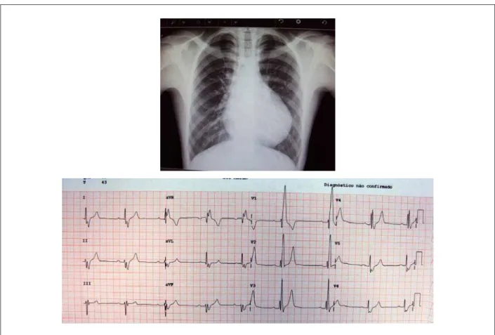
Electrocardiograms taken after surgery showed signs of trifascicular block, and complete right bundle branch, left anterosuperior and first degree atrioventricular division (PR: 0.22”), with QRS duration of 0.16 (Figure 1).
Chest radiographyshows marked increase in the cardiac area due to right chambers, excavated medial arch and increased pulmonary vascular markings (Figure 1).
Clinico Radiological Session
Edmar Atik
Arq Bras Cardiol 2011;96(5):e90-e91 Figure 1 -Chest X-ray in late postoperative period of tetralogy of Fallot shows moderate increase in right cavities, excavated medial arch and increased pulmonary vasculature, due to the presence of pulmonary valve insuficiency. Electrocardiogram revealed the usual indings of chronic volume overload of right ventricle, such as right bundle branch block, left anterosuperior divisional block and irst degree AV block.
implanted, with interposition of a pericardial patch in the right ventricle in 115 minutes of cardiopulmonary bypass, at 28o C.
The immediate outcome was good and uneventful. Heart murmurs disappeared and the patient was discharged on the fifth postoperative day.
Considerations
Pulmonary valve insufficiency is inevitable after correction of tetralogy of Fallot, especially when the narrow pulmonary annulus is to be widened. Therefore, in such cases, the outcome is more painful in view of progressive dilatation of the right heart chambers. This situation becomes serious when there is worsening of right ventricular function. Hence the need for surgical reintervention before the onset of irreversible alterations. Right ventricular dilatation at end-systole greater than 80 ml/m2 and diastole greater than 180
ml/m2, are accompanied by poor prognosis. From this data,
it is necessary to indicate earlier surgery, as occurred with the patient presented.
Figure 2 -MRI shows a sharp increase in right chambers on a two-chamber subcostal section in diastole in A, and in systole in B. There is a clear demonstration of good right ventricular function, despite the cavity dilation. Abbreviations: RV - right ventricle; LV - left ventricle.