Case Report
Abstract
In this report, we describe the case of a 64-year-old male patient, with no history of alcohol consumption, who presented the Osler’s triad, which is the association of endocarditis, pneumonia, and meningitis caused by a single agent. This syndrome is called Austrian syndrome, when the infection is caused by Streptococcus pneumoniae. We discuss the clinical manifestations, the pathophysiology, and the therapeutic approach to this condition. Given the rarity of the condition and its high morbidity and mortality, the importance of an early diagnosis and an appropriate treatment to reduce the complications associated with this disease will be emphasized.
Introduction
Pneumococcal endocarditis has become a rare disease since the introduction of penicillin in the early 40s, accounting for 1 to 3% of all cases in native valves. In recent years, however, a larger number of cases have been reported due to the emergence of penicillin-resistant Streptococcus pneumoniae strains¹. The association of pneumonia, meningitis, and endocarditis was described in 1862, by Herchl, following the autopsy of 5 patients. In 1881, Osler called it Austrian syndrome, when its etiology was Streptococcus pneumoniae²,³.
By 1882, Netter once more showed this clinical relationship, indicating a clear predisposition of the aortic valve towards the condition. In 1957, Robert Austrian reported a total of eight cases, of which 6 died: the fatal outcome of these patients was often found to be the rupture of the aortic valve4.
We report the case of a man who presented the Austrian syndrome, and who, even after having been diagnosed and
treated properly, developed the inherent complications of this rare condition.
Case Report
A farmer, male, 64 years old, was admitted to the emergency room of the HRMS with low fever, myalgia, arthralgia, and appetite loss for three weeks, which had evolved to productive cough, breathlessness, and impaired general condition in the last three days. No history of alcohol consumption, smoking habit, I.V. drug use, or previous diseases was reported. On admission, he was tachydyspneic, depleted, vomiting, confused, and agitated. His pupils were isocoric and normally reactive to light, and he had a negative plantar cutaneous reflex, no focal signs, and terminal neck stiffness. Lung auscultation detected crackling rales at right lung base. Cardiac auscultation revealed a grade 2/6 diastolic murmur in the mitral area, without irradiation, and a grade 3/6 aortic systolic murmur, with cervical irradiation. WBC showed 18,700 cells/mm³ and 6% of band cells. Chest x-ray showed alveolar opacity in the right lower lobe (Figure 1). The patient developed acute respiratory failure, requiring intubation and mechanical ventilation. A cranial computed tomography (CT) showed no changes, and CSF analysis from lumbar puncture revealed 78 cells/mm³ (87% polymorphonuclear leukocytes), low glucose concentration, and positive latex agglutination test and microscopic analysis for bacterial meningitis, confirming the diagnosis of pneumococcal meningitis.
The treatment was initiated with ceftriaxone, using an adequate dose for central nervous system infections, but the fever persisted. Blood, urine, and tracheal aspirate cultures were collected, and a transthoracic echocardiography (TTE) showed thickening and calcification of the aortic valve with stenosis and mild reflux, a mobile filament attached to the posterior mitral leaflet with mild mitral stenosis, a maximum aortic gradient of 17 mmHg, a mitral peak gradient of 9 mmHg and a mean gradient of 4 mmHg; left atrium: 36 mm; right ventricle: 20 mm; LVDD; 55 mm; LVSD: 30mm; and EF: 83%. Blood culture was positive for Streptococcus pneumoniae in two samples, and tracheal aspirate cultures were positive for the same agent. Vancomycin therapy was initiated, and ceftriaxone was replaced by meropenem with a favorable response, based on clinical and laboratory criteria.
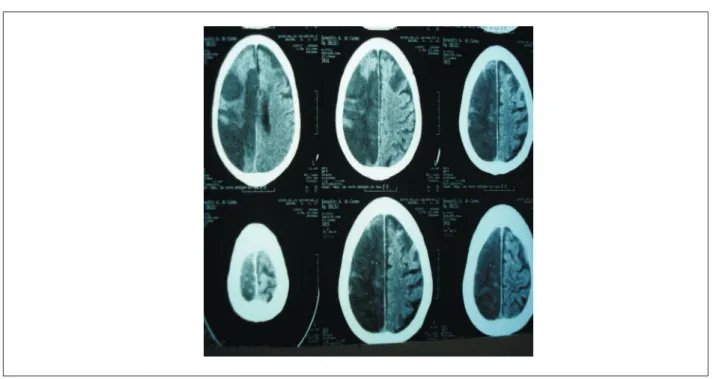
The patient was extubated after improvement in the breathing pattern, but presented a sudden left hemiparesis, with muscle strength grade II and sensory loss. A cranial CT
Palavras-chave
Austrian syndrome; endocarditis; streptococcus pneumoniae.
Austrian Syndrome
Márcio Estevão Midon, Fernando Goldoni, Sylvian Greicy Rocha Souza, Jan Norimitsu Schiemann Miyasato
Hospital Regional de Mato Grosso do Sul, Campo Grande, MS – Brazil
Mailing address: Fernando Goldoni •
Rua Flávio de Matos, 2462 - Vila Morumbi – 79051510 - Campo Grande, MS – Brasil
E-mail: [email protected]
Manuscript received November 26, 2009; revised manuscript received March 31, 2010; accepted May 25, 2010.
Case Report
Midon et als. Austrian Syndrome
(Arq Bras Cardiol 2011; 97(3) : e50-e52)
scan showed an extensive area of hypodensity in the right hemisphere (Figure 2). After 21 days of carbapenem therapy and 28 days of vancomycin therapy, the patient showed clinical and laboratory improvement, and was discharged from the hospital for home intensive physiotherapy and rehabilitation.
On a follow-up cardiac evaluation, one month after discharge, the patient remained stable, with no significant anatomic or functional heart abnormalities; only the neurological deficit persisted.
Discussion
The incidence of infective endocarditis by pneumococci was reduced to less than 3% after the use of penicillin. However, the mortality rate remains high and the incidence of pneumococcal resistance has increased worldwide over the past 10 years5.The predominant clinical presentation is acute, with a rapid and aggressive clinical course, associated with a high mortality rate. Peripheral signs and symptoms of endocarditis are often not present in the pneumococcus etiology, thus delaying diagnosis and treatment. In addition, cardiac and noncardiac complications are common, particularly hemodynamic instability, abscess formation, and systemic embolization1. Alcohol consumption and advanced age are major risk factors.
In pneumococcal endocarditis, the native aortic valve is the most frequent location of vegetations. Despite appropriate
Figure 1 - Chest X-ray showing alveolar opacity at right.
antibiotic therapy, the clinical course is usually acute and very aggressive, with a high rate of local complications (perivalvular abscesses) and systemic complications, requiring surgical treatment in most cases. A subacute clinical course is less frequent and often involves the mitral valve6. Respiratory tract infections are the main gateways to pneumococci. The aortic valve is predominantly affected.
Usually there are extensive vegetations, which lead to frequent septic embolization, as it occurred with the patient of this case report7. In a recent retrospective study on 80 cases of pneumococcal meningitis in an intensive care unit, only six patients developed endocarditis, and only two of these patients evolved to cardiogenic shock and death8. Likewise, in the largest cohort study on pneumococcal endocarditis (325 patients) recorded in literature, only three patients presented the triad9. However, Aronin et al2 reported an Osler’s triad prevalence of 42% in a review of pneumococcal endocarditis in the penicillin era, with a mortality rate greater than 50%.
A study conducted by Vuille et al10 showed that only 17% of the vegetations disappeared after antibiotic therapy, and 39% of cases eventually required corrective surgery, which was not necessary for this patient.
Currently, the choice of antibiotics for pneumococcal endocarditis has changed, not only because of the emergence of penicillin resistance, but also due to an aggressive clinical course and its association with meningitis.
Case Report
Midon et als. Austrian Syndrome
(Arq Bras Cardiol 2011; 97(3) : e50-e52)
References
1. Uemura L, Grassi NC, Cazarin L. Endocardite pneumocócica de evolução subaguda. Arq Bras Cardiol. 2001; 76 (4): 315-8.
2. Aronin SI, Mukherjee SK, West JC, Cooney EL. Review of pneumococcal endocarditis in adults in the penicillin era. Clin Infect Dis. 1998; 26 (1): 165-71.
3. Karchmer AW. Infective endocarditis. In: Braunwald E. Heart disease: a textbook of cardiovascular medicine. 5th ed. Philadelphia: WB Saunders
Company; 1997. p.1077-104.
4. Rubio JRS, Sánches MA, Domingues JCC, Moreno AR, Pavlovic D, Cortés FB, et al. Síndrome de Austrian (Endocarditis, meningitis y neumonía por Streptococcus pneumoniae): a propósito de un caso poco frecuente. Rev Esp Cardiol. 1998; 51 (12): 1006-8.
5. Campbell GD Jr, Silberman R. Drug-resistant Streptococcus pneumoniae. Clin Infect Dis. 1998; 26 (5): 1188-95.
6. du Cheyron D, Lesage A, Le Page O, Flais F, Leclercq R, Charbonneau P. Corticosteroids as adjunctive treatment in Austrian’s syndrome
(pneumococcal endocarditis, meningitis, and pneumonia): report of two cases and review of the literature. J Clin Pathol. 2003; 56 (11): 879-81.
7. Ugolini V, Pacifico A, Smitherman TC, Mackowiak PA. Pneumococcal endocarditis update: analysis of 10 cases diagnosed between 1974 and 1984. Am Heart J. 1986; 112 (4): 813s-9.
8. Auburtin M, Porcher R, Bruneel F, Scanvic A, Trouillet JL, Bedos JP, et al. Pneumococcal meningitis in the intensive care unit: prognostic factors of clinical outcome in a series of 80 cases. Am J Respir Crit Care Med. 2002; 165 (5): 713-7.
9. Gransden WR, Eykyn SJ, Phillips I. Pneumococcal bacteraemia: 325 episodes diagnosed at St Thomas’s Hospital. BMJ (Clin Res Ed). 1985; 290 (6467): 505-8.
10. Vuille C, Nidorf M, Weyman AE, Picard MH. Natural history of vegetations during successful medical tretment of endocarditis. Am Heart J. 1994; 128 (6 pt 1): 1200-9.
Figure 2 - Cranial CT showing hypodensity on the right side.
Penicillin is not the antibiotic of choice, and although there is no formal recommendation at this point, the orientation is the same as for pneumococcal meningitis, i.e., the treatment should be initiated with a third generation cephalosporin with or without vancomycin, depending on the level of cephalosporin resistance in the studied region2,3,7. Despite the low incidence of pneumococcal endocarditis, this condition presents high morbidity and mortality, especially in the rare cases in which it is associated with pneumonia and meningitis. Therefore, early identification and treatment are crucial in order to change the negative outcome.
Potential Conflict of Interest
No potential conflict of interest relevant to this article was reported.
Sources of Funding
There were no external funding sources for this study.
Study Association
This study is not associated with any post-graduation program.