Arq Neuropsiquiatr 2011;69(2-A):237-241
Role of IL-4 in an experimental
model of encephalitis induced
by intracranial inoculation of
herpes simplex virus-1 (HSV-1)
Márcia Carvalho Vilela1, Roberta Dayrell de Lima Campos1,
Daniel Santos Mansur1, David Henrique Rodrigues1,
Norinne Lacerda-Queiroz1, Graciela Kunrath Lima2,
Milene Alvarenga Rachid1, Erna Geessien Kroon2,
Marco Antônio Campos3, Antônio Lúcio Teixeira1
ABSTRACT
Herpes simplex virus-1 (HSV-1) is a pathogen that may cause severe encephalitis in humans. In this study, we aimed to investigate the role of interleukin-4 (IL-4) in a model of HSV-1 brain infection. IL-4 knockout (IL-4–/–) and wild type (WT) C57BL/6 mice were inoculated with 104 plaque-forming units of HSV-1 by the intracranial route. Histopathologic analysis revealed a distinct profile of infiltrating cells at 3 days post-infection (dpi). Infected WT mice presented mononuclear inflammatory cells while IL-4–/– mice developed meningoencephalitis with predominance of neutrophils. IL-4–/– mice had diminished leukocyte adhesion at 3 dpi when compared to infected WT animals in intravital microscopy study. Conversely no differences were found in cerebral levels of CXCL1, CXCL9, CCL3, CCL5 and TNF-α between WT and IL-4–/– infected mice. IL-4 may play a role in the recruitment of cells into central nervous system in this acute model of severe encephalitis caused by HSV-1.
Key words: herpes simplex virus type 1, IL-4, neuroinflammation.
Papel da IL-4 em modelo experimental de encefalite induzida pela inoculação intracraniana do herpes simplex vírus-1 (HSV-1)
RESUMO
O vírus herpes simplex-1 (HSV-1) é um patógeno que pode causar encefalite grave em humanos. Neste estudo, buscamos investigar o papel da interleucina-4 (IL-4) no modelo de infecção intracerebral por HSV-1. Camundongos C57BL/6 selvagens (WT) e deficientes no gene IL-4 (IL-4–/–) foram inoculados com 104 unidades formadoras de placas de HSV-1 por via intracraniana. A análise histopatológica revelou um padrão distinto de infiltrado leucocitário. Camundongos WT infectados apresentaram infiltrado de células mononucleares, enquanto camundongos IL-4–/– desenvolveram meningoencefalite com predomínio de neutrófilos 3 dias pós-infecção (dpi). Animais IL-4–/– tiveram menor adesão de leucócitos 3 dpi quando comparados aos animais WT infectados à microscopia intravital. Em contrapartida, não foram encontradas diferenças nos níveis cerebrais de CXCL1, CXCL9, CCL3, CCL5 e TNF-α entre camundongos WT e IL-4–/– infectados. Esses resultados sugerem que IL-4 pode desempenhar um papel no recrutamento de células no sistema nervoso central neste modelo agudo de encefalite grave causada pelo HSV-1.
Palavras-chave: vírus herpes simplex tipo 1, IL-4, neuroinflamação.
Correspondence
Antônio Lúcio Teixeira
Laboratório de Imunofarmacologia Depto. de Bioquímica e Imunologia Instituto de Ciências Biológicas (UFMG) Av. Antônio Carlos, 6627
31270-901 Belo Horizonte MG - Brasil E-mail: altexr@gmail.com
Support
This work was supported by Conselho Nacional de Desenvolvimento Científico e Tecnológico (CNPq) and Fundação de Amparo à Pesquisa do Estado de Minas Gerais (Fapemig), Brazil
Received 22 June 2010
Received in final form 13 October 2010 Accepted 20 October 2010
1Laboratório de Imunofarmacologia, Departamento de Bioquímica e Imunologia, Instituto de Ciências Biológicas (ICB), UFMG,
Belo Horizonte MG, Brazil; 2Departamento de Microbiologia, ICB, UFMG; 3Centro de Pesquisas René Rachou, Belo Horizonte
Interleukin-4 (IL-4) is a pleiotropic cytokine synthe-sized primarily by CD4+ T lymphocytes in response to
their activation1,2. Studies have reported that IL-4 may
have either detrimental or protective effects during viral infection3-6. For instance, the expression of IL-4 by
mousepox virus, due to the insertion of a copy of mouse IL-4 cDNA in viral genome, turns this virus lethal to micethat are usually resistant to the infection3. Similarly,
the expression of IL-4 by myxoma virus enhances viru-lence and overcomes genetic resistance of rabbits to viral infection4. IL-4 knockout (IL-4–/–) mice challenged with
herpes simplex virus type 1 (HSV-1) by ocular route had reduced virus load in their eyes when compared with wild type (WT) mice5. Conversely one study
demon-strated that a recombinant HSV-1 expressing IL-4 had a great decrease in its pathogenic potential6.
HSV-1 is a neurotropic virus known to cause infec-tion in the central nervous system (CNS). Herpes sim-plex encephalitis is a common sporadic viral disease of the brain7,8. Although antiviral treatment has greatly
re-duced mortality due to herpeticencephalitis,the majority of survivors presents residual neuropsychological deicits and/or neuropsychiatric symptoms9. Our group has
de-veloped an experimental model of severe HSV-1 enceph-alitis10-12. We observed an increase in the levels of rolling
and adhered leukocytes in meningeal vessels of infected mice in parallel with the increase of the expression of several cytokines in the CNS12. Nonetheless, the role of
IL-4 on the early inlammatory response to HSV-1 brain infection has not been investigated yet.
In the present study we aimed to investigate the pos-sible involvement of IL-4 in the inlammatory response to HSV-1, assessing the recruitment of leukocytes by in-travital microscopy, the chemokine and cytokine pro-ile and the histopathological changes in IL-4–/– and WT
mice infected with an intracerebral inoculum of HSV-1.
METHOD Mouse strains
Male C57BL/6 mice and IL-4–/– mice on a C57BL/6
background, aged 6-9 weeks, were obtained from An-imal Care Facilities of the Institute of Biological Sciences (ICB), Federal University of Minas Gerais (UFMG). All experiments were approved by the Animal Ethics Com-mittee of UFMG.
Virus
HSV-1 strain EK13 was allowed to multiply in Vero
cells and was maintained with minimal essential me-dium (GIBCO, Grand Island, NY) containing 5% fetal bovine serum (FBS) (GIBCO) and 25 μg/μL of ciprolox-acin (Cellofarm, Carapina, ES, Brazil) at 37ºC in 5% CO2.
Virus was puriied in sucrose gradient and the titers
de-termined in Vero cells as previously described14,15. he
virus titers obtained were 1.1×10⁸ plaque-forming cells
(PFU)/mL for HSV-1.
Vero cells
Vero cells were maintained in minimal essential me-dium (GIBCO) supplemented with 5% heat-inactived FBS and antibiotics in 5% CO2 at 37ºC. hese cells were
used for virus multiplication.
Infection with HSV-1
Mice were anesthetized by intraperitoneal injection of a mixture of ketamine (150 mg/kg) and xylazine (10 mg/kg). A 104 plaque-forming units (PFU) inoculum of
HSV-1 resuspended in 10 μL of phosphate-bufered sa-line (PBS) was injected intracranially in the right side of sagittal suture at the level of the eye10-12. Control mice
received PBS.
Intravital microscopy
At 1 and 3 days post-infection (dpi) intravital mi-croscopy of the mouse brain microvasculature was per-formed12,16,17. Briely, WT injected with PBS (control),
infected WT and infected IL-4–/– mice (n=4 for each
group) were anesthetized by intraperitoneal injection and the tail vein was cannulated for administration of luorescent dyes. A craniotomy was performed using a high-speed drill and the dura mater was removed to ex-pose the underlying pial vasculature. Throughout the experiment, the mouse was maintained at 37ºC with a heating pad and the exposed brain was continuously su-perfused with artiicial cerebrospinal luid bufer.
Leukocytes were luorescently labeled by intravenous administration of Rhodamine 6G-Sigma (0.5 mg/kg body weight) and were observed using a microscope (Olympus B201, 10× objective lens, corresponding to 100 µm of area) outitted with a luorescent light source (epi-illu-mination at 510-560nm, using a 590 nm emission ilter). he number of rolling and adherent leukocytes was deter-mined oline during video playback analyses. Leukocytes were considered adherent to the venular endothelium if they remained stationary for a minimum of 30s. Rolling leukocytes were deined as white cells moving at a velocity lower than that of erythrocytes within a given vessel.
Enzyme-linked immunosorbent assay (ELISA) for cytokines in the CNS
Brains from control and infected WT and IL-4–/–
homogenate was centrifuged at 3000g for 10 min at 4ºC, and the supernatants were collected and stored at −20ºC. The concentration of chemokines CXCL1, CXCL9, CCL3 and CCL5 and cytokine TNF-α was determined using ELISA. he supernatants of brain tissue were as-sayed in an ELISA setup using commercially available antibodies, according to the procedures provided by the manufacturer (R&D Systems, Minneapolis, MN).
Histopathology
For histological analysis, the right fragment of the brain was preserved in 10% bufered formalin (infected WT n=5, infected IL-4-/- n=4). Sections 5 µm thick were
cut and mounted for routine haematoxylin and eosin staining. hese sections were examined at the optical level in the Olympus microscopy. Digital images were acquired for documentation.
Statistical analysis
Data are shown as mean±standard error. A one-way ANOVA with Tukey’s correction was used for multiple comparisons. Statistical signiicance was set at p<0.05.
RESULTS
HSV-1 infection induced milder meningitis in IL-4–/– mice
WT animals infected with 104 PFU showed
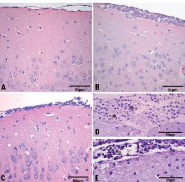
progres-sive inlammatory iniltrate in the meninges composed of neutrophils, lymphocytes and macrophages (Fig 1A). At day 3, the inlammation was more widespread and composed mainly of mononuclear cells. Inlammatory iniltrates were observed at the meninges (Fig 1B) and around some small cerebral blood vessels. Focal cere-bral degenerative changes were also visualized adjacent to these inlamed areas.
Brains of IL-4–/– infected animals presented mild to
moderate meningitis. At day 1, lymphocytes, macro-phages and rare neutrophils were detected in the me-ninges (Fig 1C). At day 3, only leptomeme-ninges were focally iniltrated by neutrophils and occasional mono-nuclear cells (Fig 1D). he inlammation was restricted to the meninges and no degenerative changes were de-tected in the brain from IL-4–/– mice. Histological
sec-tions obtained from brains of control animals showed no alteration.
IL-4–/– infected mice had a diminished adherence
of leukocytes to meningeal vessels at day 3 p.i. Both infected groups (WT and IL-4–/–) presented
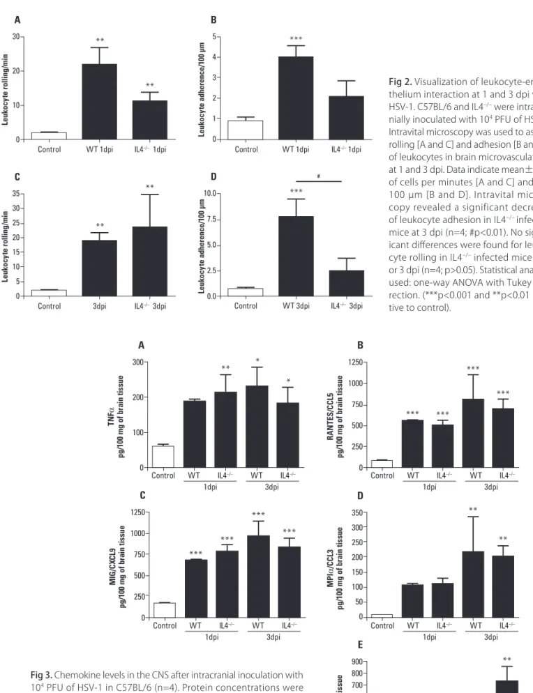
in-crease of leukocyte rolling and adhesion in meningeal vessels when compared with control group. In compar-ison with infected WT mice, lower leukocyte adhesion was observed in IL4–/– infected mice at 3 dpi (Fig 2).
Absence of IL-4 did not alter chemokines and cytokines levels in the brain
tissue of HSV-1 infected mice
Brain tissue extracts were obtained from control an-imals, infected WT and IL-4–/– mice to evaluate the
ex-pression of chemokines (CXCL1, CXCL9, CCL3 and CCL5) and cytokine (TNF-α) by ELISA. WT and IL-4–/– infected mice presented signiicant increase in these
molecules levels relative to controls at 3 dpi. However, there were no signiicant diferences in cytokines or che-mokines levels in brain tissue of infected IL-4–/– mice
when compared to the infected WT group (Fig 3).
DISCUSSION
he present study investigated the role of the cyto-kine IL-4 in the inflammatory response to HSV-1 ce-rebral infection. We found that the absence of IL-4 in-terferes in the response to HSV-1 infection in mice. Histopathological analysis revealed a delayed cellular in-iltration and predominance of distinct leukocyte types at 3 dpi. With the progression of the disease, IL-4–/–
mice showed diminished leukocyte adhesion, one rele-vant step for cellular recruitment into inlamed tissues. By contrast, there was no alteration in the levels of some chemokines and the cytokine TNF-α in brain tissue from IL4–/– when compared to controls.
Our histological analysis revealed diferences between cellular iniltrates of WT and IL-4–/– mice. A model of
Fig 2. Visualization of leukocyte-endo-thelium interaction at 1 and 3 dpi with HSV-1. C57BL/6 and IL4–/– were intracra-nially inoculated with 104 PFU of HSV-1. Intravital microscopy was used to assess rolling [A and C] and adhesion [B and D] of leukocytes in brain microvasculature, at 1 and 3 dpi. Data indicate mean±SEM of cells per minutes [A and C] and per 100 µm [B and D]. Intravital micros-copy revealed a significant decrease of leukocyte adhesion in IL4–/– infected mice at 3 dpi (n=4; #p<0.01). No signif-icant diferences were found for leuko-cyte rolling in IL4–/– infected mice at 1 or 3 dpi (n=4; p>0.05). Statistical analysis used: one-way ANOVA with Tukey cor-rection. (***p<0.001 and **p<0.01 rela-tive to control).
30 20 10 0 Leuk ocy te ro ll ing/min A 35 30 25 20 15 10 5 0 Leuk ocy te ro ll ing/min C 5 4 3 2 1 0 Leuk ocy te a dh e re nc e/100 µm B
Control WT 1dpi IL4–/– 1dpi
Control 3dpi IL4–/– 3dpi
10.0 7.5 5.0 2.5 0.0 Leuk ocy te a dh e re nc e/100 µm D
Control WT 3dpi IL4–/– 3dpi
Control WT 1dpi IL4–/– 1dpi
300 200 100 0 T N F α
pg/100 mg of b
ra
in ti
ssu
e
A
Control WT IL4–/–
1dpi 3dpi WT IL4–/–
1250 1000 750 500 250 0 R AN T E S/CCL5
pg/100 mg of b
ra
in ti
ssu
e
B
Control WT IL4–/–
1dpi 3dpi WT IL4–/–
1250 1000 750 500 250 0 M IG/CX CL9
pg/100 mg of b
ra
in ti
ssu
e
C
Control WT IL4–/–
1dpi 3dpi WT IL4–/–
350 300 250 200 150 100 50 0 M PI α /CCL3
pg/100 mg of b
ra
in ti
ssu
e
D
Control WT IL4–/–
1dpi 3dpi WT IL4–/–
900 800 700 600 500 400 300 200 100 0 Kc/CX CL1
pg/100 mg of b
ra
in ti
ssu
e
E
Control WT IL4–/–
1dpi 3dpi WT IL4–/– Fig 3. Chemokine levels in the CNS after intracranial inoculation with
HSV-1 corneal infection in mice revealed an elevated expression of IL-4 and IL-10 associated with massive CD8+ T cell iniltration in trigeminal ganglion, but not
in the cornea where inlammatory iniltrate was mainly composed of polymorphonuclear leukocytes. herefore, these authors have suggested that IL-4 and IL-10 limit the iniltration of polymorphonuclear leukocytes and the destruction of neural tissues in this model18. In line with
this, our results showed that when IL-4 is absent, the iniltration of neutrophils is delayed. Rare neutrophils were observed at 1 dpi in IL-4–/– mice brains. In contrast,
they were the predominant cell type in WT infected mice at this same time point. At 3 dpi, neutrophils prevailed amongst cellular infiltrate in IL-4-/- mice whereas
in-fected WT animals presented mainly mononuclear cells. herefore the expression of IL-4 may prevent early inil-tration of neutrophils in the brain after HSV-1 infection. he migration of leukocytes from the vessels to brain parenchyma can be caused by the presence of a pathogen at this site and is composed of a series of events. Leuko-cytes must tether and roll along the venular wall before they can attach irmly and emigrate from the vasculature. hese processes depend on multiple proteins interac-tions among leukocytes and vascular endothelial cells19-21.
Our results revealed that infected IL-4–/– mice
showed diminished leukocyte adhesion at 3 dpi when compared to infected WT mice. Previous studies have demonstrated that IL-4 regulates expression of adhesion molecules, with an upregulatory efect on vascular cell adhesion molecule-1 (VCAM-1)22,23 and possibly on
P-selectin24,25 and a downregulatory efect on E-selectin26,27.
It has also been reported that IL-4 can act synergistically with other cytokines, such as IL-1β and TNF-α, in order to promote lymphocyte binding to cultured microvas-cular endothelial cells28,29. It is possible that the absence
of IL-4 modiies the proile of adhesion molecules ex-pressed during infection causing the changes in leukocyte adhesion and cellular iniltrates observed in this study.
In conclusion, the lack of IL-4 gene in mice was as-sociated with a delayed cellular infiltration and pre-dominance of neutrophils. Overall leukocyte adhesion to brain microvasculature was diminished in the third day after infection. However, the lack of IL-4 did not abolish nor exacerbated the inlammatory response in mice brains. he results suggest that IL-4 plays a role in an acute model of severe encephalitis caused by HSV-1. Further studies are necessary to deine the involvement of IL-4 in the outcome of the disease.
REFERENCES
1. Paul WE. Interleukin-4: a prototypic immunoregulatory lymphokine. Blood 1991;77:1859-1870.
2. Seder RA, Paul WE. Acquisition of lymphokine-producing phenotype by CD4+ T-cells. Ann Rev Immunol 1994;12:635-673.
3. Jackson RJ, Ramsay AJ, Christensen CD, Beaton S, Hall DF, Ramshaw IA. Ex-pression of mouse interleukin-4 by a recombinant ectromelia virus sup-presses cytolytic lymphocyte responses and overcomes genetic resis-tance to mousepox. J Virol 2001;75:1205-1210.
4. Kerr PJ, Perkins HD, Inglis B, et al. Expression of rabbit IL-4 by recombinant myxoma viruses enhances virulence and overcomes genetic resistance to myxomatosis. Virology 2004;324:117-128.
5. Ghiasi H, Cai S, Slanina SM, Perng GC, Nesburn AB, Wechsler SL. The role of interleukin (IL)-2 and IL-4 in herpes simplex virus type 1 ocular replica-tion and eye disease. J Infect Dis 1999;179:1086-1093.
6. Ghiasi H, Osorio Y, Perng GC, Nesburn AB, Wechsler SL. Recombinant herpes simplex virus type 1 expressing murine interleukin-4 is less viru-lent than wild-type virus in mice. J Virol 2001;75:9029-9036.
7. Domingues RB, Teixeira AL. Management of acute viral encephalitis in Brazil. Braz J Infect Dis 2009;13:433-439.
8. Whitley RJ, Roizman B. Herpes simplex virus infections. Lancet 2001;357: 1513-1518.
9. McGrath N, Anderson NE, Croxson MC, Powell KF. Herpes simplex enceph-alitis treated with acyclovir: diagnosis and long term outcome. J Neurol Neurosurg Psychiatry 1997;63:321-326.
10. Vilela MC, Lima GK, Rodrigues DH, et al. TNFR1 plays a critical role in the control of severe HSV-1 encephalitis. Neurosci Lett 2010;479:58-62. 11. Vilela MC, Mansur DS, Lacerda-Queiroz N, et al. The chemokine CCL5 is
essential for leukocyte recruitment in a model of severe herpes simplex encephalitis. Ann N Y Acad Sci 2009;1153:256-263.
12. Vilela MC, Mansur DS, Lacerda-Queiroz N, et al. Traic of leukocytes in the central nervous system is associated with chemokine up-regulation in a severe model of herpes simplex encephalitis: an intravital microscopy study. Neurosci Lett 2008;445:18-22.
13. Nogueira ML, Siqueira RC, Freitas N, et al. Detection of herpesvirus DNA by the polymerase chain reaction (PCR) in the vitreos samples from patients with necrotizing retinitis. J Clin Pathol 2001;54:103-106.
14. Campos MA, Kroon EG. Critical period of irreversible block of vaccinia virus replication. Rev Microbiol 1993;24:104-110.
15. Joklik WK. The puriication of four strains of poxvirus. Virology 1962;18:9-18. 16. Lacerda-Queiroz N, Rodrigues DH, Vilela MC, et al. Inlammatory changes
in the central nervous system are associated with behavioral impairment in Plasmodium berghei (strain ANKA)-infected mice. Exp Parasitol 2010; 125:271-278.
17. Rodrigues DH, Vilela MC, Barcelos LS, Pinho V, Teixeira MM, Teixeira AL. Ab-sence of PI3K[gamma] leads to increased leukocyte apoptosis and dimin-ished severity of experimental autoimmune encephalomyelitis. J Neuro-immunol 2010;222:90-94.
18. Liu T, Tang Q, Hendricks RL. Inlammatory iniltration of the trigeminal ganglion after herpes simplex virus type 1 corneal infection. J Virol 1996; 70:264-271.
19. Ley K. Molecular mechanisms of leukocyte recruitment in the inlamma-tory process. Cardiovasc Res 1996;32:733-742.
20. Kubes P, Ward PA. Leukocyte recruitment and the acute inlammatory re-sponse. Brain Pathol 2000;10:127-135.
21. Kerfoot SM, Kubes P. Overlapping roles of P-selectin and alpha 4 integrin to recruit leukocytes to the central nervous system in experimental auto-immune encephalomyelitis. J Immunol 2002;69:1000-1006.
22. Lampinen M, Carlson M, Hakansson LD, Venge P. Cytokine-regulated accu-mulation of eosinophils in inlammatory disease. Allergy 2004;59:793-805. 23. Schleimer RP, Sterbinsky SA, Kaiser J, et al. IL-4 induces adherence of
human eosinophils and basophils but not neutrophils to endothelium. Association with expression of VCAM-1. J Immunol 1992;148:1086-1092. 24. Yao L, Pan J, Setiadi H, Patel KD, McEver RP. Interleukin 4 or oncostatin M
induces a prolonged increase in P-selectin mRNA and protein in human endothelial cells. J Exp Med 1996;184:81-92.
25. Inomata M, Into T, Nakashima M, Noguchi T, Matsushita K. IL-4 alters ex-pression patterns of storage components of vascular endothelial cell-spe-ciic granules through STAT6- and SOCS-1-dependent mechanisms. Mol Immunol 2009;46:2080-2089.
26. Lee YW, Eum SY, Chen KC, Hennig B, Toborek M. Gene expression proile in interleukin-4-stimulated human vascular endothelial cells. Mol Med 2004;10:19-27.
27. Huang H, Lavoie-Lamoureux A, Moran K, Lavoie JP. IL-4 stimulates the ex-pression of CXCL-8, E-selectin, VEGF, and inducible nitric oxide synthase mRNA by equine pulmonary artery endothelial cells. Am J Physiol Lung Cell Mol Physiol 2007;292:1147-1154.
28. Masinovsky B, Urdal D, Gallatin WM. IL-4 acts synergistically with IL-1 beta to promote lymphocyte adhesion to microvascular endothelium by induction of vascular cell adhesion molecule-1. J Immunol 1990;145: 2886-2895.