Short communication
ISSN 0102-695X
http://dx.doi.org/10.1590/S0102-695X2012005000040 Received 2 Sep 2011 Accepted 23 Jan 2012 Available online 3 Apr 2012
plants:
Piper renitens, Siparuna guianensis
and
Alternanthera brasiliana
Valdir A. Facundo,
*,1Mariangela S. Azevedo,
1Rosely V.
Rodrigues,
1Leandro F. do Nascimento,
1Júlio S. L. T. Militão,
1Gil V. J. da Silva,
2Raimundo Braz-Filho
31Departamento de Química, Universidade Federal de Rondônia, Campus UNIR,
Brazil,
2Departamento de Química, Faculdade de Filosofia, Ciências e Letras de
Ribeirão Preto, Universidade de São Paulo, Brazil, 3Pesquisador visitante emérito-FAPERJ-UENF-UFRRJ.
Abstract:Chemical study of three medicinal plants: from leaves of Piper renitens
(Miq.) Yunck, Piperaceae, and Siparuna guianensis Aubl., Siparunaceae, and from flowers of Alternanthera brasiliana (L.) Kuntze, Amaranthaceae, resulted in isolation of nine compounds: three steroids, β-sitosterol, stigmasterol from P. renitens and sitosterol-3-O-β-D-glucopyranoside from A. brasiliana, the diterpene kaurane ent-kauran-16α,17-diol from P. renitens, two derivatives kaempferol-methylether, kumatakenine (kaempferol-3,7-dimethylether) and kaempferol-3,7,3’-trimethylether from S. guianensis and three flavones, crysoeriol
(5,7,4’-trihydroxy-3’-methoxyflavone), tricin (5,7,4’-trihydroxy-3’,5’-dimethoxyflavone) and 7-O-β-D-glucopyranoside-5,4’-dihydroxy-3’-methoxyflavone from A. brasiliana.
Compounds structures were determinate using 1D and 2D 1H NMR and 13C spectral
data, mass and IR spectra, comparing with literature data. Keywords:
Piper renitens Siparuna guianensis Alternanthera brasiliana ent-kaunare diterpene kaempferol-methylether flavonoids
Introduction
Plants utilization in treatment, cure and prevention of any kind of disease is a traditional practice in the entire world. The necessity of knowledge from chemical constituents of plants becomes important as many pharmaceutical products that exist today were developed from substances of that origin (Newman et al., 2003).
Piper genus is included as one of the most important in Piperaceae family and is well distributed in tropical and sub-tropical regions of the planet. Chemical and pharmaceutical studies of this genus are related to the isolation of various secondary metabolites classes
(amides, l avonoids, lignans, terpenes, propenylphenols,
alkaloids and cyclopetenediones derivatives) and to its
use in treatment of some diseases (Parmar et al., 1997;
Facundo et al., 2003). In Mexico and Brazil, leaves from P. amalago are used to treat stomachache and some infections. Leaves and stems from P. marginatum and P. tuberculatum are used against snakebite and as sedatives
(Chaves et al., 2006; Araújo-Junior et al., 1999). P.
renitens (Miq.) Yunck is known by the Amazonian rainforest natives as “bararupyangihua” and tea from leaves is used mainly to cure fever, infant diarrhea and
skin inl ammations (Ribeiro et al., 1999). Chemical
volatile constituents from their leaves were related this
specie (Soleane et al., 2007).
Siparuna (formerly Monimiaceae) contains
about 72 species of shrubs, straggling shrubs, and trees
(Renner et al. 1997). S. guianensis Aubl., Siparunaceae,
is a small tree found in various parts of Brazilian forests, popularly known as “capituí”, “negramena”, “erva-santa”, “limão-bravo” and others (Zoghbi et al., 1998). Several species of this genus are used in traditional medicine to handling stomach disorders and skin diseases (Leitão et al., 1999). Literature reports the use of S. guianensis leaves in popular medicine to cure various diseases, including, sinusitis, fever, rheumatism, migraine, body
aches and as anti-inl ammatory (Valentini et al., 2010;
Rodrigues, 2006). In state of Rondônia, Brazil (an oriental Amazonian state), there are some reports of using leaves tea in treatment of malaria and against rheumatic pains. Chemical studies had revealed presence of volatile
compounds, alkaloids and l avonoids (Braz-Filho et al., 1976; Chiu et al., 1982; Machado et al., 1994).
treat several pathologies, including coughing, diarrhea
and for its anti-inlammatory and analgesic properties (Delaporte et al., 2005; Araújo & Onofre, 2011). In
state of Rondônia, Brazil, tea from leaf is used for its
anti-inlammatory properties. Chemical study of the
leaves of A. brasiliana had revealed the presence of six di- and triglycosyl kaempferol derivatives and quercetin (Brochado et al., 2003).
This work describes the isolation and structural characterization of the nine compounds from three
medicinal plants popularly used as anti-inlammatory: β-sitosterol 1 and stigmasterol 2 from leaves of P. renitens, sitosterol-3-O-β-D-glucopyranoside 9 from lowers of A. brasiliana, ent-kauran-16α,17-diol 3 from leaves of P. renitens, kumatakenine (kaempferol-3,7-dimethylether)
4 and kaempferol-3,7,4’-trimethylether 5, from leaves
of S. guianensis and crysoeriol
(5,7,4’-trihydroxy-3’-methoxylavone) 6, tricin
(5,7,4’-trihydroxy-3’,5’-dimethoxylavone) 7 and 7-O-β-D
-glucopyranoside-5,4’-dihydroxy-3’-methoxylavone 8, from lowers
of A. brasiliana. The structures were established by spectroscopic techniques, mainly EIMS and 1D and 2D
NMR. This is the irst report of the occurrence of these
compounds in these plants.
Materials and Methods
Melting points were obtained on a Mettler FP82HT apparatus and are uncorrected. IR spectra were recorded using a Perkin Elmer 1000 FT-IR spectrophotometer. The mass spectra were obtained on
a Hewlett-Packard 5971 mass spectrometer by electron
impact ionization (70 eV). 1H and 13C NMR where
recorded on a Bruker Avance DRX-500 (500 MHz for 1H and 125 MHz for 13C); Silica gel 60 (Merck, kiesegel 60 F254, 0.20 mm) were used for analytical TLC. Silica gel 60 (Merck, 230-240 mesh) was used for column chromatography. All compounds were visualized on TLC by spraying with AlCl3/EtOH and vanillin/perchloric acid/EtOH followed by heating.
Leaves from Siparuna guianensis Aubl., Siparunaceae, and Piper renitens (Miq.) Yunck, Piperaceae, were collected in Mirante da Serra County, Rondonia State, Brazil on April, 2010, and May,
2009, respectively. The plant material was classiied
systematically by the botanist Dr. J. Gomes from Instituto Nacional de Pesquisa da Amazônia herbarium where voucher specimens were deposited: 216220 for S. guianensis and 213523 for P. renitens. Leaves from P. renitens (1.1 kg) and S. guianensis (1.8 kg) were dried, triturated and submitted to ethanol extraction at room temperature in portions of 2 and 3 L, respectively, for three times. After solvent distillation at reduced pressure, 26.0 g and 41.0 g, respectively, of extracts were obtained. A portion of 22.0 g of the ethanol extract from P. renitens
was adsorbed in silica gel (100.3 g) and eluted under reduced pressure in a chromatographic column, using successively hexane, chloroform, ethyl acetate and methanol as solvents.
Chloroform fraction was treated with MeOH:H2O (90:10) and kept under refrigeration overnight to induce chlorophyll separation. This material was dried and re-chromatographed on a silica gel column and eluting with mixtures of hexane and chloroform of increasing polarity,
obtaining a mixture of β-sitosterol 1 and of stigmasterol 2
[32.3 mg (hexane/chloroform 40:60), 13C NMR (CDCl 3)
data, see Jácome et al., 2004] and ent-kauran-16α,17-diol
3 [22.1 mg (hexane/chloroform 50:50), 187-188 oC, 13C
NMR (CDCl3) data, see Nascimento & Lopes, 2003]. Similarly, a portion of 35.5 g of the ethanol extract from S. guianensis were adsorbed by silica gel (90.0 g) and eluted under reduced pressure in a chromatographic column, using successively hexane, chloroform, ethyl acetate and methanol as solvents. Chromatography on a silica gel column of the chloroform fraction (9.3 g) eluting with mixtures of hexane and chloroform of increasing polarity yielded 23 sub-fractions. The subfractions 32 to 54 were combined and re-chromatographed on a silica gel column and eluted with hexane and ethyl acetate mixture with increasing polarity yielded kumatakenin
(kaempferol-3,7-dimethylether) 4 [31.8 mg (hexane/ethyl acetate
60:40), 243-245 oC, 13C NMR (CDCl
3) data, see table 1,
Silva et al., 2009] and kaempferol-3,7,4’-trimethylether 5 [21.5 mg (hexane/ethyl acetate 55:45), 146-148 oC, 13C NMR (CDCl3) data, see table 1, Paula et al., 2006].
Flowers from Alternanthera brasiliana (L.)
Kuntze, Amaranthaceae, were collected in Porto Velho,
Rondônia State, Brazil on September 2008. The plant
material was classiied systematically by the botanist
Dra. Ana Cristina R. de Souza from Dr. Ary Tupinamba Pena Pinheiro Herbarium in Faculdade Sao Lucas (Porto
Velho, Rondonia State, Brazil) where may be found a voucher specie identiied by 445801 descriptor.
Flowers (0.8 kg) from A. brasiliana, were dried, triturated and submitted to ethanol extraction at room temperature in portions of 1.5 L, in three times. After solvent distillation at reduced pressure, 21.3 g of extract were obtained. All the extract were absorbed by silica gel and eluted in a chromatography column with chloroform and ethyl acetate as binary mixtures with increasing polarity yielded 104 sub-fractions. The subfraction 9
(1.2 g chloroform/ethyl acetate 90:10) yielded a solid residue which after recrystallization from acetone yielded
crysoeriol (5,7,4’-trihydroxy-3’-methoxylavone) 6
[21.9 mg, 324-326 oC, 13C NMR (DMSO-d
6) data, see
table 1, Awaad et al., 2006]. The subfractions 17 to 34
(5,7,4’-trihydroxy-3’,5’-dimethoxylavone) 7 [23.1 mg
(chloroform/ethyl acetate 82:18), 278-280 oC, 13C NMR
(DMSO-d6) data, see table 1, Jiao et al., 2007] and 7-
O-β-D-glucopyranoside-5,4’-dihydroxy-3’-methoxylavone
8 [17.4 mg (chloroform/ethyl acetate 40:60), 176-178
oC, 13C NMR (DMSO-d
6) data, see table 1, Markham
& Moore, 1980]. The subfraction 56 (chloroform/
ethyl acetate 90:10) yielded a solid residue which after recrystallization from methanol yielded
sitosterol-3-O-β-D-glucopyranoside 9 [34.2 mg, 232-235 oC, 13C NMR (DMSO-d6) data, see table 1, Macari et al., 1990].
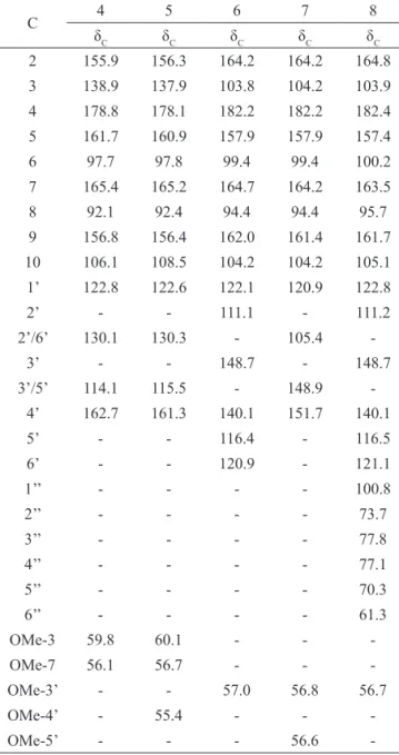
Table 1. 13C-NMR spectral data for compounds 4-8.
C 4 5 6 7 8
δC δC δC δC δC
2 155.9 156.3 164.2 164.2 164.8
3 138.9 137.9 103.8 104.2 103.9
4 178.8 178.1 182.2 182.2 182.4
5 161.7 160.9 157.9 157.9 157.4
6 97.7 97.8 99.4 99.4 100.2
7 165.4 165.2 164.7 164.2 163.5
8 92.1 92.4 94.4 94.4 95.7
9 156.8 156.4 162.0 161.4 161.7
10 106.1 108.5 104.2 104.2 105.1
1’ 122.8 122.6 122.1 120.9 122.8
2’ - - 111.1 - 111.2
2’/6’ 130.1 130.3 - 105.4
-3’ - - 148.7 - 148.7
3’/5’ 114.1 115.5 - 148.9 -4’ 162.7 161.3 140.1 151.7 140.1
5’ - - 116.4 - 116.5
6’ - - 120.9 - 121.1
1’’ - - - - 100.8
2’’ - - - - 73.7
3’’ - - - - 77.8
4’’ - - - - 77.1
5’’ - - - - 70.3
6’’ - - - - 61.3
OMe-3 59.8 60.1 - -
-OMe-7 56.1 56.7 - -
-OMe-3’ - - 57.0 56.8 56.7
OMe-4’ - 55.4 - -
-OMe-5’ - - - 56.6
-Results and Discussion
The structure of the steroids β-sitosterol 1 and
stigmasterol 2, isolated from the leaves of P. renitens, and sitosterol-3-O-β-D-glucopyranoside 9, isolated from
lowers of A. brasiliana were assigned based on the
analysis of the 1D and 2D 1H and 13C NMR spectra and by comparison with the literature data (Jácome et al.,
2004; Macari et al., 1990).
Compound 3 was obtained from the leaves of P. renitens, their EIMS spectrum showed the molecular ion peak at m/z 306, compatible with the molecular formula C20H34O2. The IR spectrum displayed absorption bands indicating the presence of OH (3430 cm-1) group. The 13C NMR spectrum of compound 3 showed twenty signals, which were attributed to three CH3 (δC 17.7, 21.5, 33.5), ten CH2 (δC 66.4, 53.4, 42.0, 42.1, 40.3, 37.3, 26.4, 20.67, 18.6, 18.3), three CH (δC 56.7, 56.2, 45.5) and four C (δC
81.9, 44.7, 39.4, 33.2). The 1H NMR spectrum showed
two doublet at δH 3.78 (J=11.0 Hz) e 3.67 (J=11.0 Hz), revealed the presence of a carbinolic methylene (CH2
O-17) and three singlets assigned to methyl groups at δH
1.03, 0.85 and 0.81. The peak at δC 81.9 and literature data
comparison with 16α,17-ent-kauranediol, was decisive to
locate the hydroxymethyl and hydroxyl groups at C-16 and consider the same structure for 3 (Nascimento &
Lopes, 2003).
Compounds 4 and 5 were isolated from the leaves of S. guianensis and were identiied as O-methylated derivatives of kaempferol. These compounds show
positive test for lavonoids using AlCl3/EtOH in TLC
plate. The IR spectrum displayed O-H stretching bands at 3350 cm-1 for 4 and 3375 cm-1 for 5, and conjugated
carbonyl group absorptions at 1675 cm-1 and 1667 cm-1,
respectively. The 1H NMR spectra of compounds 4 and 5 showed two meta-coupled doublets for A-ring, at δH 6.55 (d, J=2.2 Hz, H-6), 6.13 (d, J=2.2 Hz, H-8) for 4 and 6.35 (d, J=2.0 Hz, H-6), 6.44 (d, J=2.0 Hz, H-8) for 5 and two orto-coupled doublets, for B-ring, at δH 8.02 (d, J=8.8
Hz, H-2’/6’) and 6.99 (d, J=8.8 Hz, H-3’/5’) for 4 and
8.08 (d, J=7.9 Hz, H-2’/6’), 7.02 (d, J=7.8 Hz, H-3’/5’) for 5, commonly observed in the kaempferol nucleus (Pizzolatti et al., 2003). Also, one signal for a chelated hydroxyl (OH-5) was observed at δH 12.76 for 4 and 12.53 for 5. The 13C NMR spectra of compounds 4 and 5 showed signals for methoxyl groups at δC 59.8 and 56.1 for 4 and at δC 60.1, 56.7 and 55.4 for 5. The downield
shift of the methoxyl groups at δC 59.8 for 4 and 60.1 for
5, suggests a crowded position such as C-3 (Silva et al.,
2009); this assignment is supported by the absence of the
characteristic absorption at δH 6.80 of a vinylic hydrogen
in lavones (Fossen & Andersen, 2006). A correlation
peak in HMBC between C-7 (δC 165.4) and a methoxy
group (δH 3.77) completed the elucidation of the structure of compound 4. Therefore, compound 4 was established
as kumatakenin (kempferol-3,7-dimethylether) (Silva et
al., 2009).
The location of the two methoxyl groups at C-7
and C-4′ for compound 5, was supported by analysis
C-7 at δC 165.2 and a methoxyl group at δH 3.87 and a correlation between C-4´ at δC 161.3 and a methoxyl group at δH 3.86 were observed. On this basis the compound
5 was unambiguously identiied as
kaempferol-3,7,3’-trimethylether (Paula et al., 2006).
Compounds 6-8 were isolated from the lowers
of A. brasiliana and were identiied as three lavones.
These compounds show positive test for lavonoids using
AlCl3/EtOH in TLC plate. The IR spectra of compounds
6-8 displayed absorption bands indicating hydroxyl
and conjugated carbonyl groups at 3387 and 1673 cm-1,
respectively (Silva et al., 2006). The HMBC spectra of compounds 6 and 8 exhibited correlations between the hydrogen signal at δH 6.91 (s, H-3) for 6, 6.90 (s, H-3) for
7 and 6.90 (s, H-3) for 8, and the three quaternary carbon
resonances at δC 122.1 (C-1’), 111.1 (C-2) and 182.2 (C-4) for 6, 120.9 (C-1’), 105.4 (C-2) and 182.2 (C-4)
for 7 and 122.8 (C-1’), 111.2 (C-2) and 182,4 (C-4) for
8, respectively, which allowed us to unequivocally assign this hydrogen at position 3 of the lavone nucleous (Fossen
& Andersen, 2006). 1H NMR spectra of compounds 6-8
showed signals at δH 12.98 for 6, 12.91 for 7 and 12.93 for 8 assigned to the chelated hydroxyl group at C-5, two meta-coupled doublets at δH 6.21 (d, J=2.0 Hz, H-6) and 6.50 (d, J=2.0 Hz, H-8) for 6, 6.20 (d, J=1.9 Hz, H-6), 6.54 (d, J=1.9 Hz, H-8) for 7 and 6.46 (d, J=1.9 Hz, H-6) and 6.89 (d, J=1.9 Hz, H-8) for 8, suggesting an A-ring
substituted at positions C-5 and C-7. 1H NMR spectra of
compounds 6 and 8 showed one broad singlet and two doublets at δH 7.56 (bs, H-2’), 6.95 (d, J=8.9 Hz, H-5’)
and 7.55 (d, J=8.9 Hz, H-6’) for 6 and 7.56 (bs, H-2’),
6.96 (d, J=8.8 Hz, H-5’) and 7.58 (d, J=8.8 Hz, H-6’) for
8, suggesting a B-ring substituted at positions C-3’ and
C-4’. The 1H NMR spectra of compounds 7 showed one
signal at δH 7.56 (bs), compatible with a B-ring substituted
at positions C-3’, C-4’ and C-5’. The 1H and 13C NMR
spectra of compounds 6-8 showed signals at δH 3.91 (3H, s), for 6, 3.90 (6H, s) for 7 and 3.92 (3H, s) for 8 and δC
57.0 for 6, 56.6 and 56.8 for 7 and 56.7 for 8 assigned to methoxyl groups. For compound 8, additional signals were observed at δH 5.06 (1H, d, J=7.3 Hz, H-1’’), 3.46
(1H, m, H-2’’), 3.35 (1H, m, H-3’’), 3.30 (1H, m, H-4’’),
3.21 (1H, m, H-5’’), 3.75(1H, dd, J=11.6 and J=2.2),
3.51(1H, dd, J=11.6 and J=2.2 Hz) and δC 100.8, 77.8,
77.1, 73.7, 70.3, and 61.3, which supported the presence
of an O-β-D-glucose moiety. The HMBC spectra of compounds 6-8 exhibited unambiguous correlations between the hydrogens of OCH3-3’ (δH 3.91) and C-3’ (δC 148.6) for 6, OCH3-3’/5’ (δH 3.90) and C-3’/5’ (δC 148.8) for 7 and OCH3-3’ (δH 3.91) and C-3’ (δC 148.7) for 8. The HMBC spectrum of 8 exhibited also a correlation between the anomeric hydrogen at δH 5.06 (1H, d, J=7.3
Hz, H-1’’) and C-7 (δC 163.5).
Comparison of these data and the literature leads to the following conclusion: (a) compound 6 is crysoeriol
(5,7,4’-trihydroxy-3’-methoxylavone) (Awaad et al.,
2006), (b) compound 7 is tricin (5,7,4’-trihydroxy-3’,5’-dimethoxylavone) (Jiao et al., 2007) and (c) compound
8 is 7-O-β-D
-glucopyranoside-5,4’-dihydroxy-3’-methoxylavone (Markham & Moore, 1980).
RO
H H
22 23
122,23 dihydro; R=H
2∆22,23; R=H
922,23 dihydro; R=β-D-glucopyranoside
H H
OH CH2OH
1
2 3
4 5
18 19 6 7
8 9 20
11 12
13
14 16
17
10
3
O
R6 OR5
O R1 R3O
OR2
R4
4R
1=OCH3; R2=R4=R5=R6=H; R3=CH3 5R1=OCH3; R2=R4=R6=H; R3=R5=CH3
6R1=R2=R3=R4=R5= H; R6=OCH3
7R
1=R2=R3=R5=H; R4=R6=OCH3
8R1=R2= R4=R5=H; R6=OCH3; R3=O-β-D-glucopyranoside
2
3 4 10 5 6
7
8 1'
6' 5'
4'
3'
Acknowledgements
Authors thank the CNPq for inancial support,
Dr. J. Gomes and Dra. Ana Cristina R. de Souza for
identiication of plant material and CENAUREMN (Universidade Federal do Ceará) for the NMR spectra.
References
Araújo-Júnior JX, Chaves MCO, Cunha EVL, Gray AI 1999. Cepharanone B from Piper tuberculatum. Biochem Syst Ecol 27: 325-327.
Araújo D, Onofre SB 2011. Ação do extrato hidroalcoólico
Alternanthera brasiliana (L.) O. Kunt., (Amaranthaceae) sobre a atividade de antimicrobianos utilizados na terapêutica. Rev Saúde e Biol 6: 1-8.
Awaad AS, Maitland DJ, Soliman GA 2006. Hepatoprotective activity of Schouwia thebica Webb. Bioorg Med Chem 16: 4624-4628.
Braz-Filho R, Gabriel SJ, Gomes CMR, Gottlieb OR, Bichara MGA, Maia JGS 1976. Oxoaporphine alkaloids from Fusea longifolia and Siparuna guianensis. Phytochemistry 15: 1187-1188.
Brochado CO, Almeida AP, Barreto BP, Costa LP, Ribeiro LS, Pereira RLC, Koatz VLG, Costa SS 2003. Flavonol robinobiosides and rutinosides from Alternanthera brasiliana (Amaranthaceae) and their effects on lymphocyte proliferation in vitro. J Braz Chem Soc 14: 449-451.
Chaves MCO, Oliveira AH, Santos BVO 2006. Aristolactams from Piper marginatum Jacq. (Piperaceae). Biochem Sys Ecol 34: 75-77.
Chiu SY, Dobberstein RH, Fong HHS, Farnsworth NR 1982. Oxoporphine alkaloids from Sipaaruna guianensis. J Nat Prod 45: 229-230.
Delaporte RH, Guzenl KP, Takemura1 OS, Mello JCP 2005. Estudo mineral das espécies vegetais Alternanthera brasiliana (L.) Kuntze e Bouchea luminensis (Vell) Mold. Rev Bras Farmacogn 15: 133-136.
Facundo VA, Sá AL, Silva SAF, Morais SM, Matos CRR, Braz-Filho R 2003. Three new natural cyclopentenedione derivatives from Piper carniconnectivum. J Braz Chem Soc 15: 140-145.
Fossen T, Andersen OM 2006. Flavonoids Chemistry, Biochemistry and Applications: Spectroscopic Techniques Applied to Flavonoids; Anderson, O. M.;
Markham, K. R., eds.; Taylor & Francis: New York, p. 37-142.
Jácome RLRP, Oliveira AB, Rasian DS, Wagner H 2004. Estudo químico e peril cromatográico das cascas de
Aspidosperma parvifolium A. DC. ("pau-pereira").
Quim Nova 27: 897-900.
Jiao J, Zhang Y, Liu C, Liu J, Wu X, Zhang Y 2007. Separation and puriication of tricin from an antioxidant product derived from bamboo leaves. J Agric Food Chem 55:
10086-10092.
Leitão GG, Simas NK, Soares SSV, Brito APP, Claros BMG, Brito TBM, Monache FD 1999. Chemistry and pharmacology of Monimiaceae: a special focus on
Siparuna and Mollinedia. J Ethnopharmacol 65:
87-102.
Macari PAT, Emerenciano, VP, Ferreira ZGS 1990. Identiicação dos triterpenos de Miconia albicans Triana através de análise por microcomputador. Quim Nova 13: 260-262. Machado SMF, Ribeiro A, Facundo VA, Militão JSLT, Morais
SM, Machado MIL 1994. Seasonal variation of (E)-nerolidol in Siparuna guianensis Aublet and 13C-NMR
spectral assignments of (E)- and (Z)-nerolidol. J Essent Oil Res 13: 130-132.
Markham KR, Moore NA 1980. Comparative lavonoid glycoside biochemistry as a chemotaxonomic tool in the subdivision of the classical ‘genus’ Lycopodium. Biochem Syst Ecol 8: 17-20.
Nascimento IR, Lopes LMX 2003. Diterpene esters of aristolochic acids from Aristolochia pubescens. Phytochemistry 63: 953-957.
Newman DJ, Cragg GM, Snader KM 2003. Natural products as sources of new drugs over the period 1981-2002. J Nat Prod 66: 1022-1037.
Parmar VS, Jain SC, Bisht KS, Jain R, Taneja P, Jha A, Tyagi OD, Prasad AK, Wengel J, Olsen CE, Boll PM 1997. Phytochemistry of the genus Piper. Phytochemistry 46: 597-673.
Paula VF, Cruz MP, Barbosa LC 2006. Constituintes químicos de Bombacopsis glabra (Bombacaceae). Quim Nova 29: 213-215.
Pizzolatti MG, Cunha Jr. A, Szpoganicz B, Sousa E, Braz-Filho, R, Schripsema J 2003. Flavonoides glicosilados das folhas e lores de Bauhinia foricata (Legminosae).
Quim Nova 26: 466-469.
Renner SS, Schwarzbach AE, Lohmann L 1997. Phylogenetic position and loral function of Siparuna (Siparunaceae: Laurales). Int J Plant Sci 158 (6 Suppl.): S89-S98. Ribeiro JEL, Hopkins MJG, Vicentin A, Sothers CA, Costa
MA, Brito JM, Souza MAD, Martins LHP, Lohmann LG, Assuncão PACL, Pereira EC, Mesquita MR, Procópio LC 1999. Flora da Reserva Ducke - Guia de
identiicação das plantas vasculares de uma loresta de terra-irme na Amazônia Central. INPA-DFID, Manaus, Amazonas, Brasil, p. 181-187.
Rodrigues E 2006. Plants and Animals Utilized as Medicines in the Jaú National Park (JNP), Brazilian Amazon.
Phytother Res 20: 378-391.
Soleane H, Azevedo MS, Facundo VA, Rover M, Santos OA, Slana GBC, Barreto AS 2007. Essential oil of Piper renitens (Miq.) Yunck leaves and stems (Piperaceae) from Brazilian amazonian forest. J Essent Oil Res 19: 557-558.
e Atividade Antioxidante de Sida galheiransi Ulbr.
(Malvaceae). Quim Nova 29: 1250-1254.
Silva TMS, Carvalho MG, Braz-Filho R 2009. Estudo espectroscópico em elucidação estrutural de flavonóides de Solanum jabrense Agra & Nee e S. paludosa Moric. Quim Nova 32: 1119-1128.
Valentini CMA, Rodriguez-Ortiz CE, Coelho MFB 2010.
Siparuna guianensis Aublet (negramina): uma revisão.
Rev Bras Pl Med 12: 96-104.
Zoghbi MGB, Andrade EHA, Santos AS, Silva MHL, Maia
JGS, Silva MHL, Haia JGS 1998. Essential oils of
Siparuna guianensis Aubl. J Essent Oil Res 10: 543-544.
*Correspondence
Valdir A. Facundo
Departamento de Química, Universidade Federal de Rondônia BR 364, km 9,5, 78.900-500 Porto Velho-RO, Brazil