Radiol Bras. 2015 Jan/Fev;48(1):21–25 21
Ambient radiation levels in positron emission tomography/
computed tomography (PET/CT) imaging center
*
Níveis de radiação ambiental em serviço de tomografia por emissão de pósitrons acoplada a tomografia computadorizada (PET/CT)
Santana PC, Oliveira PMC, Mamede M, Silveira MC, Aguiar P, Real RV, Silva TA. Ambient radiation levels in positron emission tomography/computed tomography (PET/CT) imaging center. Radiol Bras. 2015 Jan/Fev;48(1):21–25.
Abstract
R e s u m o
Objective: To evaluate the level of ambient radiation in a PET/CT center.
Materials and Methods: Previously selected and calibrated TLD-100H thermoluminescent dosimeters were utilized to measure room radiation levels. During 32 days, the detectors were placed in several strategically selected points inside the PET/CT center and in adjacent buildings. After the exposure period the dosimeters were collected and processed to determine the radiation level.
Results: In none of the points selected for measurements the values exceeded the radiation dose threshold for controlled area (5 mSv/ year) or free area (0.5 mSv/year) as recommended by the Brazilian regulations.
Conclusion: In the present study the authors demonstrated that the whole shielding system is appropriate and, consequently, the workers are exposed to doses below the threshold established by Brazilian standards, provided the radiation protection standards are followed.
Keywords: Dosimetry; Thermoluminescence dosimetry; PET/CT.
Objetivo: Avaliar o nível de radiação no ambiente de um serviço de PET/CT.
Materiais e Métodos: Para a determinação dos níveis de radiação no ambiente foram utilizados dosímetros termoluminescentes TLD-100H previamente selecionados e calibrados. Estes detectores foram expostos durante 32 dias em diversos pontos estrategicamente escolhidos nas dependências do serviço e nos prédios adjacentes. Após o período de exposição, os dosímetros foram recolhidos e processados.
Resultados: Em nenhum dos pontos avaliados os valores medidos ultrapassaram os limites de restrição de dose para área controlada (5 mSv/ano) ou para área livre (0,5 mSv/ano) recomendados pelas normas brasileiras.
Conclusão: Com este trabalho foi possível demonstrar que todas as blindagens do serviço estão adequadas e que, consequentemente, os trabalhadores, desde que seguindo as normas de radioproteção, receberão doses abaixo da dose de restrição indicada no Brasil.
Unitermos: Dosimetria; Dosimetria termoluminescente; PET/CT.
* Study developed in the Department of Anatomy and Imaging, Universidade Federal de Minas Gerais (UFMG), Belo Horizonte, MG, Brazil.
1. PhDs, Associate Professors, Universidade Federal de Minas Gerais (UFMG), Belo Horizonte, MG, Brazil.
2. PhD, Full Professor, Universidade Federal de Minas Gerais (UFMG), Belo Horizonte, MG, Brazil.
3. Graduate Students, Course of Technology in Radiology, Universidade Federal de Minas Gerais (UFMG), Belo Horizonte, MG, Brazil.
4. PhD, Titular Researcher at Centro de Desenvolvimento da Tecnologia Nuclear – Comissão Nacional de Energia Nuclear (CDTN/CNEN), Belo Horizonte, MG, Brazil. Mailing Address: Dra. Priscila do Carmo Santana. Departamento de Anatomia e Imagem – UFMG. Avenida Professor Alfredo Balena, 190, Santa Efigênia. Belo Ho-rizonte, MG, Brazil, 30130-100. E-mail: [email protected].
Received June 11, 2013. Accepted after revision May 8, 2014.
Nacional de Energia Nuclear – CNEN), both for free and controlled areas, are not exceeded(1).
Ambient dosimetry is an integral part of the ambient monitoring program. Such a dosimetry is necessary to esti-mate the doses in locations where there might exist expo-sure to ionizing radiations, both for occupationally exposed individuals and patients, as well as for the public in general. The preoccupation with ambient dosimetry is observed in all radiodiagnosis modalities.
Adad et al. have determined the isodose curve in a mam-mography room and concluded that the utilization of addi-tional shielding at mammography rooms is not necessary, since at distances > 0.50 m, the measurements generated absorbed doses < 0.1 mGy per exposure(2). Vieira et al.
(De-termination of isodose curves in brachytherapy with linear radiation sources. VI Congressso Brasileiro de Física Médica; 2001 Oct 4-6; Rio de Janeiro, Brazil) have determined iso-dose curves in brachytherapy for linear radiation sources, while Goulart et al. (Determination of isoexposure curves from a digital fluoroscopy apparatus at a hemodynamics
Priscila do Carmo Santana1, Paulo Marcio Campos de Oliveira1, Marcelo Mamede2, Mariana de Castro Silveira3, Polyanna Aguiar3, Raphaela Vila Real3, Teógenes Augusto da Silva4
INTRODUCTION
room. VIII Congresso Brasileiro de Física Médica; 2003 May 13-16; Porto Alegre, Brazil) have determined isodose curves in a hemodynamics room, and Andrade et al. (Determination of isoexposure curves in patients submitted to iodine therapy. VIII Congresso Brasileiro de Física Médica; 2003 May 13-16; Porto Alegre, Brazil) have estimated isodose curves for a digital fluoroscopy apparatus and also in the room where patients received iodine therapy doses in nuclear medicine. Avila et al. have determined the ambient dose at a nuclear medicine service with TLD-100 and TLD-900 detectors. In the gamma chamber, the rate of ambient dose equivalent was approximately 0.05 µSv/h. In the other monitored locations, with the two types of detectors, the ambient dose equivalent values with TLD-900 detector were 25%–45% higher than the values observed with the utilization of the TLD-100 detector. Such a result was attributed to the production of low-energy scattered radiation that results in greater response from the TLD-900 detector and, therefore, the values found with the TLD-100 detector were considered to be more reliable(3).
PET/CT apparatuses are dedicated to the study of posi-tron emitters (for example: 18
F, 11
C, 15
O). At PET/CT-based nuclear medicine centers, the most commonly found radioiso-tope is fluorine-18-labeled fluorodeoxyglucose (18
F-FDG), whose energy released after interaction with the medium is 511 keV, with a half-life of approximately 109 minutes. CT, which was incorporated into this technology, is basically utilized for attenuation correction in organs surrounding the region of interest and to assist in the accurate anatomical localization of the molecular changes identified by PET.
Imaging diagnosis centers specialized in the PET/CT technique must comply with annual dose restriction values (5 mSv for controlled and supervised areas, and 0.5 mSv for free areas) established by the standard CNEN NN 3.01 – “Directives for radiological protection”(1).
With a view to assuring radiological protection for work-ers and general public individuals, dosimetry was carried out at Centro de Imagem Molecular (CImol) (Molecular Im-aging Center), a building located beside the Faculdade de Medicina da Universidade Federal de Minas Gerais com-plex. The dosimetry comprised the entire area occupied by CImol, as well as adjacent rooms.
According to Ordinance 453/98 from Agência Nacional de Vigilância Sanitária (Brazilian Health Surveillance Agency), “Directives for radiological protection in dental and medical radiodiagnosis” the areas should be classified into free areas and controlled areas and the dose restriction lev-els should be 0.5 mSv/year and 5.0 mSv/year, respectively(4).
On the other hand, the standard CNEN NN 3.01 – “Radio-logical Protection Basic Directives” – establishes that the services must assess the levels of dose restriction compat-ible with their activities as a limiting condition for the pro-cess of radiological protection optimization(1). In the publi-cation AAPM-108 – “PET and PET/CT shielding require-ments” –, the American Association of Physicists in Medi-cine recommends the utilization of the 5.0 mSv/year value
for the purpose of shielding calculation, in order to optimize the radiation level to which occupationally exposed individu-als are subjected(5).
The present study was aimed at demonstrating that the radiation levels in areas occupied by professionals operat-ing PET/CT equipment, workers in adjacent areas and gen-eral public individuals are compatible with the thresholds for external exposure established by more restrictive stan-dards such as Ordinance 453/98, provided appropriate ra-dioprotection measures are adopted.
MATERIALS AND METHODS
CIMol is a PET/CT imaging center that complies with the recommended radioprotection requirements, periodically performing ambient monitoring with thermoluminescent detectors in order to assure that the radiation levels are within recommended limits. As a part of Instituto Nacional de Ciência e Tecnologia em Medicina Molecular, CIMol pro-vides nuclear medicine services, occupying an area of ap-proximately 320 m2
, is equipped with a GE Discovery 690 PET/CT apparatus with Lyso detectors technology and a 64-channel (multislice) CT apparatus.
The PET/CT installation is located on the first floor, comprising two toilets for workers, a small coffee break room, meeting room and reporting room, all of these being con-sidered free areas. The controlled area comprises a source handling room, a control room, the PET/CT apparatus room, machinery room, nursing station, three exclusive toilets for patients, four activation boxes and a waste area. The access to the controlled area is only possible by means of digital iden-tification. The second floor comprises a research laboratory and offices where workers spend about eight hours per day. The entire CIMol facility and surrounding areas were monitored with the utilization of TLD-100H magnesium-, copper- and phosphor-doped (LiF:Mg,Cu,P) lithium fluo-ride thermoluminescent dosimeters calibrated for the equiva-lent dose range for photons (Hx), whose detection threshold
is 5 × 10⁻³
mSv. The dosimeters calibration was carried out at the Dosimeter Calibration Laboratory of Centro de Desenvolvimento da Tecnologia Nuclear, a research insti-tute from CNEN. The decision to utilize the TLD-100H detectors was based on some advantageous features, among them the high sensitivity to gamma radiation, 40 times greater than that of other thermoluminescent detectors(6).
The calculation of the annual dose estimates from the results in terms of dose equivalent for photons (Hx)
evalu-ated in the 32 day interval, was made according to equation, as follows:
Hx (mSv/year) = Lmean (mSv/month) × 12 (months/year)
× (8 working hours per day / 24 monitoring hours per day) × T
where: Hx is the dose equivalent for photons in mSv/year;
Lmean is the mean value of the readings in mSv; T is the area
occupation factor, based on the mean stay time of a person in that location.
The measurement results were followed by the respec-tive standard expanded uncertainty values and the main con-sidered contribution sources were the measurements repro-ducibility (type A uncertainty) and the intrinsic dosimeters’ features, such as energetic and angular dependence, besides the imported calibration uncertainties. All calculations were performed on the basis of the Brazilian recommendations(7). An average number of 12 PET/CT studies per week were performed, a number that may vary slightly on a monthly basis, but not significantly over a one-year period. It is im-portant to highlight that, after the radiopharmaceutical in-jection, the patient remains for approximately 50 minutes in an activation box, being then swiftly moved through an in-ternal circulation corridor that leads to a toilet and to the
PET/CT apparatus. The patient remains in the toilet for approximately 2 minutes and is then moved to the examina-tion room, where he or she stays for a mean period of 30 minutes for acquisition of the PET images.
For the acquisition of the CT images, the parameters of 120 kV, with modulated current (between 10 and 150 mA) are utilized according to patient size and scan type, with slice thickness varying according to the evaluated region (0.625 to 20 mm) and slice time of 0.5 to 1.0 s.
RESULTS
After the measurements were taken, the ambient dose was calculated, and Table 1 presents the estimates of exter-nal annual dose (Hx) for each monitored point with the
re-spective measurement uncertainty value.
DISCUSSION
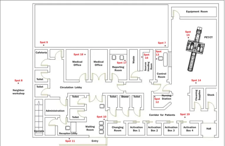
At the spots 2, 4, 6, 9 and 11, the dose estimates indi-cated values of up to 44% of the restriction level of annual dose for free areas (0,5 mSv/ano). The spots 3, 5, 7, 8, 10, 17 and 18 presented annual dose estimates reaching values between 72% and 87% of the restriction level of annual dose for free areas. Such results demonstrated that the shielding of such areas is appropriate for the current demand of the center. It is important to highlight that the CNEN standards (CNEN-NN-3.01 and CNEN-NN-3.05) do not define
am-Figure 1. Sketch of the first floor of Centro de Tecnologia em Imagem Molecular (CIMol). Spot 9
Spot 8
Spot 18
Spot 11
Spot 10
Spot 17
Spot 16
Spot 7
Spot 13
Spot 12
Spot 15
Spot 14
Spot 19
Neighbor workshop
Cafeteria
Toilet
Toilet
Upstairs
Administration
Reception Lobby
Medical Office
Medical Office
Toilet
Toilet Circulation Lobby
Waiting Room
Entry
Reporting Room
W
a
s
te
S
o
u
rc
e
s
H
a
n
d
lin
g
R
o
o
m
Control Room
PET/CT
Toilet Shower Toilet Nursing
Station Clea
n
in
g
S
u
ppl
ies
Equipment Room
Stock
Corridor for Patients
Changing Room
Activation Box 1
Activation Box 2
Activation
Box 3 Hall
Table 1—Estimate of annual equivalent dose for photons at each monitored spot. Spot 2 3 4 5 6 7 8 9 10 11 12 13 14 15 16 17 18 19 Shielding description Slab Slab Slab Slab Slab Wall Wall (several) Wall (several) Wall (several) Wall (several) Wall Wall None None Fixed screen Wall Wall (several) Wall Shielding material Reinforced concrete Reinforced concrete Reinforced concrete Reinforced concrete Reinforced concrete Reinforced concrete Reinforced concrete + masonry Reinforced concrete + masonry
Reinforced concrete Concrete + masonry Reinforced concrete Plumbiferous glass + reinforced
concrete — —
Lead + plumbiferous glass + reinforced concrete Reinforced concrete + masonry Reinforced concrete + masonry
Reinforced concrete Thickness (cm) 26.0 26.0 26.0 26.0 12.0 16.0 4 × 15.0 + 14.0 + 16.0
16.0 + 25.0 15.0 15.0 + 15.0
16.0 3.0 + 16.0
— — 2.0 + 3.0 + 16.0
15.0 + 16.0 15.0 + 16.0
16.0 T 1/4 1/2 1/6 1 1/6 1 1/2 1/4 1 1/16 1 1 1 1 1 1/2 1/2 1 Measured dose (mSv) 0.12 0.15 0.09 0.09 0.09 0.09 0.18 0.18 0.09 0.15 0.15 0.12 0.30 0.27 0.18 0.18 0.15 0.15 Hx (mSv/year) 0.12 0.30 0.06 0.36 0.06 0.36 0.36 0.18 0.36 0.04 0.60 0.48 1.2 1.08 0.72 0.36 0.30 0.60 Uncertainty (mSv/year) ± 0.02 ± 0.06 ± 0.01 ± 0.07 ± 0.01 ± 0.07 ± 0.07 ± 0.04 ± 0.07 ± 0.01 ± 0.11 ± 0.10 ± 0.24 ± 0.22 ± 0.14 ± 0.07 ± 0.06 ± 0.12 Area classification Free Free Free Free Free Free Free Free Free Free Controlled Controlled Controlled Controlled Controlled Free Free Controlled Conclusion Appropriate Appropriate Appropriate Appropriate Appropriate Appropriate Appropriate Appropriate Appropriate Appropriate Appropriate Appropriate Appropriate Appropriate Appropriate Appropriate Appropriate Appropriate
Figure 2. Sketch of the second floor of Centro de Tecnologia em Imagem Molecular (CIMol).
Spot 4
Animals Room
Experiments Room Experiments Room Animals Room Animals Room
Vivarium Vivarium Spot 2 Circulation Lobby Circulation Lobby Spot 6 Toilet Toilet A d m in is tr a ti o n Experiments Room Qu a ra n ti n e Spot 3 Office Storage Room Spot 1 Misplaced Spot Hygiene Room Circulation Lobby Circulation Lobby Upstairs
Animals Room Office Office
Spot 5
bient dose restriction values for nuclear medicine centers, thus the dose restriction levels established by the Ministry of Health (Ordinance 453/98) for medical radiodiagnosis services were utilized. Such levels were considered as pho-tons are utilized at a lower magnitude energy range, and therefore, the levels are more restrictive, thus favoring ra-diological safety.
At spots 12 to 16, and spot 19, all of them in controlled areas, the dose estimates presented values of up to 29% of the annual dose restriction level for controlled areas (5.0 mSv/ year), indicating that such spots are appropriate in terms of radiation level, and that the shielding, as applicable, are appropriate for the current demand of the center.
In all compliance evaluations with respect to dose re-striction, the results for dose equivalent for photons in one year corresponded to the sum of the mean readings from the dosimeters with their respective uncertainty values. It is important to highlight that the results were overestimated for both the Hx evaluation for the evaluation of
uncertain-ties, according to the radiological protection principles. Based on the present results, it is possible to conclude that there is no need to make any changes in the physical structure of the center, since in none of the spots the thresh-olds established by the current standards were exceeded. Also, based on the time of equipment utilization and image acqui-sition parameters, it is possible to conclude that the number of exams carried out at CIMol might be increased by 10%.
CONCLUSION
The results from the evaluation of radiation levels for both the internal and external areas of CIMol demonstrate
that the dose estimates for the areas are appropriate in rela-tion to dose restricrela-tions established by the Ministry of Health, as the CNEN regulations do not establish such values for nuclear medicine centers. The values for all the spots in the center were found to be satisfactory, demonstrating that the whole shielding system is appropriate for both the free and controlled areas. Also, it is possible to conclude that the number os exams performed at CIMol might be increased by 10%. And, considering that the center operates with un-sealed radioactive sources, one should stress the importance of strict compliance with radioprotection principles.
REFERENCES
1. Brasil. Ministério da Ciência, Tecnologia e Inovação. Comissão Nacional de Energia Nuclear. Diretrizes básicas de proteção radioló-gica. CNEN-NN-3.01, de 1º de setembro de 2011.
2. Adad MCBT, Hoff G, Streck EE, et al. Curvas de isodose no ar em uma sala de mamografia. Radiol Bras. 2008;41:255–8.
3. Avila O, Torres-Ulloa CL, Medina LA, et al. TL measurement of ambient dose at a nuclear medicine department. Radiation
Measure-ments. 2011;46:1843–6.
4. Brasil. Ministério da Saúde. Secretaria de Vigilância Sanitária. Dire-trizes de proteção radiológica em radiodiagnóstico médico e odonto-lógico. Portaria nº 453, de 1º de junho de 1998.
5. Madsen MT, Anderson JA, Halama JR, et al. AAPM Task Group 108: PET and PET-CT shielding requirements. Med Phys. 2006;33:4– 15.
6. Oliveira ML, Maia AF, Nascimento NCES, et al. Influência da de-pendência energética de dosímetros termoluminescentes na medida da dose na entrada da pele em procedimentos radiográficos. Radiol Bras. 2010;43:113–8.