Genetic potential for an acute
inflammatory response in IgA
glomerulonephritis in mice
1Laboratório de Fisiopatologia Renal, Faculdade de Medicina,
Universidade de São Paulo, São Paulo, SP, Brasil
2Laboratório de Imunogenética, Instituto Butantan, São Paulo, SP, Brasil
R.S. Kurihara1, M. Yokoo1,
W.V. Domingues1,
W.H. Cabrera2,
O.G. Ribeiro2, O.M. Ibañez2,
D.A. Malheiros1, R.T. Barros1
and E.B. de Almeida Prado1
Abstract
Mice selected on the basis of an acute inflammatory response (AIR) can provide information about the immunopathological mechanisms of glomerulonephritis. We studied the differences between mice selected for a maximal AIR (AIRmax that attract more polymorpho-nuclear cells to the site of injury) or a minimal AIR (AIRmin that attract more mononuclear cells) in an experimental model of IgA nephropathy in order to investigate the effect of genetic background on glomerular disease progression and the participation of the mono-cyte chemoattractant protein-1 (MCP-1) chemokine. IgA nephropa-thy was induced by intraperitoneal ovalbumin injection and bile duct ligation in AIRmax and AIRmin mice. Histological changes, urinary protein/creatinine ratio, serum IgA levels, immunofluorescence for IgA, IgG and complement C3 fraction, immunohistochemistry for macrophages and MCP-1, and MCP-1 levels in macerated kidney were determined. Mesangial IgA deposition was seen only in AIRmin mice, which presented more renal lesions. Increased serum IgA levels (1.5 ± 0.4 vs 0.3 ± 0.1 mg/mL, P < 0.001), high glomerular MCP-1 expression and decreased monocyte/macrophage infiltration in the interstitial area (0.3 ± 0.3 vs 1.1 ± 0.9 macrophages/field, P < 0.05) were detected in AIRmin mice compared to AIRmax mice. No glo-merular monocyte/macrophage infiltration was detected in either strain. In spite of the absence of IgA deposition, AIRmax mice presented discrete or absent mesangial proliferation. The study showed that there are differences between mice selected for AIRmax and AIRmin with respect to serum IgA levels, histological damage and MCP-1 chemokine production after ovalbumin injection in combina-tion with bile duct ligacombina-tion.
Correspondence
R.S. Kurihara R. Agostinho Cretella, 58 05337-040 São Paulo, SP Brasil
Fax: +55-11-3768-3443 E-mail: [email protected]
Research supported by CNPq.
Publication supported by FAPESP.
Received October 7, 2004 Accepted June 14, 2005
Key words
•Monocyte chemoattractant
protein-1
•IgA
•Glomerulonephritis •Acute inflammatory
response
•Chemokine
Introduction
Maximal and minimal acute inflamma-tory response (AIRmax and AIRmin) mice were obtained by bi-directional artificial
highly polymorphic and was obtained from the interbreeding of eight mouse strains (lin-eage A, DBA2, P, SWR, CBA, SJL, BALB/ c, and C57BL/6).
The phenotype characterizad was the in-flux of leukocytes and plasma protein into the inflammatory exudate 48 h after poly-acrylamide injection. The mice were selected for the ability to recruit polymorphonuclear cells at the site of injury (1,2). The F0 popu-lation presented a predominance of poly-morphonuclear cells (71%) and 29% mono-cytes. In the nineteenth generation (F19), mononuclear cells corresponded to 55% of the cells in the exudate of AIRmin mice and polymorphonuclear cells corresponded to 95% of the cells in the exudate of AIRmax mice (2).
Some differences were detected between these two mouse strains. First, the hemato-poietic study of bone marrow demonstrated that AIRmin mice produced predominantly more monocyte colony-forming units than AIRmax mice (Ribeiro OG, personal com-munication). Second, AIRmax mice pro-duced a larger amount of basal serum IgE and prostaglandin E (PGE) in the exudate compared to AIRmin mice (1). Finally, spleen cells from AIRmax mice with chronic arthri-tisproduced a larger amount of IFN-γ (3).
In the present study, we determined the differences between mouse strains genetically selected for AIRmax or AIRmin in terms of the development of IgA glomerulonephritis, the influence of monocyte chemoattractant protein-1 (MCP-1), and monocyte/macrophage infiltration. To this end, we used the experi-mental IgA glomerulonephritis model de-scribed by Emancipator et al. (4), modified, with intraperitoneal ovalbumin injections and bile duct ligation in mice, which resulted in mesangial IgA deposits.
Monocytes/macrophages are immune system cells which participate in the transi-tion from acute to chronic glomerular injury (5). These cells are known to be able to secrete products in response to different
stimuli such as cytokines, growth factors, lysosomal enzymes, protease inhibitors, fi-bronectin, cyclooxygenase products, lipoxy-genase products, reactive oxygen products, and reactive nitrogen products (6). Macro-phages are detected in the presence of both glomerular and non-glomerular injury and are related to the progression of renal dis-ease in the glomerular and interstitial com-partment (7). In human IgA nephropathy there is a correlation between glomerular macrophage infiltration and crescents and proteinuria (8).
One of the most important chemokines involved in monocyte/macrophage attrac-tion to the site of injury is MCP-1 (9). Expe-rimental models using rodents have shown an association between glomerular expres-sion of MCP-1 and monocyte infiltration in these areas (10,11).
Material and Methods
Animals
Male and female AIRmax and AIRmin mice aged 6 to 8 weeks, F26 generation, and BALB/c mice aged 6 to 8 weeks were ob-tained from the animal facilities of the Im-munogenetics Laboratory, Instituto Butan-tan, São Paulo, SP, Brazil.
Experiment
metabolic cages for 4 h for urine collection. The animals were then sacrificed and blood and kidneys were collected for analysis.
A preliminary study with BALB/c mice was also carried out. The animals were di-vided into four groups: control (N = 5), mice that did not receive ovalbumin injections and were not submitted to bile duct ligation; ovalbumin (N = 6), mice that received only ovalbumin injections; bile duct ligation (N = 6), mice only submitted to bile duct ligation; ovalbumin and bile duct ligation (N = 5), mice that received ovalbumin injections and bile duct ligation.
Urinary protein/creatinine ratio
The urine collected for 4 h was centri-fuged and the protein contained in the super-natant was measured by the method of Bradford (12).Next, urine was deproteinated with an equal part of 20% trichloroacetic acid. Creatinine was determined by the Heinegard-Tiderstrom method, modified, and the result was multiplied by 2 (13). The data obtained were used to calculate the urinary protein/creatinine ratio.
Histology
One of the kidney poles was first fixed in Bouin solution and then in buffered formalin, and embedded in paraffin. Four-micrometer paraffin sections were stained with hematoxy-lin-eosin. The other pole was frozen, and fro-zen 4-µm sections were cut, fixed in acetone for 5 min and stored at -70ºC until use.
Immunofluorescence. The frozen sections were washed with Tris-buffered saline (TBS) for 10 min and then incubated with primary antibody (1:50 goat anti-mouse IgA; or 1:100 goat anti-mouse IgG, both from Sigma) for 60 min. The sections were washed again with TBS for 10 min and then incubated with secondary antibody (rabbit anti-goat IgG di-luted 1:100; ICN, Aurora, OH, USA) for 30 min. After washing with TBS, the material
was incubated with 1:40 FITC anti-rabbit IgG and 1:20 Evans blue for 45 min.
Immunohistochemistry for C3 ( comple-ment C3 fraction). Endogenous peroxidase was blocked in the frozen material with 1% hydrogen peroxide diluted in methanol. The material was washed with TBS and incu-bated with 1:10 bovine serum albumin for 60 min. The sections were then incubated with 1:200 C3/HRP (ICN) for 30 min at 37ºC and left to stand at 4ºC overnight. The color was developed with diaminobenzidine diluted in TBS and hydrogen peroxide and then counterstained with Mayer’s hematoxy-lin for 8 min and a semi-quantitative analy-sis was conducted.
Immunohistochemistry for F4/80 ( mac-rophage staining). Paraffin-embedded sec-tions were used. The immunohistochemical StreptABComplex/AP method (Dako A/S, Glostrup, Denmark), a biotin-streptavidin complex stained with alkaline phosphatase, was adopted. The material was dewaxed, placed in sodium citrate buffer, pH 6.0, and heated 2 x 8 min in a microwave oven. Next, the material was washed in distilled water and TBS. Avidin and biotin were blocked for 20 min each and the sections were then incubated with 1:10 fetal calf serum for 60 min. The primary antibody was 1:20 rat anti-mouse F4/80 (Serotec, Oxford, England) in-cubated overnight. On the subsequent day, the secondary antibody, 1:300 biotinylated rabbit anti-rat IgG (Dako), was added. After washing, the specimens were incubated with StreptABComplex/AP for 30 min, devel-oped with Fast Red and counterstained with Mayer’s hematoxylin. Positive staining/glo-merulus and positive staining/field were counted at 400X magnification.
CA, USA). A semi-quantitative analysis was carried out.
ELISA for MCP-1
The right kidney was weighed and mac-erated in phosphate-buffered saline in a Pot-ter Elvehjem apparatus. The maPot-terial was centrifuged at 2000 rpm at 4ºC for 10 min and the supernatant was stored at -70ºC. MCP-1 was measured by ELISA using the Quantikine kit, Mouse JE/MCP-1 Immu-noassay (R&D Systems, Minneapolis, MN, USA) and detected with a microplate reader at 450 nm. Sensitivity was 2 pg/mL and the results were reported as pg/g kidney.
Serum IgA
Serum IgA levels were determined by ra-dial immunodiffusion (14). Thirty microliters anti-mouse IgA (Sigma) was placed in 1% agarose and Tris barbital, pH 8.8. The stand-ard solution was 5 µg/µL mouse IgA (Sigma). The samples were diluted 1:2 in Tris barbital and 6 µL was added to each well and incubated for 48 h. The material was washed with saline for 72 h, stained with 25% Coomassie blue, 54.4% methanol and 13.3% acetic acid for 5 min, and destained with 5% acetic acid. The diameter of the halo was measured and con-centration was plotted vs diameter2.
Statistical analysis
Data were analyzed statistically by the Mann-Whitney test for nonparametric pa-rameters and by the Student t-test for para-metric parameters, with the level of signifi-cance set at P < 0.05.
Results
Preliminary study in BALB/c mice
BALB/c mice were divided into four groups: control (N = 5), only ovalbumin
injections (N = 6), only bile duct ligation (N = 6), and ovalbumin injections with bile duct ligation (N = 5).
There were difficulties in performing the surgery in mice. The mortality after bile duct ligation was 80% due to surgical procedures and all of these animals were icteric at the time of sacrifice.
Control mice and mice receiving ovalbu-min injections did not present any histologi-cal changes, but bile duct ligation caused a mesangial expansion in some glomeruli in three of six animals and ovalbumin injec-tions with bile duct ligation increased the intensity of mesangial expansion in three of five animals.
There were no alterations in the intersti-tial area and no differences in urinary pro-tein/creatinine ratio among the groups (con-trol: 1.4 ± 0.4; ovalbumin: 3.9 ± 0.8; bile duct ligation: 11.8 ± 19.9; ovalbumin with bile duct ligation: 55.1 ± 108.1; P > 0.05).
The production of serum IgA antibody was the same for control and for the group submitted to ovalbumin injections with bile duct ligation (1.6 ± 0.7 and 2.1 ± 0.4 mg/mL, respectively); however, 60% of control mice had IgA deposition in the glomerular area, as opposed to 100% deposition in mice with bile duct ligation or ovalbumin injection.
MCP-1 levels were higher in the group submitted to ovalbumin injection and bile duct ligation (612 ± 147 pg/g) than in control (76 ± 31 pg/g). MCP-1 staining was ob-served in the mesangial area.
Histology of AIRmin and AIRmax mice
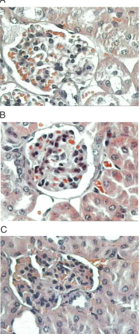
The following results were obtained for AIRmin mice: no histological alterations (N = 1), discrete mesangial expansion (N = 1), synechiae (N = 1), mesangial expansion and proliferation (N = 1), and mesangial expan-sion and synechiae (N = 2; Figure 1C).
observed erythrocytes in tubular space and in 2 mice of AIRmax group, with no histo-logical alteration, we observed erythrocytes in Bowman’s space.
No tubulo-interstitial alterations or dif-ferences in urinary protein/creatinine ratio (AIRmax: 4.9 ± 5.6; AIRmin: 15.4 ± 22.4; P > 0.05) were observed between strains.
Serum IgA and IgA, IgG and C3 deposition in renal tissue
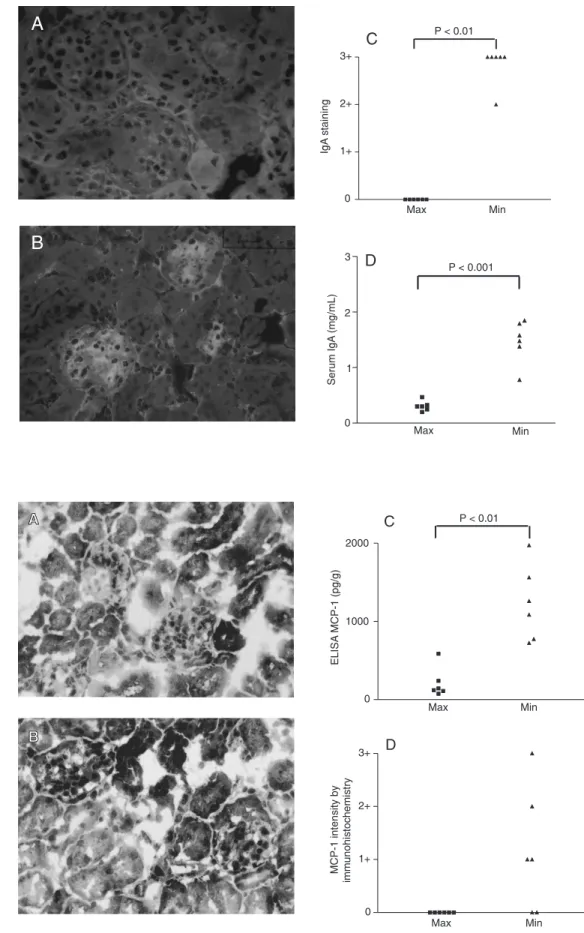
Serum IgA antibody production was higher in AIRmin than in AIRmax mice, with 1.5 ± 0.4 mg/mL serum in AIRmin mice and 0.3 ± 0.1 mg/mL in AIRmax mice (P < 0.001; Figure 2D). IgA production was lower in AIRmax than BALB/c mice (2.1 ± 0.4 mg/mL).
IgA deposition was observed only in the mesangial region of AIRmin mice (Figure 2B), with a tendency to IgG deposition in these mice and with no difference in C3 deposition between AIRmax and AIRmin mice. There was no IgA deposition in AIRmax mice.
MCP-1 and macrophages
MCP-1 levels determined by ELISA in whole macerated kidney were 1230 ± 480 pg/g in AIRmin mice and 213 ± 192 pg/g in AIRmax mice (P < 0.01; Figure 3C). BALB/ c mice showed an intermediate level of MCP-1 (6MCP-12 ± MCP-147 pg/g).
Immunohistochemistry was carried out to investigate the MCP-1 production site that showed predominant MCP-1 staining in the mesangial area of AIRmin mice. This chemokine was not detected in AIRmax mice (Figure 3 A, B and C).
Macrophages were found in the tubulo-interstitial region of AIRmax and AIRmin mice, predominantly in the periglomerular region. No staining for macrophages was observed in the glomerular area of AIRmax or AIRmin mice. AIRmax mice presented
Figure 1. A, Glomerulus from a normal maximal acute inflam-matory response (AIRmax) mouse. Hematoxylin-eosin (HE),
360X. B, Glomerulus from an
AIRmax mouse with mesangial proliferation. HE, 360X. C, Glo-merulus from a minimal AIR (AIRmin) mouse with synechiae and mesangial expansion. HE, 360X.
1.1 ± 0.9 macrophages/field and AIRmin mice 0.3 ± 0.3 macrophages/field (P < 0.05).
Discussion
Figure 2. A, Negative staining for IgA by immunofluorescence in maximal acute inflammatory response (AIRmax) mice (360X).
B, Positive staining for IgA by immunofluorescence in minimal AIR (AIRmin) mice (360X). C, Glomerular deposition of IgA in AIRmax and AIRmin mice with bile duct ligation: IgA deposition was increased in AIRmin. D, IgA production in AIRmax and AIRmin mice with bile duct liga-tion: serum IgA levels were higher in AIRmin.
Figure 3. A, Negative staining for monocyte chemoattractant protein-1 (MCP-1) in maximal acute inflammatory response (AIRmax) mice (360X). B, Posi-tive staining for MCP-1 in mini-mal AIR (AIRmin) mice (360X).
C, MCP-1 levels in macerated
kidney from AIRmax and AIRmin mice with bile duct ligation: the levels were higher in AIRmin
mice. D, MCP-1 intensity by
which are an inbred strain of mice, not se-lected for an acute inflammatory response, and then we applied this model to AIRmax and AIRmin mice to investigate the influ-ence of genetic potential on the acute in-flammatory response in the pathogenesis of glomerulonephritis.
The preliminary study in BALB/c mice showed that bile duct ligation with ovalbu-min immunization can induce discrete mes-angial expansion, in the absence of protein-uria.
The renal alterations were very discrete, probably due to the short time elapsed from bile duct ligation to sacrifice. However, if this interval were increased, other related factors such as surgery or hepatic damage would be more likely to have an effect.
The experiment in mice selected for maxi-mal or minimaxi-mal genetic acute inflammatory response showed that AIRmin mice pre-sented more histological damage such as mesangial matrix expansion, synechiae and mesangial proliferation. There was IgA depo-sition in mesangial area and MCP-1 levels were higher in macerated kidney compared to AIRmax and BALB/c mice.
These facts might be related to IgA depo-sition in the mesangial region but it was observed that AIRmax mice have mesangial proliferation even in the absence of IgA or IgG deposition in renal tissue. Melvin et al. (15) demonstrated that when bile duct liga-tion was performed in rats there was an increase in glomerular diameter. This glo-merular hypertrophy may cause a stretch in mesangial cells and it is known that expo-sure to stretch/relaxation stimulates mesan-gial cell proliferation in vitro (16). In the puromycin aminonucleoside model, the in-crease in glomerular volume can lead to segmental and focal sclerotic lesions (17).
Nevertheless, there were differences in histological lesions between the two strains. Many studies have associated mesangial pro-liferation and matrix expansion in both im-mune and non-imim-mune renal diseases
(18-20). Johnson et al. (21) have demonstrated dissociation between these two phenomena. When IFN-γ is administered to rats in the
anti-Thy 1 model, mesangial proliferation is partially inhibited and matrix expansion is maintained. In pristane-induced arthritis, IFN-γ was increased in AIRmin mice (3), a fact that may explain a tendency to mesangi-al expansion in this strain.
The reduced serum IgA levels of AIRmax animals may reflect an increase in endoge-nous regulatory factors in this mouse strain in the presence of surgical stress or bacterial translocation (22). A previous study has shown that intraperitoneal polyacrylamide injection before ovalbumin immunization reduces the anti-ovalbumin response of AIRmax, but not AIRmin, mice. This dem-onstrates that inflammation before immuni-zation decreases antibody production in AIRmax mice (Araújo LMM, personal com-munication). Glucocorticoids are known to be increased in stress situations and PGE is increased in the acute inflammatory exudate of AIRmax animals (Ribeiro OG, personal communication), and both could influence immunoglobulin production (23-26).
con-stitution, such as production as glucocorti-coids or PGE in response to stress. Dexa-methasone inhibits MCP-1 production in fi-broblast culture(27) and PGE decreases MCP-1 mRNA expression in anti-thymocyte glomerulonephritis (28). Another factor could be IFN-γ. This cytokine is increased in AIRmin mice and can stimulate human mes-angial cells to produce MCP-1 in vitro, as reported by Grandaliano et al. (29). The dissociation between MCP-1 expression and macrophage infiltration demonstrates that
other factors can be involved in macrophage recruitment in this model.
The present study showed the impor-tance of pro-inflammatory genetic modula-tion interfering with the response pattern in this model, with glomerular IgA deposition causing significant lesions such as synechiae and matrix mesangial expansion in AIRmin mice and the absence of IgA deposition and discrete mesangial proliferation in AIRmax mice.
References
1. Ibañez OM, Stiffel C, Ribeiro OG et al. (1992). Genetics of nonspe-cific immunity: I. Bidirectional selective breeding of lines of mice endowed with maximal or minimal inflammatory responsiveness.
European Journal of Immunology, 22: 2555-2563.
2. Araújo LMM, Ribeiro OG, Siqueira M et al. (1998). Innate resistance to infection by intracellular bacterial pathogens differs in mice se-lected for maximal or minimal acute inflammatory response. Euro-pean Journal of Immunology, 28: 2913-2920.
3. Vigar ND, Cabrera WHK, Araújo LMM et al. (2000). Pristane-in-duced arthritis in mice selected for maximal or minimal acute inflam-matory reaction. European Journal of Immunology, 30: 431-437. 4. Emancipator SN, Gallo GR, Razaboni R et al. (1983). Experimental
cholestasis promotes the deposition of glomerular IgA immune com-plexes. American Journal of Pathology, 113: 19-26.
5. Schreiner GF (1991). The role of the macrophage in glomerular injury. Seminars in Nephrology, 11: 268-275.
6. Nathan CF (1987). Secretory products of macrophages. Journal of Clinical Investigation, 79: 319-326.
7. van Goor H, Ding G, Kees-Folts D et al. (1994). Biology of disease: macrophages and renal disease. Laboratory Investigation, 71: 456-464.
8. Arima S, Nakayama M, Naito M et al. (1991). Significance of mono-nuclear phagocytes in IgA nephropathy. Kidney International, 39: 684-692.
9. Rollins BJ, Walz A & Baggiolini M (1991). Recombinant human MCP-1/JE induces chemotaxis, calcium influx, and the respiratory burst in human monocytes. Blood, 78: 1112-1116.
10. Rovin BH, Rumancik M, Tan L et al. (1994). Glomerular expression of monocyte chemoattractant protein-1 in experimental and human glomerulonephritis. Laboratory Investigation, 71: 536-542. 11. Tam FWK, Karkar AM, Smith J et al. (1996). Differential expression
of macrophage inflammatory protein 2 and monocyte chemoattrac-tant protein-1 in experimental glomerulonephritis. Kidney Interna-tional, 49: 715-721.
12. Bradford MM (1976). A rapid and sensitive method for the quantita-tion of microgram quantities of protein utilizing the principle of pro-tein-dye binding. Analytical Biochemistry, 72: 248-254.
13. Lopes HJJ, Sousa MO, Ratton VLA et al. (1989). New formulation for the preparation of the alkaline buffer used in the determination of
blood’s and urine’s creatinine - modified Heinegard-Tiderstrom’s method. Revista Brasileira de Análises Clínicas, 21: 121-125. 14. Mancini G, Carbonera AO & Heremans JF (1965). Immunochemical
quantitation of antigens by single radial immunodiffusion. Immu-nochemistry, 2: 235-254.
15. Melvin T, Burke B, Michael AF et al. (1983). Experimental IgA nephropathy in bile duct ligated rats. Clinical Immunology and Im-munopathology, 27: 369-377.
16. Harris RC, Akai Y, Yasuda T et al. (1994). The role of physical forces in alterations of mesangial cell function. Kidney International, 45 (Suppl): S17-S21.
17. Cahill MM, Ryan GB & Bertram JF (1996). Biphasic glomerular hypertrophy in rats administered puromycin aminonucleoside. Kid-ney International, 50: 768-775.
18. Johnson RJ, Raines EW, Floege J et al. (1992). Inhibition of mesan-gial cell proliferation and matrix expansion in glomerulonephritis in the rat by antibody to platelet-derived growth factor. Journal of Experimental Medicine, 175: 1413-1416.
19. Floege J, Eng E, Young BA et al. (1993). Heparin suppresses mesangial cell proliferation and matrix expansion in experimental mesangioproliferative glomerulonephritis. Kidney International, 43: 369-380.
20. Fukui M, Nakamura T, Ebihara I et al. (1993). Low protein diet attenuates increased gene expression of platelet-derived growth factor and transforming growth factor-beta in experimental glomeru-lar sclerosis. Journal of Laboratory and Clinical Medicine, 121: 224-234.
21. Johnson RJ, Lombardi D, Eng E et al. (1995). Modulation of experi-mental mesangial proliferative nephritis by interferon-γ. Kidney In-ternational, 47: 62-69.
22. Deitch EA, Sittig K, Li M et al. (1990). Obstructive jaundice promotes bacterial translocation from the gut. American Journal of Surgery, 159: 79-84.
23. del Rey A, Besedowsky H & Sorkin E (1984). Endogenous blood levels of corticosterone control the immunologic cell mass and B cell activity in mice. Journal of Immunology, 133: 572-575.
Investi-gation, 47: 147-152.
25. Kelley VE, Winkelstein A & Izui S (1979). Effect of prostaglandin E on immune complex nephritis in NZB/W mice. Laboratory Investiga-tion, 41: 531-537.
26. Zurier RB, Damjanov I, Sayadoff DM et al. (1977). Prostaglandin E1 treatment of NZB/NZW F1 hybrid mice. II. Prevention of glomerulo-nephritis. Arthritis and Rheumatism, 20: 1449-1456.
27. Mukaida N, Zachariae CCO, Gusella GL et al. (1991). Dexametha-sone inhibits the induction of monocyte chemotactic-activating fac-tor production by interleukin-1 or tumor necrosis facfac-tor. Journal of
Immunology, 146: 1212-1215.
28. Jocks T, Zahner G, Freudenberg J et al. (1996). Prostaglandin E1 reduces the glomerular mRNA expression of monocyte chemoat-tractant prote1 in anti-thymocyte antibody-induced glomerular in-jury. Journal of the American Society of Nephrology, 7: 897-905. 29. Grandaliano G, Valente AJ, Rozek MM et al. (1994). Gamma