Gap junctio n mo dulatio n by
e xtrace llular signaling mo le cule s:
the thymus mo de l
1Departamento de Imunologia, Instituto O swaldo Cruz,
Fundação O swaldo Cruz, Rio de Janeiro, RJ, Brasil
2Instituto de Biofísica, Universidade Federal do Rio de Janeiro,
Rio de Janeiro, RJ, Brasil L.A. Alves1,
O .K. Nihei1,2,
P.C. Fonseca1,2
A.C. Campos-de-Carvalho2
and W. Savino1
Abstract
Gap junctions are intercellular channels which connect adjacent cells
and allow direct exchange of molecules of low molecular weight
between them. Such a communication has been described as
funda-mental in many systems due to its importance in coordination,
prolif-eration and differentiation. Recently, it has been shown that gap
junctional intercellular communication (GJIC) can be modulated by
several extracellular soluble factors such as classical hormones,
neu-rotransmitters, interleukins, growth factors and some paracrine
sub-stances. Herein, we discuss some aspects of the general modulation of
GJIC by extracellular messenger molecules and more particularly the
regulation of such communication in the thymus gland. Additionally,
we discuss recent data concerning the study of different neuropeptides
and hormones in the modulation of GJIC in thymic epithelial cells. We
also suggest that the thymus may be viewed as a model to study the
modulation of gap junction communication by different extracellular
messengers involved in non-classical circuits, since this organ is under
bidirectional neuroimmunoendocrine control.
Co rre spo nde nce
L.A. Alves
Laboratório de Pesquisas sobre o Timo
Departamento de Imunologia Instituto O swaldo Cruz, FIO CRUZ Av. Brasil, 4365
21045-900 Rio de Janeiro, RJ Brasil
Fax: + 55-21-280-1589 E-mail:
alveslaa@ gene.dbbm.fiocruz.br
Presented at the Meeting “Gap Junctions in the Nervous and Cardiovascular Systems: Clinical Implications”, Rio de Janeiro, RJ, Brazil, June 6-11, 1998.
Research supported by FAPERJ, CNPq, FINEP/BID, FUJB, PADCT/CNPq and PRO NEX/CNPq to L.A. Alves, A.C. Campos-de-Carvalho and W. Savino.
Received July 30, 1999 Accepted November 23, 1999
Ke y words
·Thymus
·Thymic epithelial cells
·Connexin 43
·Hormones
·Interleukins
Intro ductio n
The establishment of cellular
communi-cation was a milestone for the origin and
evolution of multicellular organisms. Such a
communication can be mediated by 1) soluble
factors, i.e., hormones, cytokines, growth
factors and neurotransmitters, 2) membrane
surface molecules, i.e., members of
immu-noglobulin gene superfamily, integrins,
cad-herins and other classes of recognition
mol-ecules, and 3) ion channels.
Ion channels have been recognized as a
fundamental component involved in the
mechanisms that most animals use to
per-ceive the external world and the internal
milieu (mechanoception, nociception, vision,
taste, motion and hearing). Gap junction is a
particular kind of ion channel that mediates
direct transfer of molecules between
adja-cent cells by a diffusion process.
con-nexin according to its predicted molecular
weight deduced from cDNA sequences and
species of origin (1).
These channels permit direct cell-cell
exchange of small molecules such as ions,
sugars and second messengers between
ad-jacent cells (1,2). It was thought that these
channels are permeable to molecules of up
to 1 kDa in mammals. However, several
studies have demonstrated that many
con-nexins present selectivity for both charge
and size. For instance, connexin 43 allows
the passage of lucifer yellow while Cx45
does not (3). In addition, the functionality of
heterologous gap junctions has been shown
to depend on selective compatibility among
different connexins (3,4). In agreement with
this, Cx43 can form functional gap junctions
with Cx37, 43, 45 and 46, but not with 26,
31, 32, 33, 40 or 50 (5).
Gap junction channels can be gated, i.e.,
“opened” or “closed”, by several agents
in-cluding Ca
2+, H
+, lipophilic substances,
volt-age gradients, and hormones (1), the latter
generally acting via activation of protein
kinases that phosphorylate the cytoplasmic
regions of several connexins (6,7), or in
some cases by phosphatases (6,7). The
pres-ence of connexins has been demonstrated in
distinct lympho-hematopoietic organs where
their function is not clear (reviewed in Ref.
8). In these organs cell-to-cell
communica-tion is a key event to tune performance.
In this review, we will focus on the
modu-lation of gap junctions by extracellular
mes-sengers in a primary lymphoid organ, the
thymus gland. In this organ, bone
marrow-derived lymphoid precursors undergo a
pro-cess of differentiation and maturation,
cul-minating with the migration of mature T
cells to the periphery. In this process more
than 90% of thymocytes die intrathymically,
whereas some are rescued from programmed
cell death and are positively selected to
ulti-mately generate the vast majority of the T
cell repertoire (9).
Such a differentiation and maturation
pro-cess is quite complex, involving a dynamic
molecular cross-talk between T cells and the
thymic microenvironment, a tridimensional
network composed of distinct cell types
in-cluding epithelial cells, macrophages and
dendritic cells, as well as extracellular
ma-trix (ECM) elements (10).
The thymic epithelium is the major
com-ponent of the thymic microenvironment and
determines thymocyte maturation through
cell-cell contacts and secretion of a variety
of polypeptides including thymic hormones
and cytokines (11). Thymic epithelial cells
(TEC) can bind to and interact with
thy-mocytes by means of ECM ligands and
re-spective receptors as well as by classical
adhesion molecules (12) and, most
impor-tantly, by major histocompatibility complex
gene products, which present endogenous
peptides to the T cell receptors expressed on
the cell membrane of differentiating
thy-mocytes. Recently, we demonstrated the
ex-istence of a novel form of cell-to-cell
com-munication in TEC which is mediated by gap
junction channels (13).
It is also noteworthy that the thymus is an
important component of
neuroimmunoen-docrine circuits, synthesizing several
classi-cal hormones (for example, growth hormone,
prolactin and glucocorticoids) which act
pleiotropically upon the thymic epithelium
(11). Furthermore, the intrathymic
produc-tion of various neuropeptides, including the
neurohypophyseal hormones, oxytocin and
vasopressin (11), and vasoactive intestinal
peptide (VIP) has been demonstrated (11).
In this intriguing network, the data
concern-ing influence of extracellular messenger
molecules on gap junction mediated
com-munication begin to be analyzed.
Ge ne ral aspe cts of gap junction
modulation by e xtrace llular signaling
m o le cule s
possibility of multiple connexin isoform
ex-pression (1). All members of the connexin
family have the same putative structure:
cy-tosolic amino and carboxyl termini, four
transmembrane domains, and extracellular
(two) and cytoplasmic (one) loops. In the
primary sequence of these proteins there are
several consensus sites for phosphorylation
and dephosphorylation, which represent one
of the most frequent mechanisms for
hor-monal modulation of gap junctions and other
ion channels.
The first evidence of gap junction
modu-lation by hormones can be credited to Hax et
al. (14), who demonstrated the regulation of
electrical coupling by cAMP in insect cell
cultures, a finding that was confirmed by
Loewenstein and co-workers (15). As
ex-pected, several extracellular signaling
mol-ecules that raised cAMP levels could
modu-late gap junction communication in different
cell types.
Since then, regulation of these channels
by hormones and other extracellular
signal-ing molecules such as neurotransmitters,
growth factors and cytokines has been
dem-onstrated at the level of transcription, mRNA
stability, translation, cytoplasmic traffic and
gating (1,6,7). So far, the best studied
mech-anism operating during these regulatory
pro-cesses is the
phosphorylation/dephosphory-lation (6) of connexins, transcription factors
and other regulatory proteins. Except for
Cx26, all connexins cloned so far can be
regulated by
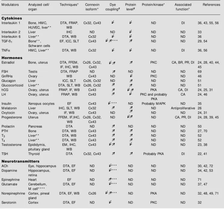
phosphorylation/dephosphory-lation. Table 1 illustrates some hormones
and other extracellular messengers that
modu-late gap junctions in several cell types. Most
of these effects are mediated by protein
ki-nases A and C, mitogen-activated protein
kinase and tyrosine kinase. These kinases
can increase or decrease junctional
commu-nication depending on the cell type studied.
It is not clear why this occurs but perhaps
different mixes of connexin isoforms and/or
selective activation of other intracellular
messenger systems that act on gap junctions
might explain the diversity of biological
re-sponses. Most of the effects studied thus far
are on Cx43 but several other connexins can
be modulated by such substances.
In endocrine glands, a rise in gap
junc-tion communicajunc-tion increases the cell
re-sponse to a given stimulus, increasing
hor-monal secretion. In Beta cells in Langerhan’s
islets, the overexpression of Cx43 leads to
an augmented secretion of insulin (16),
whereas in adrenal cells an inhibitor of gap
junction communication diminishes cortisol
secretion (17).
Conversely, in exocrine glands, a rise of
gap junction communication in general leads
to a decrease in secretion as seen in salivary
glands and in the exocrine pancreas (18).
Re gulation of thymic gap junctions
by e xtrace llular me sse nge rs
We demonstrated that mouse and human
TEC are coupled by gap junctions (13), a
concept that had been previously postulated
by Kendall in the 1980’s (19). We also
pro-vided evidence for the possible existence of
heterologous gap junctions between TEC
and thymocytes as well as for the
modula-tion of TEC hormonal secremodula-tion by gap
junc-tions.
As shown in Table 1, the majority of the
soluble messengers and the respective target
cell/organs involved in gap junction
modu-lation studies are those associated with
clas-sical endocrine, immunological and nervous
circuits such as TSH:thyroid, epinephrine:
heart, T
3,T
4:liver, oxytocin:uterus, FSH:testis,
neurotransmitters:neuronal electric coupling,
IL-1:liver, and so on. Looking for a more
integrative view, the thymus gland could be
an interesting model to study gap junction
modulation by nonclassical circuits since it
is under bidirectional
neuroimmunoendo-crine control.
Table 1 - Gap junction modulation by neuroimmunoendocrine soluble products.
ACh, Acetylcholine; BR, bone remodeling; CA, contractile activity; Cx, connexin; DI, differentiation; DTA, dye transfer assay; EF, electrophysiology; EM , electron microscopy; FFEM , freeze-fracture electron microscopy; FRAP, fluorescence recovery after photobleaching method; FSH, follicle-stimulating hormone; GnRHa, gonadotropin-releasing hormone analogue; hCG, human chorionic gonadotropin; HM C, human myoendothelial co-culture; HUVEC, human umbilical vein endothelial cells; ICC, immunocytochemistry; IHC, immunohistochemistry; IF, immuno-fluorescence; LH, luteinizing hormone; LH-RH, luteinizing hormone-releasing hormone; M APK, mitogen-activated protein kinase; NB, Northern blotting; ND: not determined; PKA, protein kinase-A; PKC, protein kinase-C; PR, proliferation; PTH, parathyroid hormone; T3, 3,3' , 5-triiodo-L-thyronine; SLT, scrape-loading technique; T4, L-thyronine; TGF-ß1, tumor grow th factor ß1; TNFa, necrosis factor a; TSH, thyrotropin stimulating hormone; WB, Western blotting. * All these parameters depended on the cell type analyzed; * * based on cell lines; * * * the modulation of the junctional conductance (Gj) w as also described (in some cases it involved exclusively Gj); * * * * based on goldfish M authner cells (M cells); #modulation of dye coupling and protein expression w as represented by the follow ing arrow s: (positive modulation), (negative modulation) and (both effects w ere described, depending on experimental conditions, cell type or concentration tested, or even the connexin isoform analyzed).
M odulators Analyzed cell/ Techniques* Connexin Dye Protein Protein/kinase* Associated References organ isoform* coupling# level# function*
Cytokines
Interleukin 1 Bone, HM C, DTA, FRAP, Cx32, Cx43 ND DI 36, 43, 55, 56 HUVEC, liver* * WB
Interleukin 2 Liver IHC ND ND ND ND 33
Interleukin 6 Liver* * DTA, WB Cx32 ND ND 36 TGF-ß1 Bone* * , EF, ICC, SLT Cx43 * * * ND ND 29, 54
Schw ann cells
TNFa HM C, Liver* * DTA, WB Cx32 ND DI 36, 56 Hormones
Estradiol Bone, uterus DTA, FFEM , Cx26, Cx32, PKC CA, BR, PR, DI 24, 28, 40, 44,
IF, IHC, WB Cx43 45
FSH Testis DTA, FRAP ND ND ND ND 69
GnRHa Ovary WB Cx43 ND PKC ND 46
Glucagon Liver ICC, SLT Cx26, Cx32 ND ND ND 51 Glucocorticoid Liver* * DTA, SLT, WB Cx26, Cx32 ND ND 51 hCG Ovary, uterus FRAP, IF, WB Cx43 PKA CA, DI 24, 25, 31 LH Ovary, uterus FRAP, WB Cx43 PKC and probably CA 24, 46
PKA
Insulin Xenopus oocytes EF Cx43 * * * ND Probably M APK ND 35 M elatonin Liver IHC, SLT, WB Cx32 ND Antiproliferative 26 Oxytocin Ovary, uterus IF, WB Cx43 ND ND ND 24, 31 Progesterone Uterus FFEM , IF,IHC, Cx26, Cx32, ND ND CA, PR, DI 24, 28, 39, 45
WB Cx43
Prolactin Pancreas DTA ND ND ND ND 50
PTH Bone DTA, WB Cx43 ND ND 27, 70
T3 Liver* * DTA, WB Cx43 ND ND 52
T4 Liver* * DTA, WB Cx43 ND ND 52
Testosterone Epididymis, EM , IHC, Cx43 ND ND 23, 38 pituitary gland WB
TSH Thyroid DTA Cx32, Cx43 Probably PKA DI 22, 41 Neurotransmitters
ACh Eye, hippocampus DTA, EF ND * * * ND ND ND 30, 42, 72 Dopamine Hippocampus, DTA, EF ND * * * ND ND ND 34, 42, 53
retina
Epinephrine Heart EF ND * * * ND ND ND 71
Glutamate Cerebellum, DTA, EF ND * * * ND ND ND 37, 47 M cell* * * *
Norepinephrine Cortex, pineal DTA, EF, WB Cx26 * * * ND PKA ND 32, 48, 49, 71 gland
Serotonin Cortex DTA, EF ND ND PKC ND 32
j j
j j
j
j
j
j
j
j j
j
j j jj
j
j j
j
j j j j
j j j
j j
j
j j
j j
j
j j
j j
j j
j j
j
j j
j j j
j j
corticotropin, growth hormone,
interleukin-1
a
, interleukin-1ß,
g
-aminobutyric acid
(GABA), neuropeptide Y, vasoactive
intes-tinal peptide, substance P, and histamine
reduce dye coupling whereas acetylcholine
and the ß-adrenergic agonist isoproterenol
have no effect (20,21; see Table 2).
Yet, in contrast to some of these data, we
found that VIP and vasopressin increase dye
coupling in one mouse TEC line and primary
cultures of thymic nurse cells. Supporting
this idea, 8Br-cAMP, a permeable analog of
cAMP (secondary messenger of VIP and
vasopressin), can also increase the degree of
inter-TEC dye coupling (Alves LA, Figueira
G, Savino W and Campos-de-Carvalho AC,
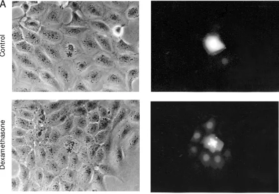
unpublished results). Additionally, we found
an increase in dye coupling in a rat cell line
(clone IT45-R1, provided by Dr. Tsumeroshi
Itoh, Tohoku University, Sendai, Japan) when
cells were treated with dexamethasone
(Fig-ure 1). Much work is still necessary in order
to obtain a clear view of the possible role of
gap junctions in thymus tissue. Furthermore,
it is essential to understand the multiple
levels of regulation of intercellular
commu-nication by way of gap junctions in the
thy-mus gland. Presently, the mechanisms by
which dye coupling increases or decreases
in thymic epithelial cells when these cells
are treated with extracellular messenger
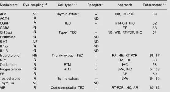
molecules are not known. Table 2
summa-rizes the effects of several extracellular
mol-ecules on inter-TEC dye coupling, as well as
the expression of the respective receptors.
One can see that receptors for some putative
modulators have not yet been formally
char-acterized in TEC, and that several hormones
for which receptors are expressed have not
been studied for their potential effects on
Table 2 - Neuroendocrine modulators of inter-TEC gap junctions.
ACh, Acet ylcholine; ACTH, adrenocort icot ropin horm one; AR, aut o-radiography; CGRP, calcit onin gene-related peptide; EF, electrophysiology; GABA, g-aminobutyric acid; GH, grow th hormone; HT, 5-hydroxytryptamine; IHC, immunohistochemistry; IL1-a, interleukin-1a; IL1-ß, interleukin-1ß; LM , light micros-copy; NB, Northern blotting; NE, no effect; NPY, neuropeptide Y; PA, pharmacological analysis; RT-PCR, reverse transcription-polym erase chain reaction; RTM , reticuloepithelial cells of thym us m edulla; SPA, Scatchard plot analysis; SP, substance P; TEC, thymic epithelial cells; VIP, vasoactive intestinal peptide; WB, Western blotting. * Summarized from Refs. 20 and 21; * * the receptor expression is repre-sented by the follow ing signals: + (positive expression), - (not found) or ND (not determined); * * * related to receptor characterization studies; #modulation of dye coupling is represented by the arrow s: (negative modulation).
M odulators* Dye coupling*# Cell type* * * Receptor* * Approach References* * * ACh NE Thymic extract + NB, RT-PCR 59
ACTH ND
CGRP TEC + RT-PCR, IHC 62
GABA - EF 68
GH (rat) Type-1 TEC + NB, WB, RT-PCR, IHC 61
Histamine ND
5-HT NE ND
IL1-a ND
IL1-ß ND
Isoproterenol NE Thymic extract, TEC + PA, NB, RT-PCR 66, 67
NPY - LM , IHC 63
Oestrogen RTM + IHC 58
Progesterone RTM + SPA, IHC 57, 58
SP - AR 60
Testosterone Thymic extract + SPA 64, 65
Thymulin NE ND
VIP Cortical/medullar TEC + RT-PCR, IHC, AR 60, 62 j
j j j j
j j
j j j j j
j
TEC gap junctions. For instance, in human
TEC the GABA receptor was not found. Yet,
when this neurotransmitter is applied to rat
Figure 1 - Increase in gap junc-tional communication induced by dexamethasone in a rat epi-thelial cell line. TEC w ere treated w ith 10-6 M dexamethasone for 48 h, and intercellular communi-cation w as evaluated by dye transfer assay using the fluoro-chrome lucifer yellow (LY). A, M icroscopy fields (phase con-trast and fluorescence, respec-tively, in the left and right pan-els) depicting the injected cell (* ) and those that w ere coupled w hen LY w as injected (magnifi-cat ion 320X). B, Hist ogram s show ing the pattern of coupling degree of control and dexameth-asone-treated cells. The analysis comprises 100 microinjections per group.
TEC, a decrease in dye coupling is observed,
indicating a possible species-specific
modu-lation or even an epiphenomenon.
C
o
n
tr
o
l
A
B
In
je
c
te
d
c
e
lls
(
%
)
75
50
25
0
Control
Dexamethasone (10-6 M )
D
e
x
a
m
e
th
a
s
o
n
e
0 1-2 3-4 5-6 7-8
Re fe re nce s
1. Bruzzone R, White TW & Paul DL (1996). Connections w ith connexins: the molecu-lar basis of direct intercellumolecu-lar signaling. European Journal of Biochemistry, 238: 1-27.
2. Sáez JC, Connor JA, Spray DC & Bennett M VL (1989). Hepatocyte gap junctions are permeable to the second messengers in-ositol 1,4,5-triphosphate and calcium ions. Proceedings of the National Academy of Sciences, USA, 86: 2708-2712.
3. St einberg TH, Civit elli R, Geist ST, Robertson AJ, Hick E, Veenstra RD, Wang HZ, Warlow PM , Westphale EM , Laing JG & Beyer EC (1994). Connexin43 and connexin45 form gap junctions w ith dif-ferent molecular permeabilities in osteo-blastic cells. EM BO Journal, 15: 744-750. 4. White TW, Paul DL, Goodenough DA & Bruzzone R (1995). Functional analysis of selective interactions among rodent con-nexins. M olecular Biology of the Cell, 6: 459-470.
5. Elfgang C, Eckert R, Lichtenberg-Frate H, Butterw eck A, Traub O, Klein RA, Hulser DF & Willecke K (1995). Specific perme-ability and selective formation of gap junc-tion channels in connexin-transfected HeLa cells. Journal of Cell Biology, 129: 805-817.
6. Sáez JC, M artinez AD, Branes M C & Gonzalez HE (1998). Regulation of gap junctions by protein phosphorylation. Bra-zilian Journal of M edical and Biological Research,31: 593-600.
7. Stagg RB & Fletcher WH (1990). The hor-mone-induced regulation of contact-de-pendent cell-cell communication by phos-phorylation. Endocrine Review s, 192: 302-325.
8. Alves LA, Cam pos-de-Carvalho AC & Savino W (1998). Gap junctions: a novel route for cell-cell communication in the immune system? Immunology Today, 19: 269-275.
9. Fow lkes BJ & Pardoll DM (1989). M olecu-lar and celluolecu-lar events of T cell develop-ment. Advances in Immunology, 44: 207-264.
10. Anderson G, M oore NC, Ow en JJT & Jenkinson EJ (1996). Cellular interactions in thymocyte development. Annual Re-view s in Immunology, 14: 73-99. 11. Dardenne M & Savino W (1994). Control
of thymus physiology by peptide hor-mones and neuropeptides. Immunology Today, 15: 518-523.
12. Savino W, Villa-Verde DM S & Lannes-Vieira J (1993). Extracellular matrix pro-teins in intrathymic T cell migration and differentiation? Immunology Today, 14: 158-161.
13. Alves LA, Campos-de-Carvalho AC, Cirne-Lima EO, Rocha-e-Souza CM , Dardenne M , Spray DC & Savino W (1995). Func-tional gap junctions in thymic epithelial cells are formed by connexin 43. Euro-pean Journal of Immunology, 25: 431-437. 14. Hax W M A, van-Venrooij GEPM & Vossenberg JBJ (1974). Cell communica-tion: a cyclic-AM P mediated phenome-non. Journal of M embrane Biology, 19: 253-266.
15. Flagg-New ton JL, Dahl G & Loew enstein WR (1981). Cell junction and cyclic AM P: 1. Up regulation of junctional membrane permeability and junctional membrane particles by administration of cyclic nucle-ot ide or phosphodiest erase inhibit or. Journal of M embrane Biology, 63: 105-121.
16. Vozzi C, Bosco D, Dupont E, Charollais A & M eda P (1997). Hyperinsulinemia-in-duced hypoglycemia is enhanced by over-expression of connexin 43. Endocrinol-ogy, 138: 2879-2885.
17. M unari-Silem Y, Lebrethon M C, M orand I, Rousset B & Saez JM (1995). Gap junc-tion-mediated cell-to-cell communication in bovine and human adrenal cells. A
pro-cess w hereby cells increase their respon-siveness to physiological corticotropin concentrations. Journal of Clinical Investi-gation, 95: 1429-1439.
18. M eda P, Pepper M S, Traub O, Willecke K, Gros D, Beyer E, Nicholson B, Paul D & Orci L (1993). Differential expression of gap junction connexins in endocrine and exocrine glands. Endocrinology, 133: 2371-2378.
19. Kendall M D (1986). The syncytial nature of epithelial cells in the thymic cortex. Journal of Anatomy, 147: 95-106. 20. Head GM , M ent lein R, von-Pat ay B,
Dow ning JEG & Kendall M D (1998). Neu-ropeptides exert direct effects on rat thy-mic epithelial cells in culture. Develop-mental Immunology, 6: 95-104.
21. Head GM , M entlein R, Kranz A, Dow ning JEG & Kendall M D (1997). M odulation of dye-coupling and proliferation in cultured rat thymic epithelium by factors involved in thymulin secretion. Journal of Anatomy, 191: 355-365.
22. M unari-Silem Y, Audebet C & Rousset B (1991). Hormonal control of the cell com-munication: Regulation by thyrotropin of the gap junction-mediated dye transfer betw een thyroid cells. Endocrinology, 128: 3299-3308.
23. Cyr DG, Hermo L & Laird DW (1996). Immunocytochemical localization and reg-ulation of connexin 43 in the adult rat epididymis. Endocrinology, 137: 1474-1484.
24. Ambrus G & Rao CV (1994). Novel regula-t ion of pregnanregula-t hum an m yom eregula-t rial smooth muscle cell gap junctions by hu-man chorionic gonadotropin. Endocrinol-ogy, 135: 2772-2779.
25. Cronier L, Bastide B, Herve JC, Deleze J & Malassine A (1994). Gap junctional commu-nication during human trophoblast differ-entiation: Influence of human chorionic go-nadotropin. Endocrinology, 135: 402-408.
Conclusion and pe rspe ctive s
Taken together, the data discussed herein
indicate that inter-TEC gap junctions in the
thymus gland can be modulated by several
extracellular messenger molecules, which
may influence the putative functions of gap
junctions in the thymic epithelium. It
re-mains to be defined at what level this
modu-lation occurs and to what extent it influences
thymus physiology.
Ackno wle dgm e nts
26. Kojim a T, M ochizuki C, M it aka T & M ochizuki Y (1997). Effects of melatonin on proliferation, oxidative stress and Cx32 gap junction protein expression in primary cultures of adult rat hepatocytes. Cell Structure and Function, 22: 347-356. 27. Civitelli R, Ziambaras K, Warlow PM ,
Lecanda F, Nelson T, Harley J, Atal N, Beyer EC & Steinberg TH (1998). Regula-tion of connexin 43 expression and func-tion by prostaglandin E2 (PGE2) and par-athyroid hormone (PTH) in osteoblastic cells. Journal of Cellular Biochemistry, 68: 8-21.
28. Lye SJ, Nicholson BJ, M ascarenhas M , M ackenzie L & Petrocelli T (1993). In-creased expression of connexin-43 in the rat myometrium during labor is associ-ated w ith an increase in the plasma es-trogen:progesterone ratio. Endocrinology, 132: 2380-2386.
29. Chiba H, Saw ada N, Oyamada M , Kojima T, Iba K, Ishii S & M ori M (1994). Hor-monal regulation of connexin 43 expres-sion and gap junctional communication in human osteoblastic cells. Cell Structure and Function, 19: 173-177.
30. Stelling JW & Jacob TJ (1997). Functional coupling in bovine ciliary epithelial cells is modulated by carbachol. American Jour-nal of Physiology, 273: C1876-C1881. 31. Klan-Daw ood FS, Yang J & Daw ood M Y
(1998). Hormonal regulation of connexin-43 in baboon corpora lutea. Journal of Endocrinology, 157: 405-414.
32. Rorig B & Sutor B (1996). Serotonin regu-lates gap junction coupling in the develop-ing rat somatosensory cortex. European Journal of Neuroscience, 8: 1685-1695. 33. Wadamori K, Oka M , Tokuda N, Fujikura
Y, Hazama S, Fukumoto T & Suzuki T (1996). Influence of continuous interleu-kin-2 administration via the portal vein on liver regeneration follow ing partial hepa-tectomy in rats. Hepatology, 23: 1578-1583.
34. M cmahon DG, Knapp AG & Dow ling JE (1989). Horizontal cell gap junctions: sin-gle-channel conductance and modulation by dopamine. Proceedings oftheNational
Academy of Sciences, USA, 86:
7639-7643.
35. Hom m a N, Alvarado JL, Coom bs W , Stergiopoulos K, Taffet SM , Lau AF & Delmar M (1998). A particle-receptor mo-del for the insulin-induced closure of con-nexin43 channels. Circulation Research, 83: 27-32.
36. Temme A, Traub O & Willecke K (1998). Dow nregulation of connexin32 protein and gap-junctional intercellular
communi-cation by cytokine-mediated acute-phase response in immortalized mouse hepato-cytes. Cell and Tissue Research, 294: 345-350.
37. Pereda AE & Faber DS (1996). Activity-dependent short-term enhancement of intercellular coupling. Journal of Neuro-science, 16: 983-992.
38. Soji T & Herbert DC (1990). Intercellular communication w ithin the rat anterior pi-tuitary gland. II. Castration effects and changes after injection of luteinizing hor-mone-releasing hormone (LH-RH) or tes-tosterone. Anatomical Record, 226: 342-346.
39. Orsino A, Taylor CV & Lye SJ (1996). Con-nexin-26 and connexin-43 are differentially expressed and regulated in the rat myo-metrium throughout late pregnancy and w ith the onset of labor. Endocrinology, 137: 1545-1553.
40. Doualla-Bell F, Lye SJ, Labrie F & Fortier M A (1995). Differential expression and regulation of connexin43 and cell-cell cou-pling in myocytes from the circular and longitudinal layers of bovine myometrium. Endocrinology, 136: 5322-5328. 41. M unari-Silem Y, Guerrier A, Fromaget C,
Rabilloud R, Gros D & Rousset B (1994). Differential control of connexin-32 and connexin-43 expression in thyroid epithe-lial cells: evidence for a direct relationship betw een connexin-32 expression and his-tiotypic morphogenesis. Endocrinology, 135: 724-734.
42. Velazquez JLP, Han D & Carlen PL (1997). Neurotransmitter modulation of gap junc-tional communication in the rat hippocam-pus. European Journal of Neuroscience, 9: 2522-2531.
43. Dorshkind K, Green L, Godw in A & Fletcher WH (1993). Connexin-43-type gap junctions mediate communication be-tw een bone marrow stromal cells. Blood, 82: 38-45.
44. M assas R, Korenstein R & Benayahu D (1998). Estrogen modulation of osteoblas-tic cell-to-cell communication. Journal of Cellular Biochemistry, 69: 282-290. 45. Risek B, Klier G, Phillips A, Hahn DW &
Gilula NB (1995). Gap junction regulation in the uterus and ovaries of immature rats by estrogen and progesterone. Journal of Cell Science, 108: 1017-1032.
46. Granot I & Dekel N (1994). Phosphoryla-tion and expression of connexin-43 ovar-ian gap junction protein are regulated by luteinizing hormone. Journalof Biological Chemistry, 269: 30502-30509.
47. M uller T, M oller T, Neuhaus J & Kettenmann H (1996). Electrical coupling
among Bergmann glial cells and its modu-lation by glutamate receptor activation. Glia, 17: 274-284.
48. Saez JC, Berthoud VM , Kadle R, Traub O, Nicholson BJ, Bennett M V & Dermietzel R (1991). Pinealocytes in rats: connexin identification and increase in coupling caused by norepinephrine. Brain Re-search, 24: 265-275.
49. Rorig B, Klausa G & Sutor B (1995). Beta-adrenoceptor activation reduces dye-cou-pling betw een immature rat neocortical neurones. NeuroReport, 6: 1811-1815. 50. M ichaels RL, Sorenson RL, Parsons JA &
Sheridan JD (1987). Prolactin enhances cell-to-cell communication among ß-cells in pancreatic islets. Diabetes, 36: 1098-1103.
51. Kojima T, M itaka T, Shibata Y & M ochizuki Y (1995). Induction and regulation of con-nexin26 by glucagon in primary cultures of adult rat hepatocytes. Journal ofCell Science, 108: 2771-2780.
52. Stock A, Sies H & Stahl W (1998). En-hancement of gap junctional communica-tion and connexin 43 expression by thy-roid hormones. Biochemical Pharmacolo-gy, 55: 475-479.
53. Hampson ECG, Vaney DI & Weiler R (1992). Dopaminergic modulation of gap junction permeability betw een amacrine cells in mammalian retina. Journalof Neu-roscience, 12: 4911-4922.
54. Chandross KJ, Chanson M , Spray DC & Kessler JA (1995). Transforming grow th factor-ß1 and forskolin modulate gap junc-tional communication and cellular pheno-type of cultured Schw ann cells. Journal of Neuroscience, 15: 262-273.
55. Hu VW & Xie H (1994). Interleukin-1a sup-presses gap junction-mediated intercellu-lar communication in human endothelial cells. Experimental Cell Research, 213: 218-223.
56. Hu J & Cotgreave IA (1997). Differential regulation of gap junctions by proinflam-matory mediators in vitro. Journal of Clini-cal Investigation, 99: 2312-2316. 57. Fujii-Hanamoto H, Grossman CJ, Roselle
GA, M endenhall CL & Seiki K (1990). Nuclear progestin receptors in rat thymic tissue. Thymus, 15: 31-45.
58. Kaw ashima I, Sakabe K, Seiki K, Fujii-Hanamoto H, Akatsuka A & Tsukamoto H (1991). Localization of sex steroid recep-tor cells, w ith special reference to thymu-lin (FTS)-producing cells in female rat thy-mus. Thymus, 18: 79-93.
normal and myasthenia gravis thymus. Journal of Immunology, 151: 6517-6524. 60. Reubi JC, Horisberger U, Kappeler A & Laissue JA (1998). Localization of recep-tors for vasoactive intestinal peptide, so-matostatin and substance P in distinct com-partments of human lymphoid organs. Blood, 92: 191-197.
61. Hull KL, Thiagarajah A & Harvey S (1996). Cellular localization of grow th hormone receptors/binding proteins in immune tis-sues. Cell and Tissue Research, 286: 69-80.
62. M arie JC, W akkach A, Coudray AM , Chastre E, Berrih-Aknin S & Gespach C (1999). Functional expression of recep-tors for calcitonin gene-related peptide, calcitonin, and vasoactive intestinal pep-tide in the human thymus and thymomas from myasthenia gravis patients. Journal of Immunology, 162: 2103-2112. 63. Weihe E, M uller S, Fink T & Zentel HJ
(1989). Tachykinins, calcitonin gene-re-lat ed pept ide and neuropept ide Y in nerves of the mammalian thymus:
inter-actions w ith mast cells in autonomic and sensory neuroimmunomodulation? Neu-roscience Letters, 100: 77-82.
64. Grossman CJ, Nathan P, Taylor BB & Sholiton LJ (1979). Rat thymic dihydrotes-tosterone receptor: preparation, location and physiochemical properties. Steroids, 34: 539-553.
65. Grossman CJ, Sholiton LJ & Helmsw orth JA (1983). Characteristics of the cytoplas-mic and nuclear dihydrotestosterone re-ceptors of human thymic tissue. Steroids, 42: 11-22.
66. Kurz B, Feindt J, von-Gaudecker B, Kranz A, Loppnow H & M entlein R (1997). Beta-adrenoceptor-mediated effects in rat cul-tured thymic epithelial cells. British Jour-nal of Pharmacology, 120: 1401-1408. 67. M archetti B, M orale M C, Paradis P &
Bou-vier M (1994). Characterization, expres-sion, and hormonal control of a thymic ß2 -adrenergic receptor. American Journal of Physiology, 265: E718-E731.
68. Siara J, Rudel R & M arx A (1991). Ab-sence of acetylcholine-induced current in
epithelial cells from thymus glands and thymomas of myasthenia gravis patients. Neurology,41: 128-131.
69. Pluciennik F, Joffre M & Délèze J (1994). Follicle-stimulating hormone increases gap junction communication in Sertoli cells from immature rat testis in primary culture. Jour-nal of Membrane Biology, 139: 81-96. 70. M assas R & Benayahu D (1998).
Parathy-roid hormone effect on cell-to-cell com-munication in stromal and osteoblastic cells. Journal of Cellular Biochemistry, 69: 81-86.
71. De-M ello WC (1997). Influence of a -adre-nergic-receptor activation on junctional conductance in heart cells: interaction w ith ß-adrenergic agonists. Journal of Car-diovascular Pharmacology,29: 273-277. 72. Chanson M , M ollard P, M eda P, Suter S &