Gho st pro te in dam age by pe ro xynitrite
and its pro te ctio n by m e lato nin
1Departamento de Bioquímica, Instituto de Q uímica, and 2Departamento de Fisiologia, Instituto de Biociências,
Universidade de São Paulo, São Paulo, SP, Brasil P. Di Mascio1,
B. Dewez1 and
C.R.S. Garcia2
Abstract
We have studied the effect of peroxynitrite (ONOO-) on the
mem-brane cytoskeleton of red blood cells and its protection by melatonin. Analysis of the protein fraction of the preparation by SDS-PAGE revealed a dose-dependent (0-600 µM ONOO-) disappearance at pH
7.4 of the main proteins: spectrin, band 3, and actin, with the concomi-tant formation of high-molecular weight aggregates resisconcomi-tant to reduc-tion by ß-mercaptoethanol (2%) at room temperature for 20 min. These aggregates were not solubilized by 8 M urea. Incubation of the membrane cytoskeleton with ONOO- was characterized by a marked
depletion of free sulfhydryl groups (50% at 250 µM ONOO-).
How-ever, a lack of effect of ß-mercaptoethanol suggests that, under our conditions, aggregate formation is not mediated only by sulfhydryl oxidation. The lack of a protective effect of the metal chelator diethyl-enetriaminepentaacetic acid confirmed that ONOO--induced
oxida-tive damage does not occur only by a transition metal-dependent mechanism. However, we demonstrated a strong protection against cytoskeletal alterations by desferrioxamine, which has been described as a direct scavenger of the protonated form of peroxynitrite. Desfer-rioxamine (0.5 mM) also inhibited the loss of tryptophan fluorescence observed when the ghosts were treated with ONOO-. Glutathione,
cysteine, and Trolox®
(1 mM), but not mannitol (100 mM), were able to protect the proteins against the effect of ONOO- in a
dose-depend-ent manner. Melatonin (0-1 mM) was especially efficidose-depend-ent in reducing the loss of spectrin proteins when treated with ONOO- (90% at 500
µM melatonin). Our findings show that the cytoskeleton, and in particular spectrin, is a sensitive target for ONOO-. Specific
antioxi-dants can protect against such alterations, which could seriously impair cell dynamics and generate morphological changes.
Co rre spo nde nce
P. Di Mascio
Departamento de Bioquímica Instituto de Q uímica, USP Caixa Postal 26077 05599-970 São Paulo, SP Brasil
Fax: + 55-11-815-5579 E-mail: pdmascio@ iq.usp.br Departmental website: http:// www.iq.usp.br/bioquimica/index.html
Research supported by FAPESP, CNPq and PRO NEX/FINEP. B. Dewez was the recipient of a FAPESP fellowship.
Received February 24, 1999 Accepted O ctober 25, 1999
Ke y wo rds
·Peroxynitrite
·Melatonin
·Spectrin
·Protein
·Ghost
·Thiols
Intro ductio n
Peroxynitrite anion (ONOO-), a highly reactive and biologically important species, is produced under physiological conditions
and in vivo by the reaction of superoxide
anion radical (O2·-) with nitric oxide (·NO),
(·NO + O2·-® ONOO-) (1). Nitric oxide, identified as endothelium-derived relaxing
factor, is formed during the conversion of L-arginine to L-citrulline by an NO-synthase. Endothelial cells, macrophages, neutrophils and neuronal cells have been shown to pro-duce ·NO (2,3).
Peroxy-nitrite can also react directly with sulfhydryl groups (7). The lifetime of this anion is sufficient for diffusion through the mem-brane and interaction with hydrogen perox-ide (8) and cellular constituents (9). Peroxy-nitrite oxidizes low molecular weight sulf-hydryls to disulfides that can be partially recovered by the glutathione-glutathione re-ductase system but leads to higher sulfur oxidation states of protein sulfhydryls (7). This damage also occurs in the absence of transition metals (1,6,9). The peroxynitrite anion reacts rapidly with carbon dioxide at a rate constant of 5.8 x 10⁴
M/s at 37o
C, ducing an adduct whose structure is pro-posed to be nitrosoperoxocarboxylate
(ONOOCO2-) and may modulate various
biological peroxynitrite-mediated processes (10-12). Peroxynitrites are able to react with lipids, DNA, proteins and small antioxidant molecules such as glutathione (13,14). These rapid and specific reactions are likely to inactivate important cellular targets. It has been shown that ONOO- promotes lipoper-oxidation, protein nitration and a decrease of intracellular reduced glutathione in human erythrocyte (15).
Red blood cell (RBC) shape and deform-ability are regulated by a submembrane cy-toskeleton whose major proteins responsible for regulating the membrane topography are: spectrin, actin, ankyrin, band 4.1, band 4.9, and tropomyosin. These proteins are arranged in a network connected to integral mem-brane proteins along the bilayer memmem-brane through associations between ankyrin and band 3 and between band 4.1 and glycophorin (16). Structural alterations in these mem-brane proteins can lead to a loss of deforma-bility, which is essential for RBC passage into small blood vessels of specific organs and tissues.
Red blood cell membrane ghosts offer a good model for studying protein damage
induced by ONOO- because their protein
composition is well known and they lack organelles, which makes them a simple and
suitable biological system.
Mate rial and Me tho ds
Che micals
Diethylenetriaminepentaacetic acid (DTPA), 5,5'-dithiobis-(2-nitrobenzoic acid) (DTNB), reduced glutathione (GSH), man-nitol, manganese dioxide, hydrogen perox-ide, sodium nitrite, acrylamide and pre-stain-ed molecular weight markers were obtainpre-stain-ed from Sigma Chemical Co. (St. Louis, MO, USA). All other solvents and chemicals used were of analytical grade and were purchased from Merck (Darmstadt, Germany). Water was purified on a Milli-Q system (Millipore, Bedford, MA, USA).
Synthe sis o f pe ro xynitrite
Peroxynitrite was synthesized in a quenched flow reactor as described by Beckman et al. (1). Excess H2O2 was
re-moved by passage over MnO2. The solution
was then filtered twice and frozen at -20o
C for two days. Peroxynitrite forms a dark yellow top layer due to freeze fractionation, which was removed and stored at -80o
C for further experiments (17). This solution
con-tained 200-400 mM ONOO- as determined
by absorbance at 302 nm (e302 = 1,670 M/
cm).
Pre paratio n o f re d blo o d ce ll gho sts and
SD S-PAGE
addition of 20 volumes of lysis buffer (5 mM phosphate, pH 8.0, and 0.1 mM ethylenedia-minetetraacetate (EDTA)). After lysis, the ghosts were sedimented at 19,000 g for 10 min and the pellet was washed in lysis buffer until hemoglobin was eliminated. The final pellet was resuspended in PBS.
Expo sure o f the m e m brane cyto ske le to n to
pe ro xynitrite
Peroxynitrite concentration was meas-ured by the increase in absorbance at 302 nm in 1.2 M NaOH. The membrane cytoskel-eton (~200 µg proteins/ml) was incubated
with synthesized ONOO- in 50 mM
potas-sium phosphate buffer, pH 7.4. The reaction was performed by placing a small aliquot of ONOO- (2-6 µl) on the side of a tube con-taining the membrane cytoskeleton solution immediately followed by vigorous vortexing. As a control for the potential effects of ni-trite, nitrate and H2O2, traces of which can be
present in synthesized peroxynitrite, ONOO -was allowed to decompose for 5 min in potassium phosphate buffer, pH 7.4, before the addition of the cytoskeleton. To insure
that ONOO- was decomposed, we measured
the absorbance of ONOO- in 50 mM potas-sium phosphate buffer, pH 7.4, at 302 nm. After 5 min, the absorbance at 302 nm of the decomposed ONOO- was the same as that of the buffer alone. The membrane cytoskel-eton was incubated in the presence of per-oxynitrite for 20 min at room temperature. After incubation, erythrocyte ghosts were sedimented, the degradation products con-tained in the buffer were eliminated, and the final pellet was dissolved in 8 M urea/1% SDS for protein and thiol measurement or in electrophoresis sample buffer containing 10% SDS and 2% mercaptoethanol.
SD S-po lyacrylam ide ge l e le ctro pho re sis
The samples were solubilized in electro-phoresis sample buffer and submitted to
SDS-PAGE in a 3.5-12% polyacrylamide gradi-ent gel (20). The gels were stained with Coomassie brilliant blue R-250 and the pro-tein bands were scanned with a Schimadzu (C-9000, Japan) densitometer.
Pro te in m e asure m e nt
Membrane cytoskeleton protein was measured by the method of Lowry et al. (21) and/or according to the method of Bradford (22) (Biorad assay; Biorad Laboratories, Hercules, CA, USA).
Pro te in sulfhydryl gro up de te rminatio n
Sulfhydryl groups were measured using the DTNB procedure described by Di Monte et al. (23).
Fluo re sce nce m e asure m e nt
Oxidation of tryptophan (excitation l =
295 nm and emission l = 310-450 nm) or
melatonin (excitation l = 272 nm and
emis-sion l = 290-450 nm) was monitored by
changes in fluorescence. Fluorescence spec-tra were recorded on a Spex 1681 spectrom-eter (Spex, Edison, NJ, USA) coupled to a 386DX personal computer.
Re sults and D iscussio n
Exposure of RBC ghost membrane
cyto-skeletons to ONOO- for 20 min induced
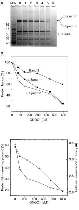
dis-Figure 1 - SDS-polyacrylamide gel electrophoresis of red blood cell ghosts (A) and percentage of the a spectrin, ß spectrin and band 3 (B) after treatment w ith different concentrations of per-oxynitrite (lanes 0, 1, 2, 3, 4, 5, 6 are 0, 74, 148, 296, 444, 592, 740 µM peroxynitrite, respec-tively), for 20 min at room tem-perature. The first band on the gel (M W) contains a mixture of proteins of different molecular masses (kDa) as reference.
all polypeptides (Figure 1A, Band 6). The disappearance of these proteins is consistent with an increase in the size of the pellet obtained prior to electrophoresis (data not shown). Loss of polypeptides through a hy-drolytic mechanism of reactive oxygen spe-cies as described by Davies and Delsignore (24), and particularly of ONOO- (25) can be excluded by measuring the total amount of ghost proteins and comparing this with the solubilized pellet content. No difference in the quantity of proteins was detected in the control or treated samples. Mallozzi et al. (26) investigated the effects of ONOO- on human erythrocytes and obtained two differ-ent responses. At low concdiffer-entration (<100 µM) ONOO- stimulated a metabolic response of phosphorylation activity and at high con-centration (>200 µM) it caused cross-link-ing of membrane proteins, nitration of ty-rosines and massive methemoglobin pro-duction.
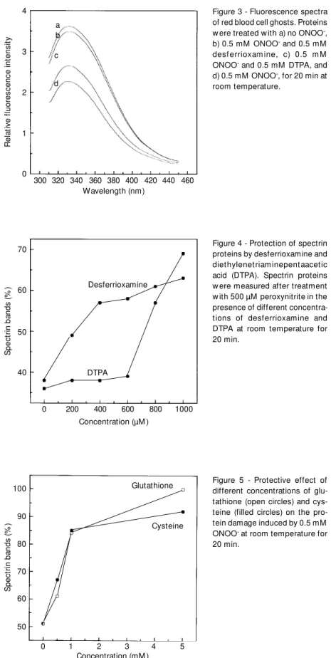
Among the major targets of ONOO- are R-SH groups (7). Figure 2 shows a dose-dependent decrease of R-SH groups (50% at
250 µM ONOO-) in ghost proteins when
treated with ONOO-. The extent of lesion to the spectrin bands was slightly different when ONOO- treatment was performed in the pres-ence of urea or under reducing conditions using ß-mercaptoethanol. This result sug-gests that crosslinking reactions other than disulfide formation contribute to aggregate formation. Thiol groups and tryptophan have
been shown to be susceptible to ONOO
-attack (7). Similarly, tryptophan residues from the ghost proteins are affected by ONOO-. The loss of R-SH shown in Figure 2 is paralleled by a decrease in fluorescence intensity at 330 nm after excitation at 295 nm after treatment with different ONOO- con-centrations. Analysis of the fluorescence spectra after treatment in the presence of 0.5 mM desferrioxamine (Figure 3, b) or 0.5 mM DTPA (Figure 3, c) showed that desfer-rioxamine has a protecting effect compared to 0.5 mM ONOO- alone. The loss of tryp-198
® ®
®
a Spectrin ß Spectrin
Band 3 116
86 66 56 40
0 1 2 3 4 5
A M W
R
e
la
ti
v
e
f
lu
o
re
s
c
e
n
c
e
i
n
te
n
s
it
y
(
)
3.5
3.0
P
ro
te
in
-S
H
(
n
m
o
l/
m
g
p
ro
te
in
)
(
) 60
50
40
30
20
0 200 400 600 800 1000 ONOO- (µM )
2.5
2.0 Figure 2 - M easurement of
sulf-hydryl groups (open circles) and tryptophan fluorescence inten-sity (filled circles) after reaction w ith different concentrations of peroxynitrite for 20 min at room temperature.
appeared at higher ONOO-concentrations. The main effect was a dose-dependent de-crease in the amount of a spectrin, ß spectrin
and band 3 (Figure 1B). At low ONOO
-concentrations, other protein bands remain intact, possibly indicating a certain specific-ity of ONOO- for the ghost proteins. Expo-sure to 740 µM peroxynitrite induced a dras-tic effect, with the disappearance of almost
P
ro
te
in
b
a
n
d
s
(
%
)
100
80
60
40
20
ß Spectrin
a Spectrin Band 3
0 100 200 300 400 500 600 ONOO- (µM )
B
·
· ·
· ·
tophan fluorescence induced by ONOO- is due to the formation of nitrotryptophan, vis-ible with the presence of a yellow color (27). These experiments were confirmed also us-ing tryptophan (data not shown).
Desferrioxamine (0.5 mM) is able to pro-tect against the disappearance of the spectrin bands (Figure 4). This confirms the results obtained for tryptophan fluorescence with 0.5 mM desferrioxamine (Figure 3), which can be explained by the scavenging effect of desferrioxamine on trans-peroxynitrite (28). Diethylenetriaminepentaacetic acid (0.5 mM) was a weak inhibitor of the ONOO- reaction (Figure 4), chelating transition metals in-volved in the process (1,10), but otherwise having no direct effect. Mannitol at 100 mM was used in the presence of ONOO- to evalu-ate the contribution of hydroxyl radicals to ghost damage. Only a 10% protective effect on the disappearance of spectrin bands could be observed (data not shown). This result shows that hydroxyl radicals are not the main reactive species in this system (11,12,28,29). However, nitrogen dioxide
formed by the decomposition of ONOO
-may also be involved in this damage (30). Glutathione and cysteine (1 mM) were highly effective in protecting spectrin against ONOO--mediated damage (Figure 5). It is known that glutathione is very important for the maintenance of erythrocyte integrity. Similarly, plasmid DNA damage caused by
ONOO- was inhibited by these compounds
in vitro (12,14).
Melatonin has been demonstrated to be a very efficient scavenger of reactive oxygen species, especially hydroxyl radicals, in bio-logical systems (31). Pieri et al. (32) demon-strated that melatonin is a better scavenger of hydroxyl radicals than a-tocopherol. In contrast, Barsacchi et al. (33) showed that vitamin E consumption by red blood cells is enhanced by melatonin under oxidative stress induced by cumene hydroperoxide or H2O2.
In other experiments, melatonin reduced the levels of DNA adduct induced by the
car-R
e
la
ti
v
e
f
lu
o
re
s
c
e
n
c
e
i
n
te
n
s
it
y
4
3
2
1
0
300 320 380 400 420 Wavelength (nm)
S
p
e
c
tr
in
b
a
n
d
s
(
%
)
100
90
80
70
60
0 1 2 3 4 5
Concentration (mM )
340 360 440 460
a b
c
d
Figure 3 - Fluorescence spectra of red blood cell ghosts. Proteins w ere treated w ith a) no ONOO-, b) 0.5 mM ONOO- and 0.5 mM desf errioxam ine, c) 0.5 m M ONOO- and 0.5 mM DTPA, and d) 0.5 mM ONOO-, for 20 min at room temperature.
50
Cysteine
Glutathione Figure 5 - Protective effect of different concentrations of glu-tathione (open circles) and cys-teine (filled circles) on the pro-tein damage induced by 0.5 mM ONOO- at room temperature for 20 min.
S
p
e
c
tr
in
b
a
n
d
s
(
%
)
70
60
50
40 DTPA
Desferrioxamine
0 200 400 600 800
Concentration (µM )
1000
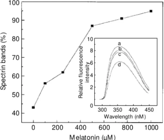
cinogen safrol (34,35). The use of this hor-mone in our model revealed that this is a very good protector of erythrocyte proteins against ONOO--induced damage. In fact, with 500 µM melatonin, the protective effect was al-most 90% (Figure 6). At lower concentra-tions, melatonin was more efficient than glu-tathione or cysteine. At physiological con-centrations (50-1000 pg/ml plasma) (36), melatonin was also able to control the quan-tity of ·NO generated via the NO-synthase reaction (37). Since melatonin has an indole moiety, like tryptophan, we also measured the fluorescence spectra of melatonin in the presence of different ONOO- concentrations (Figure 6, inset). As in the tryptophan ex-periments, a loss of fluorescence intensity
was observed, showing that melatonin scav-enges peroxynitrite.
Trolox®
(1 mM), the more water-soluble
a-tocopherol analog, was also able to pro-tect against protein damage, in a manner similar to melatonin (data not shown). Per-oxynitrite-induced erythrocyte hemolysis was also demonstrated to be partially suppressed by Trolox®
and other antioxidants (38). In-terestingly, another class of antioxidants, the pyrrolopyrimidines, prevented nitrotyrosine formation in peroxynitrite-treated red blood cell membranes, but had little effect on mem-brane cross-linking (39).
The present data show that the erythro-cyte cytoskeleton is a sensitive target for
ONOO-. Spectrin is the main target of
ONOO-, and can be protected by melatonin and thiol antioxidants. Such alterations of spectrin could seriously impair cell dynam-ics and generate morphological changes, and can be prevented by specific antioxidants.
Ackno wle dgm e nts
We thank Chuck Saker Farah and Brian Bandy, Departamento de Bioquímica, Instituto de Química, Universidade de São Paulo, for reading the manuscript.
Figure 6 - Red blood cell ghosts cytoskeleton protection by mel-atonin in the presence of per-oxynitrite. Inset: Fluorescence spect ra of 10 µM m elat onin w hen treated w ith ONOO-: a) no ONOO-, b) 0.5 mM ONOO-, c) 1 m M ONOO- and d) 2.5 m M ONOO- , for 20 min at room tem-perature.
Re fe re nce s
1. Beckm an JS, Beckm an TW , Chen J, M arshall PA & Freeman BA (1990). Appar-ent hydroxyl radical production by peroxy-nitrite: implication for endothelial injury from nitric oxide and superoxide. Proceed-ings of the National Academy of Sciences, USA, 87: 1620-1624.
2. Ischiropoulos H, Zhu L & Beckman JS (1992). Peroxynitrite formation from mac-rophage-derived nitric oxide. Archives of
Biochemistry and Biophysics, 298:
446-451.
3. Kooy NW & Royall JA (1994). Agonist-induced peroxynitrite production from en-dothelial cells. Archives of Biochemistry
and Biophysics, 310: 352-359.
4. Pryor WA, Jin X & Squadrito GL (1994).
One- and tw o-electron oxidations of me-thionine by peroxynitrite. Proceedings of
the National Academy of Sciences, USA,
91: 11173-11177.
5. van der Vliet A, O’Neill CA, Halliw ell B, Cross CE & Kaur H (1994). Aromatic hy-droxylation and nitration of phenylalanine and tyrosine by peroxynitrite. Evidence for hydroxyl radical production from per-oxynitrite. FEBS Letters, 339: 89-92. 6. Augusto O, Gatti RM & Radi R (1994).
Spin-trapping studies of peroxynitrite de-composition and of 3-morpholinosydnon-imine N-ethylcarbamide autoxidation: di-rect evidence for metal-independent for-mation of free radical intermediates.
Ar-chives of Biochemistry and Biophysics,
310: 118-125.
7. Radi R, Beckman JS, Bush KM & Free-man BA (1991). Peroxynitrite oxidation of sulfhydryls: the cytotoxic potential of su-peroxide and nitric oxide. Journal of
Bio-logical Chemistry, 266: 4244-4250.
8. Di M ascio P, Bechara EJH, M edeiros M HG, Briviba K & Sies H (1994). Singlet molecular oxygen production in the reac-tion of peroxynitrite w ith hydrogen perox-ide. FEBS Letters, 355: 287-289. 9. Beckman JS, Chen J, Ischiropoulos H &
Crow JP (1994). Oxidative chemistry of peroxynitrite. M ethods in Enzymology, 233: 229-240.
10. Lymar SV & Hurst JK (1996). Carbon diox-ide: physiological catalyst for
peroxyni-S
p
e
c
tr
in
b
a
n
d
s
(
%
)
100
90
80
70
60
50
40
0 200 400 600 800 1000
M elatonin (µM )
R
e
la
ti
v
e
f
lu
o
re
s
c
e
n
c
e
in
te
n
s
it
y
10
8
6
4
2
0
300 350 400 450 Wavelength (nM )
a b c
trite-mediated cellular damage or cellular protectant? Chemical Research in
Toxi-cology, 9: 845-850.
11. Sharov VS, Driomina ES, Briviba K & Sies H (1998). Sensitization of peroxynitrite chemiluminescence by triplet carbonyl sensitizer coumarin-525. Effect of CO2.
Photochemistry and Photobiology, 68:
797-801.
12. Bonini M G, Radi R, Ferrer-Suet a G, Ferreira AM , Da C & Augusto O (1999). Direct EPR detection of the carbonate radical anion produced from peroxynitrite and carbon dioxide. Journal of Biological
Chemistry, 274: 10802-10806.
13. Salgo M G, Bermudez E, Squadrito GL & Pryor WA (1995). Peroxynitrite causes DNA damage and oxidation of thiols in rat thymocytes. Archives of Biochemistry and
Biophysics, 322: 500-505.
14. Douki T, Cadet J & Ames BN (1996). An adduct betw een peroxynitrite and 2' -de-oxyguanosine: 4,5-dihydro-5-hydroxy-4-(nitrosooxy)-2' -deoxyguanosine.
Chemi-cal Research in Toxicology, 9: 3-7.
15. Soszynski M & Bartosz G (1996). Effect of peroxynitrite on erythrocytes. Archives of
Biochemistry and Biophysics, 1291:
107-114.
16. Bennet V (1985). The membrane skeleton of human erythrocytes and its implication for more complex cells. Annual Review of
Biochemistry, 54: 273-304.
17. Di M ascio P, Briviba K, Bechara EJH, M edeiros M HG & Sies H (1996). The re-action of peroxynitrite and hydrogen per-oxide produces singlet molecular oxygen
(1Dg). M ethods in Enzymology, 269:
395-400.
18. Homew ood CA & Neame KD (1976). A comparison of methods used for removal of w hite cells from malaria infected blood. Annals of Tropical and M edical Parasitol-ogy, 70: 249-251.
19. Tyler TM , Hargraves WR & Branton D (1980). Associations of erythrocyte mem-brane protein. Proceedings of the National
Academy of Sciences, USA, 70:
5192-5196.
20. Laemmli UK (1970). Cleavage of
struc-tural proteins during the assembly of the head of bacteriophage T4. Nature, 227: 680-685.
21. Low ry O, Rosebrough NJ, Farr AL & Randall RJ (1951). Protein measurement w ith the Folin phenol reagent. Biological
Chemistry, 183: 265-275.
22. Bradford M M (1976). A rapid and sensi-tive method for the quantification of mi-crogram quantities of protein utilizing the principle of protein dye binding. Analytical
Biochemistry, 7: 248-254.
23. Di M onte D, Ross D, Bellomo G, Eklöw L & Orrenius S (1984). Alterations in intra-cellular thiol homeostasis during the me-tabolism of menadione by isolated rat hepatocytes. Archives of Biochemistry
and Biophysics, 235: 334-342.
24. Davies RJ & Delsignore M E (1987). Pro-tein damage and degradation by oxygen radicals. Journal of Biological Chemistry, 262: 9908-9913.
25. Ischiropoulos H & Al-M ehdi AB (1995). Peroxynitrite-mediated oxidative protein modifications. FEBS Letters, 364: 279-282.
26. M allozzi C, Di Stasi AM & M inetti M (1997). Peroxynitrite modulates tyrosine-dependent signal transduction pathw ay of human erythrocyte band 3. FASEB Jour-nal, 11: 1281-1290.
27. Alvarez B, Rubbo H, Kirk M , Barnes S, Freeman BA & Radi R (1996). Peroxyni-trite-dependent tryptophan nitration.
Chemi-cal Research in Toxicology, 9: 390-396.
28. Denicola A, Souza JM , Gatti RM , Augusto O & Radi R (1995). Desferrioxamine inhi-bition of the hydroxyl radical-like reactivity of peroxynitrite: role of the hydroxamic groups. Free Radical in Biology and M edi-cine, 19: 11-19.
29. Augusto O, Radi R, Gatti RM & Vasquéz-Vivar J (1996). Detection of secondary radicals from peroxynitrite-mediated oxi-dations by electron spin resonance. M
eth-ods in Enzymology, 269: 346-354.
30. Zhu L, Gumm C & Beckman JS (1992). Bactericidal activity of peroxynitrite.
Ar-chives of Biochemistry and Biophysics,
289: 452-457.
31. Reiter RJ, M elchiorri D, Sew erynek E, Poeggeler B, Barlow -Walden L, Chuang JL, Ortiz GG & Acunacastroviejo D (1995). A review of the evidence supporting mel-atonin role as an antioxidant. Journal of
Pineal Research, 18: 1-11.
32. Pieri C, M arra M , M oroni F, Recchioni R & M archeselli F (1994). M elatonin: A peroxyl radical scavenger more effective than vi-tamin E. Life Sciences, 55: 271-276. 33. Barsacchi R, Kusmic C, Damiani E, Carloni
P, Greci L & Donato L (1998). Vitamin E consumption induced by oxidative stress in red blood cells is enhanced by melato-nin and reduced by N-acetylserotomelato-nin.
Free Radical in Biology and M edicine, 24:
1187-1192.
34. Tan DX, Poeggeler B, Reiter RJ, Chen LD, Chen S, M anchester LC & Barlow w alden LR (1993). The pineal hormone melatonin inhibits DNA-adduct formation induced by the chemical carcinogen safrole in vivo.
Cancer Letters, 70: 65-71.
35. Tan DX, Reiter RJ, Chen LD, Poeggeler B, M anchest er LC & Barlow w alden LR (1994). Both physiological and pharmaco-logical levels of melatonin reduce DNA adduct formation induced by the carcino-gen safrole. Carcinogenesis, 15: 215-218. 36. Covaci A, Doneanu C, Aboul-Enein HY & Schepens P (1999). Determination of mel-atonin in pharmaceutical formulations and human plasma by gas chromatography-electron impact mass spectrometry.
Bio-medical Chromatography, 13: 431-436.
37. Pozo D, Reiter RJ, Calvo JO & Guerrero JM (1997). Inhibition of cerebellar nitric oxide synthase and cyclic GM P produc-tion by melatonin via complex formaproduc-tion w ith calmodulin. Journal of Cell Biology, 65: 430-442.
38. Kondo H, Takahashi M & Nikin E (1997). Peroxynitrite-induced hemolysis of hu-man erythrocytes and its inhibition by an-tioxidants. FEBS Letters, 413: 236-238. 39. Rohn TT & Quinn M T (1998). Inhibition of
peroxynitrite-mediated tyrosine nitration by a novel pyrrolopyrimidine antioxidant.
European Journal of Pharmacology, 353: