Effects of melatonin on DNA damage
induced by cyclophosphamide in rats
S.G. Ferreira
1, R.A. Peliciari-Garcia
1, S.A. Takahashi-Hyodo
2, A.C. Rodrigues
3, F.G. Amaral
1,
C.M. Berra
4, S. Bordin
1, R. Curi
1and J. Cipolla-Neto
1 1Departamento de Fisiologia e Biofı´sica, Instituto de Cieˆncias Biome´dicas I, Universidade de Sa˜o Paulo, Sa˜o Paulo, SP, Brasil 2A´rea de Cieˆncias da Sau´de, Universidade Braz Cubas, Mogi das Cruzes, SP, Brasil 3Departamento de Ana´lises Clı´nicas e Toxicolo´gicas, Faculdade de Cieˆncias Farmaceˆuticas, Universidade de Sa˜o Paulo,Sa˜o Paulo, SP, Brasil 4Departamento de Microbiologia, Instituto de Cieˆncias Biome´dicas II, Universidade de Sa˜o Paulo, Sa˜o Paulo, SP, Brasil
Abstract
The antioxidant and free radical scavenger properties of melatonin have been well described in the literature. In this study, our objective was to determine the protective effect of the pineal gland hormone against the DNA damage induced by cyclophosphamide (CP), an anti-tumor agent that is widely applied in clinical practice. DNA damage was induced in rats by a single intraperitoneal injection of CP (20 or 50 mg/kg). Animals received melatonin during the dark period for 15 days (1 mg/kg in the drinking water). Rat bone marrow cells were used for the determination of chromosomal aberrations and of formamidopyrimidine DNA glycosylase enzyme (Fpg)-sensitive sites by the comet technique and ofXpfmRNA expression by qRT-PCR. The number (mean ± SE) of chromosomal aberrations in pinealectomized (PINX) animals treated with melatonin and CP (2.50 ± 0.50/100 cells) was lower than that obtained for PINX animals injected with CP (12 ± 1.8/100 cells), thus showing a reduction of 85.8% in the number of chromosomal aberrations. This melatonin-mediated protection was also observed when oxidative lesions were analyzed by the Fpg-sensitive assay, both 24 and 48 h after CP administration. The expression ofXpfmRNA, which is involved in the DNA nucleotide excision repair machinery, was up-regulated by melatonin. The results indicate that melatonin is able to protect bone marrow cells by completely blocking CP-induced chromosome aberrations. Therefore, melatonin administration could be an alternative and effective treatment during chemotherapy.
Key words: Melatonin; Cyclophosphamide; Chromosomal aberration; DNA fragmentation; Comet assay;Xpfexpression
Introduction
Melatonin, the mammalian pineal gland hormone, is essential for the entrainment of the circadian and seasonal rhythms to the light/dark cycle since its synthesis depends on photic information (1,2). Melatonin participates in the regulation of several physiological processes, acting both centrally and peripherally in a wide variety of target systems (3-5). Its nocturnal synthesis is mainly regulated by the norepinephrine released from sympathetic nerve endings, triggering the transcription and translation of arylalkylamine-N-acetyltransferase, the most important enzyme involved in melatonin synthesis (6).
Due to its amphiphilicity, melatonin can be found in any cellular compartment (7-9). However, studies suggest that the pineal gland hormone is preferentially localized inside the nucleus and can protect nuclear DNA from oxidative damage by interacting with double-stranded DNA and promoting its stability (10). Moreover, melatonin
exerts a powerful antioxidant action acting either directly on free radical species or by modulating the gene expression of antioxidant enzymes such as glutathione peroxidase, catalase and superoxide dismutase (10). It was shown that the antioxidant effect of melatonin involves DNA repair, and that the hormone can repair the oxidation induced by the guanosine (GN) radical (11).
Melatonin treatment is also effective in protecting tissues from the oxidative damage caused by glutathione deple-tion and ischemia-reperfusion injury (12,13). In addideple-tion to its antioxidant property, melatonin has been investigated as a potential antitumor agent (14-16). Indeed, the study of the effects of melatonin in chemotherapy has become an interesting area of investigation (17).
The main aim of chemotherapy is to destroy tumor cells while preserving normal ones. However, most antitumor agents act in a nonspecific way, destroying
Correspondence: J. Cipolla-Neto, Departamento de Fisiologia e Biofı´sica, Instituto de Cieˆncias Biome´dicas I, Universidade de Sa˜o Paulo, Av. Prof. Lineu Prestes, 1524, 05508-900 Sa˜o Paulo, SP, Brasil. E-mail: [email protected]
both normal and malignant cells. Cyclophosphamide (CP) is one of the most frequently used antitumor agents in clinical practice (18). Nevertheless, research on its mechanisms of action has shown that CP alkylates nucleophilic macromolecules, including DNA. It is also capable of inducing depurination, depyrimidation, mono-adduct formation, and DNA-DNA and DNA-protein cross-links (19). Likewise, CP induces gene mutations, DNA-strand breaks, chromosome aberrations, micronuclei and sister chromatid exchanges, apoptosis and generation of free radicals (20). Based on these observations, the International Agency for Research on Cancer concluded that there is sufficient evidence to classify CP as a carcinogenic agent for animals and humans (21).
Therefore, considering the known protective proper-ties of melatonin, the aim of the present study was to evaluate the effect of this hormone on CP-induced chromosomal aberrations, increase of formamidopyrimi-dine DNA glycosylase (Fpg)-sensitive sites and Xpf
expression in bone marrow cells of intact and pine-alectomized rats.
Material and Methods
Animals
Male Wistar rats aged 4-5 weeks (90-110 g) were obtained from the animal facility of the Instituto de Cieˆncias Biome´dicas, Universidade de Sa˜o Paulo, SP, Brazil. Animals were kept under a 12-h light/dark cycle (light: fluorescent light, 200/300 lux at cage level; dark: red filter Kodak 1A, 0.5 to 1 lux, lights on at 6:00 am, 21 ± 26C) with food and waterad libitum. The protocol is in accordance with the Ethics Principles for Animal Research adopted by the Brazilian College of Animal Experimentation (COBEA) and was approved by the Ethics Committee for Animal Research of the Instituto de Cieˆncias Biome´dicas (protocol
#048/03).
Experimental design
The present study includes the following experimental groups: control (intact animals); MEL (control animals supplemented with daily nocturnal melatonin administra-tion); PINX (pinealectomized rats); PINX+MEL (pine-alectomized rats treated with melatonin in the drinking water consumed at night); CP (intact animals treated with cyclophosphamide); CP+MEL (intact animals injected with cyclophosphamide and supplemented with melato-nin); CP+PINX (pinealectomized rats treated with cyclophosphamide); CP+PINX+MEL (pinealectomized rats treated with melatonin and injected with cyclophos-phamide).
Experimental and surgical procedures
Animals were anesthetized with pentobarbital (40 mg/kg) and then submitted to pinealectomy by the method of Roffman and Reiter (22). Briefly, anesthetized
animals were placed in a stereotaxic apparatus (David Kopf Instruments, USA) and a sagittal opening was made in the scalp. The skin and muscles were pulled apart to expose the lambda confluence for skull suturing. A disc-shaped opening was made around the lambda with a circular drill, whereupon the pineal gland (which is located just below the posterior venous sinus confluence) was removed with a thin forceps. After brief homeostasis, the disc-shaped piece of bone was replaced and the scalp sutured with cotton thread.
Melatonin administration
Melatonin (Sigma, USA) was administered orally in the drinking water during the dark period at 1 mg/kg body weight for 15 days. During the light period, tap water was available to the animals. Plain water bottles were replaced with melatonin-containing water bottles from the begin-ning to the end of the dark period.
Administration of cyclophosphamide
A single intraperitoneal injection of CP (20 or 50 mg/kg, ASTA Me´dica Ltda., Brazil) was administered on the 15th day after pinealectomy and/or at the beginning of melatonin supplementation (23). The latter concentra-tion was used solely in DNA fragmentaconcentra-tion studies.
Chromosome analysis
Determination of Fpg-sensitive sites by the Fpg-comet assay
The alkaline comet assay at pH 13 was carried out as previously described (29). Briefly, one or two drops of blood were collected from the animals’ tails 24 and 48 h after CP treatment (50 mg/kg). Seven microliters of cell/heparin mixture was then embedded in 93mL LMP agarose (0.50 g/100 mL PBS). The resulting mixture was spread over a pre-coated microscope slide for 5 min at 46C to allow gel solidification. The cells were then lysed (2.5 M NaCl, 100 mM EDTA, 10 mM Tris, pH 10, plus 1% Triton-X 100 and 10% dimethyl sulfoxide added just before use), and kept at 46C for 1 h. The Fpg enzyme was used in the comet assay because it recognizes the 8-OH guanine and formami-dopyrimidine that occur in spontaneous breaks in damaged purines (30). Fpg is involved in the first step of the base excision repair by removing AP-generating modified DNA bases at a site that is cut by AP-lyase activity, thereby resulting in a gap in the DNA strand detectable by the comet assay. When the comet assay was combined with bacterial Fpg, the slices were washed with an enzyme buffer (40 mM HEPES, 100 mM KCl, 0.5 mM EDTA, 0.2 mg/mL BSA, pH 8.0), covered with 60mL of buffer alone or buffer with Fpg protein (1:2000), sealed with a coverslip, and finally incubated for 30 min at 376C. Slices with and without Fpg post-treatment were denatured for 20 min and submitted to electrophoresis for a further 20 min. The slices were coded and images of 50 randomly selected cells stained with ethidium bromide were obtained using a fluorescent microscope Nikon Eclipse E1000 (USA) attached to a Nikon FDX35 video camera equipped with excitation (528/553 nm) and barrier (590 nm) filters. The parameter used to express the DNA damage was tail moment. Tail moment (numerical measurement of DNA damage) is the product of the length of the ‘‘tail’’ of DNA trailing the nucleus and the percentage of total DNA in the tail. Higher tail moments indicate greater DNA damage (30). The comets (tail moment) were analyzed using the Scion Image Corporation software (Comet 1.3 application).
RNA isolation and cDNA synthesis
Total RNA was extracted from bone marrow cells using TrizolH Reagent (InvitrogenH Corporation, USA) according to manufacturer instructions. RNA was dis-solved in DEPC water and the concentration and purity of each sample was determined with A260/A280
measure-ments (mQuant, BioTeK Instruments, Inc., USA). A 5-mg aliquot of total RNA was reverse-transcribed using SuperScript III Reverse Transcriptase (InvitrogenH
Corporation; 200 U; 256C for 10 min, 426C for 75 min, 706C for 15 min) and random primers (656C, 5 min). cDNA samples of bone marrow cells were stored at -206C prior to the analysis.
Quantitative PCR
The best concentrations of primers and samples, as well as the most appropriate annealing temperature were established prior to qPCR. The reactions were carried out using 12.5mL PlatinumH SyBrGreen qPCR Super Mix-UDG (InvitrogenH), 42.5 ng cDNA and 200 nM of each primer (Table 1). The amplification conditions for qPCR consisted of an initial step at 956C for 10 min followed by 40 cycles, each at 956C for 15 s, 606C for 30 s and 726C for 30 s. Primer specificity was assessed by analysis of the melting curve that consisted of heating the samples from 606 to 996C (incremental changes of 16C at 5-s intervals) after the 40th cycle. All sample measurements were performed in duplicate. Data were generated by the Rotor-Gene Real-Time Analysis Software 6.0 (Rotor Gene 3000 Real-Time PCR System, Corbett Research, Australia).
All quantifications were normalized to the house-keeping genes beta-2-microglobulin (B2m) and histone deacetylase 1 (Hdac1). The relative quantification value of each target gene was analyzed using the comparative CT
method (31). 2-DDCT was used to calculate the relative amount of transcript in the sample, normalized to B2m
andHdac1, whereDCTis the difference between the CT
of the gene of interest and the CTof both housekeeping
genes and DDCT for the sample = mean DCT of the
sample - mean DCT of the control sample (used as
calibrator).
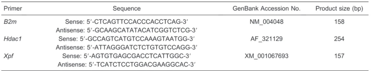
Table 1. Primer sequences for ratXpf,Hdac1andB2mused in qRT-PCR assays.
Primer Sequence GenBank Accession No. Product size (bp)
B2m Sense: 59-CTCAGTTCCACCCACCTCAG-39 NM_004048 158
Antisense: 59-GCAAGCATATACATCGGTCTCG-39
Hdac1 Sense: 59-GCCAGTCATGTCCAAAGTAATGG-39 AF_321129 254
Antisense: 59-ATTAGGGATCTCTGTGTCCAGG-39
Xpf Sense: 59-AGTGTGAGCGACCTCATTGGC-39 XM_001067693 157
Antisense: 59-TCATCTCCTGGACGAAGGCAC-39
Statistical analysis
Data are reported as means ± SE and were analyzed statistically by one- or two-way analysis of variance (ANOVA), followed by the Bonferroni multiple compar-isons post hoc test (GraphPad Software Inc., USA). Differences were considered to be significant when P,
0.05.
Results
Effect of melatonin on mitotic index and chromosomal aberrations induced by cyclophosphamide
The slide analysis of a rat bone marrow smear from control animals revealed a frequency of 7 types of chromosomal aberrations/600 analyzed cells (1.20 ± 0.40/100 cells), while MEL supplementation did not change the spontaneous incidence of chromosomal aberrations, presenting 4 types/600 analyzed cells (0.67 ± 0.21/100 cells). Cells from PINX animals presented 27 types of chromosomal aberrations/600 analyzed cells (4.50 ± 0.89/100 cells). This result represents a significant increase in comparison to the spontaneous mutations detected in intact animals from the control group. On the contrary, bone marrow cells from pine-alectomized animals that received melatonin (PINX+MEL) daily, beginning the first night after surgery, presented reduced levels of spontaneous aberrations that were similar to control levels, showing 6 types of aberrations/ 600 analyzed cells (1.00 ± 0.26/100 cell), which represents a 75% reduction compared to the PINX group. CP injection performed 24 h before sacrifice induced 70 types of chromosomal aberrations/600 cells (11.00 ± 1.30/100 cells). On the other hand, a complete blockage of CP-induced mutations was observed when the animals
were supplemented with melatonin (CP+MEL), showing 8 aberrations/600 cells (1.30 ± 0.21/100 cells). This result reveals a reduction of 83.8% in the number of aberrant chromosomes. PINX animals injected with CP (CP+PINX) presented 71 types of chromosomal aberrations (12.00 ± 1.80/100 cells). As expected, the reposition of melatonin in the CP+PINX group (CP+PINX+MEL) was able to inhibit the development of chromosomal aberrations, reducing the total number of aberrant cells to values similar to those observed in control animals (2.50 ± 0.50), i.e., a reduction of 85.8% (Tables 2 and 3).
Regarding the rate of cells that are in division (MI), MEL supplementation was able to increase the MI compared to the control group (2.52 ± 0.09vs 1.74 ±
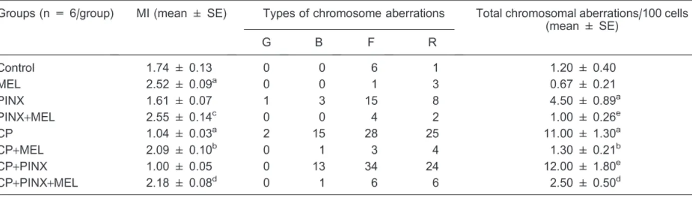
Table 2. Effect of melatonin on the mitotic index (MI) and frequency of cells with chromosome aberrations induced by cyclophosphamide (CP, 20 mg/kg).
Groups (n = 6/group) MI (mean ± SE) Types of chromosome aberrations Total chromosomal aberrations/100 cells (mean ± SE)
G B F R
Control 1.74 ± 0.13 0 0 6 1 1.20 ± 0.40
MEL 2.52 ± 0.09a 0 0 1 3 0.67 ± 0.21
PINX 1.61 ± 0.07 1 3 15 8 4.50 ± 0.89a
PINX+MEL 2.55 ± 0.14c 0 0 4 2 1.00 ± 0.26e
CP 1.04 ± 0.03a 2 15 28 25 11.00 ± 1.30a
CP+MEL 2.09 ± 0.10b 0 1 3 4 1.30 ± 0.21b
CP+PINX 1.00 ± 0.05 0 13 34 24 12.00 ± 1.80e
CP+PINX+MEL 2.18 ± 0.08d 0 1 6 6 2.50 ± 0.50d
G = gaps; B = breaks; F = fragments; R = rearrangements; MEL = melatonin; PINX = pinealectomized rats; CP = intact animals treated with CP.aP,0.05vscontrol group;bvsCP;cvsPINX;dvsPINX+CP;evsPINX. For MI, 1000 cells were analyzed per animal (a total of 6000 cells/treatment). For chromosomal aberrations, 600 cells were analyzed per group. Statistical significance between means of cells with chromosome aberrations and the MI was analyzed using one-way ANOVA followed by the Newman-Keuls multiple comparisonspost hoctest.
Table 3. Analysis of chromosomal aberrations after melatonin treatment.
Treatments Observed Expected Decrease (%)
Control 7 -
-MEL 4 -
-PINX 27 -
-PINX+MEL 6 24 75
CP 68 -
-CP+MEL 8 65 83.8
CP+PINX 71 88 19.3
CP+PINX+MEL 13 92 85.8
0.13) while the CP group presented a clear reduction (1.04 ± 0.03). Melatonin treatment promoted an increase in MI in the PINX and CP groups (PINX+MEL and CP+MEL) compared to the respective controls (2.55 ± 0.14vs1.61 ± 0.07 and 2.09 ± 0.10vs1.04 ± 0.03). Pinealectomy (PINX group)per sedid not change the MI (1.61 ± 0.07) compared to the control group. Melatonin reposition also increased the MI of the CP+PINX+MEL
group compared to its control (2.18 ± 0.08 vs 1.00 ± 0.05).
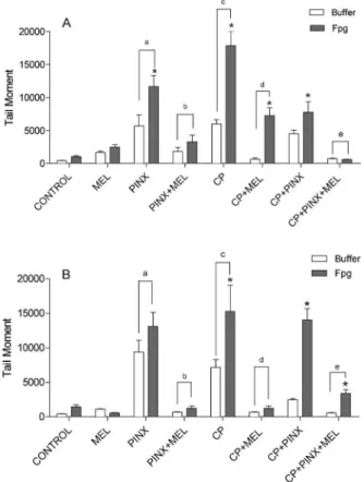
Determination of Fpg-sensitive sites by the comet assay
An increase in DNA migration (tail moment) was observed under both conditions (buffer and Fpg-sensitive sites) 24 and 48 h after CP administration. Melatonin
treatment elicited a considerable decrease in oxidative damage (more than 50%), promoting the reduction not only of Fpg-sensitive sites but also of the buffer condition to their control values. This melatonin-mediated reduction effect was better observed 48 h after CP administration, although a significant reduction was already seen 24 h
after CP injection. Melatonin supplementation alone did not induce DNA damage or the appearance of Fpg-sensitive sites. The absence of circulating melatonin in PINX animals induced an increase in the tail moment of buffer and Fpg-sensitive sites compared to the control groups. Moreover, when PINX animals were supplemented with melatonin (PINX+MEL), a complete prevention of DNA oxidative damage was observed. The positive effect of melatonin was also seen in PINX animals injected with CP compared to CP+PINX+MEL animals (Figures 1A and B and 2A and B).
Melatonin treatment and Xpf mRNA expression
Besides being involved in the DNA nucleotide excision repair (NER) machinery, theXpfgene is also responsible for the cleavage in DNA lesions. The present results show that melatonin supplementation associated with a highly oxidative damage condition such as CP+PINX, PINX or CP was able to increase XpfmRNA expression in bone marrow cells, while individual conditions such as PINX, MEL, CP, or even CP+PINX did not cause modifications ofXpfmRNA expression (Figure 3).
Discussion
In this study, we aimed to investigate the protective effect of melatonin on CP-induced chromosomal damage in pinealectomized rats. The present melatonin treatment regimen gave us support to determine the undeniable protective effects of the pineal gland hormone against induced DNA damage. The ablation of plasma melatonin
Figure 2. A, Quantification of sensitive sites by the Fpg-comet assay 24 h after treatment with 50 mg/kg cyclophos-phamide (CP). The damage measured by alkaline comet assay was estimated as the percentage of DNA in the tail after treatment with Fpg (filled bars) and buffer without Fpg (open bars). Tail moment (numerical measurement of DNA damage) is the product of the length of the ‘‘tail’’ of DNA trailing the nucleus and the percentage of total DNA in the tail. Fpg = formamido-pyrimidine DNA glycosylase enzyme; MEL = melatonin; PINX = pinealectomized animals. Data are reported as means ± SE for n = 5 animals per group. *P,0.05vstheir respective control;aP
,0.05vscontrol and MEL;bP,0.05vsPINX;cP,0.05vs control, MEL and PINX+MEL;dP,0.05vsCP;eP
,0.05vs CP+PINX (two-way ANOVA followed by the Bonferroni multiple comparisons post hoc test).B, Quantification of Fpg-sensitive sites by Fpg-comet assay 48 h after treatment with 50 mg/kg CP. The damage measured by alkaline comet assay was estimated as the percentage of DNA in the tail after treatment with Fpg (filled bars) and buffer without Fpg (open bars). Data are reported as means ± SE for n = 5 animals per group. *P,0.05vstheir respective control;aP,0.05vscontrol and MEL;bP,0.05vs PINX;cP,0.05vscontrol, MEL and PINX+MEL;dP,0.05vs CP;eP,0.05vsCP
+PINX (two-way ANOVA followed by the Bonferroni multiple comparisonspost hoctest).
due to pinealectomy also revealed the ineffectiveness of the cells in overcoming oxidative lesions.
The reduction of blood melatonin levels caused by pinealectomy has been demonstrated by many investiga-tors, and this results in a complete ablation of the circadian pattern of melatonin release by the pineal gland (32). Although sham-operated animals (a similar surgical procedure without excising the pineal gland) showed urinary excretion of a melatonin metabolite (33), sham surgery was not performed in the present study because the unintentional damage of peripheral innervations of the gland could compromise the well known modulatory effect of melatonin synthesis exerted by these afferents (9).
We demonstrated here that melatonin is able to prevent the spontaneous formation of chromosomal aberrations, which was found to be higher in PINX animals than in melatonin-treated PINX animals. These results agree with data presented by De Salvia et al. (34), who showed that melatonin treatment promotes a reduc-tion in the number of abnormal CHO cells induced by H2O2 and CP. Our data indicate that melatonin at the
concentration tested is not cytotoxic and may confer anti-mutagenic activity against CP-induced chromosome aberrations. Melatonin also induced a significant increase in the MI of bone marrow cells in mitotic metaphase, indicating an increase in the rate of cell division. Thus, our findings are in agreement with the reported protective effects of melatonin on bone marrow of rats exposed to cytotoxic drugs (16).
The mechanism of action of melatonin on bone marrow cells has been previously investigated. Some authors have discussed that the protective effect of melatonin is due to its antioxidant capacity, which prevents bone marrow damage by stimulating cell growth and increasing glutathione levels (8,35). The stimulatory effect of melatonin on cell growth in lymphatic tissues was also described in the literature (36). In addition, since melatonin is able to enter the cell nucleus and interact with chromatin, this could explain its action against CP alkylating damage, which can promote the distortion of the DNA double helix (10). Moreover, melatonin could also release IL-2 from granulocytes and stimulate bone marrow cells, acting as a growth factor (37).
To the best of our knowledge, this is the first study that used the comet assay to evaluate cells from PINX animals treated with melatonin, although the role of melatonin in reducing oxidative injury has already been demonstrated
by the use of the same technique in other models (38,39). Our results demonstrated a significant CP-induced DNA damage in peripheral lymphocytes, additionally increased in the presence of Fpg. In addition to melatonin being able to reduce these damages 24 h after CP injection, with complete normalization after 48 h, a reduction of the spontaneous increase in oxidative damage induced by pinealectomy was also observed. These results indicate that melatonin, in addition to its well-known antioxidant activity, could be effective in mobilizing DNA repair mechanisms. A previous study suggested that melatonin can activate either DNA repairing enzymes or those genes responsible for initiating new protein kinase C-mediated DNA synthesis (35).
Moreover, the components of the NER machinery were evaluated. Among several genes studied, Xpfwas the one that revealed a more consistent increase in its expression in response to melatonin. Since Xpf partici-pates in the final steps of the NER pathway involving nucleotide excision repair around the lesion, it is possible that mechanisms involved in DNA repair might be mediated by theXpfgene and should be further analyzed. The expressions of other genes involved in the repair mechanisms (Top1, Csb) or in the cell cycle (P53,P21) or even in DNA fragmentation (Bcl-2, Bax) were not consistently altered in response to the presence or absence of melatonin (data not shown).
Our results demonstrated that melatonin treatment alone is neither mutagenic nor toxic. Pinealectomyper se
leads to oxidative DNA lesions, which were reduced by melatonin treatment. Melatonin was capable of protecting bone marrow cells by completely blocking CP-induced chromosome aberrations. In addition, the melatonin-induced Xpf gene expression facilitated the repair of CP-induced DNA damage, as shown by the comet assay. According to the present data, it is possible to consider the benefits of the therapeutic use of melatonin in addition to chemotherapy medication.
Acknowledgments
The authors thank Prof. Dr. Carlos Frederico Martins Menck (ICB/USP), Prof. Dr. Kayo Okazaki (IPEN) and Julieta S. Falca˜o for technical assistance. Research supported by CAPES (to S.G. Ferreira) and FAPESP (#04/06767-2 to J. Cipolla-Neto).
References
1. Ganguly S, Coon SL, Klein DC. Control of melatonin synthesis in the mammalian pineal gland: the critical role of serotonin acetylation. Cell Tissue Res2002; 309: 127-137, doi: 10.1007/s00441-002-0579-y.
2. Goldman BD. Mammalian photoperiodic system: formal properties and neuroendocrine mechanisms of
photoper-iodic time measurement.J Biol Rhythms2001; 16: 283-301, doi: 10.1177/074873001129001980.
4. Reiter RJ, Tan DX, Sainz RM, Mayo JC, Lopez-Burillo S. Melatonin: reducing the toxicity and increasing the efficacy of drugs. J Pharm Pharmacol2002; 54: 1299-1321, doi: 10.1211/002235702760345374.
5. Afeche SC, Amaral FG, Villela DCM, Abrahao MV, Peres R, Cipolla-Neto J. Melatonin and pineal gland. In: Romano E, De Luca S (Editors), New research on neurosecretory systems. Hauppauge: Nova Science Publishers; 2008. p 151-177.
6. Klein DC. Arylalkylamine N-acetyltransferase: ‘‘the Timezyme’’. J Biol Chem 2007; 282: 4233-4237, doi: 10.1074/jbc.R600036200.
7. Reiter RJ, Tan DX, Manchester LC, Tamura H. Melatonin defeats neurally-derived free radicals and reduces the associated neuromorphological and neurobehavioral damage.J Physiol Pharmacol2007; 58 (Suppl 6): 5-22. 8. Rodriguez C, Mayo JC, Sainz RM, Antolin I, Herrera F,
Martin V, et al. Regulation of antioxidant enzymes: a significant role for melatonin. J Pineal Res2004; 36: 1-9, doi: 10.1046/j.1600-079X.2003.00092.x.
9. Simonneaux V, Ribelayga C. Generation of the melatonin endocrine message in mammals: a review of the complex regulation of melatonin synthesis by norepinephrine, pep-tides, and other pineal transmitters.Pharmacol Rev2003; 55: 325-395, doi: 10.1124/pr.55.2.2.
10. Tan D, Reiter RJ, Chen LD, Poeggeler B, Manchester LC, Barlow-Walden LR. Both physiological and pharmacological levels of melatonin reduce DNA adduct formation induced by the carcinogen safrole.Carcinogenesis1994; 15: 215-218, doi: 10.1093/carcin/15.2.215.
11. Mahal HS, Sharma HS, Mukherjee T. Antioxidant properties of melatonin: a pulse radiolysis study.Free Radic Biol Med 1999; 26: 557-565, doi: 10.1016/S0891-5849(98)00226-3. 12. Vijayalaxmi, Meltz ML, Reiter RJ, Herman TS. Melatonin
and protection from genetic damage in blood and bone marrow: whole-body irradiation studies in mice. J Pineal Res 1999; 27: 221-225, doi: 10.1111/j.1600-079X.1999. tb00618.x.
13. Oz E, Ilhan MN. Effects of melatonin in reducing the toxic effects of doxorubicin.Mol Cell Biochem2006; 286: 11-15, doi: 10.1007/s11010-005-9003-8.
14. Blask DE. Melatonin, sleep disturbance and cancer risk. Sleep Med Rev 2009; 13: 257-264, doi: 10.1016/j.smrv. 2008.07.007.
15. Reiter RJ. Melatonin: clinical relevance.Best Pract Res Clin Endocrinol Metab2003; 17: 273-285, doi: 10.1016/S1521-690X(03)00016-2.
16. Martins E Jr, Fernandes LC, Bartol I, Cipolla-Neto J, Costa Rosa LF. The effect of melatonin chronic treatment upon macrophage and lymphocyte metabolism and function in Walker-256 tumour-bearing rats.J Neuroimmunol1998; 82: 81-89, doi: 10.1016/S0165-5728(97)00191-4.
17. Messina G, Lissoni P, Marchiori P, Bartolacelli E, Brivio F, Magotti L. Enhancement of the efficacy of cancer che-motherapy by the pineal hormone melatonin and its relation with the psychospiritual status of cancer patients. J Res Med Sci2010; 15: 225-228.
18. Salvadori DM, Ribeiro LR, Oliveira MD, Pereira CA, Becak W. The protective effect of beta-carotene on genotoxicity induced by cyclophosphamide.Mutat Res1992; 265: 237-244, doi: 10.1016/0027-5107(92)90052-4.
19. Hengstler JG, Hengst A, Fuchs J, Tanner B, Pohl J, Oesch F. Induction of DNA crosslinks and DNA strand lesions by cyclophosphamide after activation by cytochrome P450 2B1.Mutat Res1997; 373: 215-223, doi: 10.1016/S0027-5107(96)00200-X.
20. S el v ak umar E , Pra ha la th an C, Va r al aks hm i P, Kumarasamy P, Saravanan R. Modification of cyclopho-sphamide-induced clastogenesis and apoptosis in rats by alpha-lipoic acid.Mutat Res2006; 606: 85-91, doi: 10.1016/ j.mrgentox.2006.03.005.
21. Franke SI, Pra D, Erdtmann B, Henriques JA, da Silva J. Influence of orange juice over the genotoxicity induced by alkylating agents: an in vivoanalysis. Mutagenesis2005; 20: 279-283, doi: 10.1093/mutage/gei034.
22. Hoffman RA, Reiter RJ. Rapid pinealectomy in hamsters and other small rodents.Anat Rec1965; 153: 19-21, doi: 10.1002/ar.1091530103.
23. Anderson D, Bishop JB, Garner RC, Ostrosky-Wegman P, Selby PB. Cyclophosphamide: review of its mutagenicity for an assessment of potential germ cell risks.Mutat Res1995; 330: 115-181, doi: 10.1016/0027-5107(95)00039-L. 24. Ford CE, Hamerton JL. A colchicine, hypotonic citrate,
squash sequence for mammalian chromosomes. Stain Technol1956; 31: 247-251.
25. Preston RJ, Dean BJ, Galloway S, Holden H, McFee AF, Shelby M. Mammalianin vivocytogenetic assays. Analysis of chromosome aberrations in bone marrow cells. Mutat Res 1987; 189: 157-165, doi: 10.1016/0165-1218(87) 90021-8.
26. Savage JR. Classification and relationships of induced chromosomal structual changes. J Med Genet 1976; 13: 103-122, doi: 10.1136/jmg.13.2.103.
27. Krishna G, Kropko ML, Ciaravino V, Theiss JC. Simultaneous micronucleus and chromosome aberration assessment in the rat. Mutat Res1991; 264: 29-35, doi: 10.1016/0165-7992(91)90042-3.
28. Swierenga SH, Heddle JA, Sigal EA, Gilman JP, Brillinger RL, Douglas GR, et al. Recommended protocols based on a survey of current practice in genotoxicity testing labora-tories, IV. Chromosome aberration and sister-chromatid exchange in Chinese hamster ovary, V79 Chinese hamster lung and human lymphocyte cultures.Mutat Res1991; 246: 301-322, doi: 10.1016/0027-5107(91)90050-X.
29. Hartmann A, Herkommer K, Gluck M, Speit G. DNA-damaging effect of cyclophosphamide on human blood cells in vivoandin vitrostudied with the single-cell gel test (comet assay). Environ Mol Mutagen 1995; 25: 180-187, doi: 10.1002/em.2850250303.
30. Collins AR. Investigating oxidative DNA damage and its repair using the comet assay.Mutat Res2009; 681: 24-32, doi: 10.1016/j.mrrev.2007.10.002.
31. Livak KJ, Schmittgen TD. Analysis of relative gene expression data using real-time quantitative PCR and the 2(-Delta Delta C(T)) Method.Methods2001; 25: 402-408, doi: 10.1006/meth.2001.1262.
32. Agez L, Laurent V, Guerrero HY, Pevet P, Masson-Pevet M, Gauer F. Endogenous melatonin provides an effective circadian message to both the suprachiasmatic nuclei and the pars tuberalis of the rat.J Pineal Res2009; 46: 95-105, doi: 10.1111/j.1600-079X.2008.00636.x.
obligatory for the regulation of the rat sleep-wake cycle. Sleep2010; 33: 833-840.
34. De Salvia R, Fiore M, Aglitti T, Festa F, Ricordy R, Cozzi R. Inhibitory action of melatonin on H2O2- and cyclopho-sphamide-induced DNA damage. Mutagenesis 1999; 14: 107-112, doi: 10.1093/mutage/14.1.107.
35. Vijayalaxmi, Reiter RJ, Meltz ML, Herman TS. Melatonin: possible mechanisms involved in its ‘radioprotective’ effect. Mutat Res 1998; 404: 187-189, doi: 10.1016/S0027-5107(98)00112-2.
36. Maestroni GJ. The immunoneuroendocrine role of melato-nin.J Pineal Res1993; 14: 1-10, doi: 10.1111/j.1600-079X. 1993.tb00478.x.
37. Lissoni P, Pittalis S, Brivio F, Tisi E, Rovelli F, Ardizzoia A, et al.
In vitromodulatory effects of interleukin-3 on macrophage activation induced by interleukin-2.Cancer 1993; 71: 2076-2081, doi: 10.1002/1097-0142(19930315)71:6, 2076::AID-CNCR2820710624.3.0.CO;2-I.
38. Musatov SA, Anisimov VN, Andre V, Vigreux C, Godard T, Gauduchon P, et al. Modulatory effects of melatonin on genotoxic response of reference mutagens in the Ames test and the comet assay. Mutat Res 1998; 417: 75-84, doi: 10.1016/S1383-5718(98)00094-1.