ABSTRACT: Enteric methane (CH4) emissions in ruminants have attracted considerable atten-tion due to their impact on greenhouse gases and the contribuatten-tion of agricultural practices to global warming. Over the last two decades, a number of approaches have been adopted to mitigate CH4 emissions. However, the mechanisms of methanogenesis have still not been fully defined. According to the genome sequences of M. ruminantium in the rumen and of M. AbM4 in the abomasum, the pathways of carbon dioxide (CO2) reduction and formate oxidation to CH4 have now been authenticated in ruminants. Furthermore, in the light of species or genera description of methanogens, the precursors of methanogenesis discovered in the rumen and research advances in related subjects, pathways of acetate dissimilation via Methanosarcina and Methanosaeta as well as metabolism of methanol to CH4 might be present in the rumen, although neither process has yet been experimentally demonstrated in the rumen. Herein the research advances in methanogenesic mechanisms including existing and potential mechanisms are reviewed in detail. In addition, further research efforts to understand the methanogenesis mechanism should focus on isolation and identification of more specific methanogens, and their genome sequences. Such increased knowledge will provide benefits in terms of improved dietary energy utilization and a reduced contribution of enteric CH4 emissions to total global greenhouse gas emissions from the ruminant production system.
Keywords: methanogenesis, carbon dioxide, formate, acetate, methanol
Introduction
It is predicted that the global surface temperature will increase between 1 oC to 6 oC during the
twenty-first century, primarily due to the increased emissions of greenhouse gases in the atmosphere. With over 21 times more heat per molecule than carbon dioxide (CO2), meth-ane (CH4) is a particularly potent greenhouse gas and accounts for 16 % of total global greenhouse gas emis-sions. The CH4 formation is a microbial-driven process, mainly dominated by methanogens, which are members of the Archaea domain and inhabit certain anaerobic environments, such as freshwater sediments abundant in organic matter, swamps and waterlogged soils, sew-age treatment plants as well as the rumen of ruminants (Woese et al., 1978). Nowadays, ruminants can produce globally more than 80 million tons of CH4, which annu-ally, accounts for 28 % of anthropomorphic emissions, and has drawn attention to the contribution of animal agriculture to global warming (Beauchemin et al., 2008). In ruminants, enteric CH4 emissions not only contribute to global climate warming, but also account for 2 ~ 12 % of the ingested energy. In particular, for high-produc-ing lactathigh-produc-ing animals, at least 6 % of gross energy intake is lost by way of CH4 emissions. Therefore, mitigating CH4 emissions in ruminants will not only assist in the achievement of international commitments under the Kyoto Protocol but also in the improvement of energy utilization efficiency and the performance of the host animal. Any mitigation option should be undertaken on the basis of a clear understanding of the mechanism of
methanogenesis in order to ensure a specific interven-tion in methanogens that maintains the normal digestive functions of other microbes in the rumen. This review will comprehensively describe the species or genera of methanogens and all the pathways of methanogenesis in ruminants.
Methanogens in the rumen
Methanogens belong to the phylum Euryarchaeota of the domain Archaea, and are divided into the fol-lowing five orders, namely Methanosarcinales, Metha-nomicrobiales, Methanobacteriales, Methanococcales and Methanopyrales. In the rumen, methanogens are a large and diverse group of Archaea. Usually, the populations of methanogens range from 107 to 109 g−1 of rumen
con-tents in concentrate-fed ruminants and are up to 109
to 1010 g−1 of rumen contents in pasture-fed ruminants
(Attwood et al., 2011). To date, methanogens, such as Methanobrevibacter ruminantium, Methanobrevibacter smithii, Methanobrecibacter millerae, Methanobrevibacter olleyae, Methanobacterium formicicum, Methanobacterium bryantii, Methanosarcina barkeri, Methanosarcina mazai and Methanomicrobium mobile have been isolated by cul-ture methods (Janssen and Kirs, 2008; Sirohi et al., 2010; Kumar et al., 2012).
The above-mentioned isolated species have been proven to utilize limited substrates to produce CH4 (Ta-ble 1). However, application of molecular technologies has further revealed that there is considerable genetic diversity in methanogens in the rumen. Methanosphaera stadtmanae, Methanobrevibacter thaueri, Methanobrevi-1The Chinese Academy of Sciences/Institute of Subtropical
Agriculture − Key Lab. for Agro-Ecological Processes in Subtropical Region/South-Central Experimental Station of Animal Nutrition and Feed Science in Ministry of Agriculture – 410125 – Hunan, PR – China.
2University of the Chinese Academy of Sciences – 100000 – Beijing, PR – China.
*Corresponding author <[email protected]>
Edited by: Concepta Margaret McManus Pimentel
Potential and existing mechanisms of enteric methane production in ruminants
Junyi Qiao1,2, Zhiliang Tan1*, Min Wang1
bacter gottschalkii and others have also been identified by the 16S rRNA PCR (Pei et al., 2010; Franzolin et al., 2012). In general, the majority of rumen methanogens de-tected can be mainly classified into three genera by the analysis of pooled data from several surveys; these are Methanobrevibacter, Methanomicrobium and rumen un-cultured cluster C (Tymensen and McAllister, 2012). Of these, Methanobrevibacter accounts for nearly two-thirds (62 %) of rumen methanogens, Methanomicrobium and ru-men uncultured cluster C are roughly equal and account for 15 % and 16 %, respectively (Janssen and Kirs, 2008; St-Pierre and Wright, 2013). Nonetheless, the proportion of these groups is reported to differ greatly (Morgavi et al., 2010; St-Pierre and Wright, 2013); these differences may be due to methodological differences or result from effects of animal species and/or diets. The remaining gen-era of methanogens should also include Methanosphagen-era, Methanobacterium and Methanosarcina. In our opinion, the fast development of molecular technologies allows for identification, diversity and colonization of more specific and functional methanogens in the rumen.
Pathways of methanogenesis
In the rumen, methanogens produce CH4 from a limited amount of substrates, namely H2 + CO2, for-mate, methanol and acetate (Oppermann et al., 1961; Hungate et al., 1970; Neumann et al., 1999; Rea et al., 2007). The H2 + CO2, formate and acetate are derived from carbohydrate fermentation, whereas methanol comes from pectin fermentation. As of today, the path-ways of methanogenesis from all above-mentioned pre-cursors have yet to be fully defined in the rumen. As rumen methanogens are difficult to culture, it is useful to gain a better understanding of their metabolism and physiology and to define the pathways of CH4 produc-tion in the rumen with the help of genome sequencing technology. Through the application of such technol-ogy, M. ruminantium and Methanobrevibacter. AbM4, two prominent methanogens found in the rumen and abomasum respectively, have been confirmed to contain two complete methanogenic pathways for reduction of
CO2 and oxidation of formate to CH4 (Leahy et al., 2010; Leahy et al., 2013). In the light of species or genera of methanogens plus the precursors found in the rumen, other potential pathways (i.e., either acetate or methanol metabolism) might be proposed, even though they are not yet supported by direct experimental data in rumi-nants.
Pathway of CO2 reduction
The combination of H2+CO2 is the most common substrate of methanogens for methanogenesis, and the detailed pathway for the formation of CH4 has been shown previously (Liu and Whitman, 2008). In this pathway, CO2 is reduced successively to CH4 by H2 as the primary electron donor through formyl, methenyl, methylene and methyl intermediates. The reduction of the carbon moiety involves several steps catalyzed by a number of unique cofactors and enzymes (Figure 1).
CO2 reduction to formyl-MFR
The step of CO2 reduction to formyl-MFRconsists of two processes, the binding of CO2 with methanofuran (MFR) as well as its H2-dependent reduction to formyl-MFR. During the process, ferredoxin has the ability to directly accept an electron to form a reduced state. The MFR, as the C1 carrier, is composed of a C4-substituted furfurylamine ring (Figure 2) and is the only cofactor known to contain a furan moiety. It is present in all known methanogens at the level of 0.5 - 2.5 mg kg−1
of cell dry weight (Sirohi et al., 2010). The formation of formyl-MFR is catalyzed by formylmethanofuran dehy-drogenase (a molybdenum enzyme) which has five dis-tinct subunits when derived from M. barkeri. It contains molybdopterin guanine dinucleotide, 30 non-heme iron and 30 acid-labile sulfide molecules.
The genes encoding the five subunits form a tran-scription unit (fmdEFACDB) and are co-transcribed with an additional gene, fmdF, encoding a polyferredoxin that possibly contains eight [4Fe-4S] clusters (Vorholt et al., 1996). The sequence, deduced from the subunit FmdB, includes a molybdopterin cofactor and indicates FmdB
Table 1 − The genera and species of methanogens and methanogenesis substrates in ruminants.
Animal Genera Representative species Methanogenesis substrates Reference
Cattle;
ovine Methanobrevibacter
M.ruminantium; M.smithii
CO2/H2; formate
(Zhou et al., 2010); (Liu and Whitman, 2008); (Kumar et al., 2012)
Cattle Methanosphaera M.stadtmanae CO2/H2;
methanol
(Zhou et al., 2010); (Liu and Whitman, 2008)
Cattle Methanomicrobium M.mobile CO2/H2;
formate
(Tymensen and McAllister, 2012); (Liu and Whitman, 2008); (Sirohi et al., 2010)
Cattle;
ovine Methanobacterium
M.formicicum; M.bryantii
CO2/H2; formate
(Tymensen and McAllister, 2012); (Liu and Whitman, 2008); (Sirohi et al., 2010) Cattle;
goat; ovine
Methanosarcina M.barkeri; M.mazai
CO2/H2; methanol;
acetate
is the catalytic subunit. In addition, FmdB has the poten-tial ability to bind one [4Fe-4S] cluster. M. thermoautotro-phicum is shown to include two forms of formylmetha-nofuran dehydrogenases, one a molybdenum enzyme and the other a tungsten enzyme. The former consists of three distinct subunits (FmdABC) and encodes fm-dECB operon that is only transcribed in the presence of molybdenum,while the latter consists of four differ-ent subunits (FwdABCD). The genes of FwdABCD are co-transcribed with three other genes (fwdEFG) to form the fwdEFGDACB operon that is transcribed in the pres-ence of either molybdenum or tungsten. In the fwdEF-GDACB, the fwdE and fwdG embody two [4Fe-4S] clus-ters, whereas the fwdF possesses eight [4Fe-4S] clusters (Hochheimer et al., 1995). The FwdB harboring the ac-tive site between FmdABC and FwdABCD has only 47 % identical amino acid homology and demonstrates that the two dehydrogenases are very different.
Transfer of the formyl group from formyl-MFR to formyl-H4MPT
During the transfer processes of formyl-MFR to formyl-H4MPT, both a coenzyme and an enzyme are involved. The coenzyme H4MPT, as a C1 carrier, has an electron-donating methylene group in conjugation to N10
via the aromatic ring (Figure 2). The formyl-transferase (Ftr), which has the ability to transfer a formyl group
being dependent on the salt concentration, consists of a subunit with a molecular mass of approximately 32 kDa but without the prosthetic group, and exists in a mono-mer/dimer/tetramer association equilibrium. Of these, the tetramers are thermostable, but the monomers and dimers are the active forms (Mamat et al., 2002). The ftr gene directing the synthesis of functional formyl-trans-ferase in E.coli does not produce an operon and is mono-cistronic. The amino acid sequences of formyl-transfer-ases from M. barkeri and M. thermoautotrophicum are 64 % identical. The ftr genes from M. barkeri and M. thermoautotrophicum encode proteins with isoelectric points of 4.9 and 4.5, respectively, and their guanine + cytosine (G+C) contents are 42 mol % and 48 mol %, re-spectively (Kunow et al., 1996). Two ftr genes are found in the genomic sequence of M. thermoautotrophicum, but only one of them is essential.
Formation of methenyl-H4MPT
The process from formyl-H4MPT to methenyl-H4MPT is catalyzed by methenyl-H4MPT cyclohydrolase (Mch). The Mch has a molecular mass of approximately 35 kDa and lacks a chromophoric prosthetic group. It is present in a homotrimeric state and is stable under aerobic conditions. The enzymatic process of reversible
Figure 1 − Pathway of methanogenesis from H2+CO2 and methanol. F420H2, reduced form of coenzyme F420; Fdred, reduced form of ferredoxin; MFR, methanofuran; H4MPT, tetrahydromethanopterin; HS-CoM, coenzyme M; HS-CoB, coenzyme B. Enzymes: 1. Ech hydrogenase; 2. formylmethanofuran dehydrogenase; 3. formyl-MFR:H4MPT formyltransferase; 4. methenyl-H4MPT cyclohydrolase; 5. methylene-H4MPT dehydrogenase; 6. F42O -reducing hydrogenase; 7. methylene-H4MPT redutase; 8. methyl-H4MPT:HS-CoM methyltransferase; 9. methyl-CoM reductase; 10.
Mch reaction consists of a nucleophilic addition of an activated water molecule followed by a ring-cleavage elimination step (Upadhyay et al., 2012). The mch gene encoding the enzyme from M. thermoautotrophicum is apparently transcribed monocistronically. By comparing the mch gene sequence with the deductive mch genes determined from the genomic sequence of M. ruminan-tium, M. smithii, M. barkeri and M. AbM4, the length of the mch gene is approximately 960 bp(Aufhammer et al., 2005; Samuel et al., 2007; Leahy et al., 2010; Leahy et al., 2013), the identity of the genes encoding the four methenyl-H4MPT cyclohydrolases is 38 % based on mul-tiple sequence comparison. This degree of identity is large when the different genera and phylogenetic devel-opment are considered.
Reduction of methenyl-H4MPT to methyl-H4MPT The methenyl-H4MPT is subsequently reduced to methylene-H4MPT and further to methyl-H4MPT. In both reactions, F420, a deazaflavine derivative, acts as a coenzyme for hydride transfer, and serves as the reduc-tant (Figure 2) (Hendrickson and Leigh, 2008). It has blue green fluorescence in the oxidized state, but not in the reduced state. The F420 in all methanogens usu-ally ranges from 1.2 to 65 mg kg−1 of dry cell weight.
During the process of methanogenesis, coenzyme F420 is reduced and catalyzed to F420H2 (reduced form of coenzyme F420) by H2 and F420-reducing hydrogenase, respectively. An alternative pathway is where F420 -de-pendent methylene-H4MPT dehydrogenase (Mtd) and H2-forming methylene-H4MPT dehydrogenase (Mth) reduce F420 with H2 to form F420H2, this is called the Mtd-Mth cycle. The hydrogenase consists of three dif-ferent subunits and contains flavin adenine dinucleo-tide, nickel, iron, acid-labile sulfide, but not selenium (Vogt et al., 2008). The process from methenyl-H4MPT to methylene-H4MPT is catalyzed by the methylene-H4MPT dehydrogenase system which consists of Mtd and Mth. The former catalyzes the reversible reduction of methenyl-H4MPT and F420H2 to methylene-H4MPT
whereas the latter catalyzes the reversible reduction of methenyl-H4MPT and molecular hydrogen to meth-ylene-H4MPT. The Mtd consists of subunits each with an approximate molecular mass of 32 kDa and exists as either a hexamer or an ancotamer lacking of a chromo-phoric prosthetic group.
The stability of Mtd is relatively long under aero-bic conditions (24 h, 4 oC), but this declines markedly
in the presence of Mth in a strictly anaerobic environ-ment. Consequently, Mtd is usually hard to detect under anaerobic conditions. Because of the strictly anaerobic environment in the rumen, the Mth can directly cata-lyze the reduction of methenyl-H4MPT and molecular hydrogen to methylene-H4MPT. That is to say, only Mth is involved in the conversion of methenyl-H4MPT to methylene-H4MPT in the rumen, hydrogen taking part in this process as a molecular form rather than as F420H2. The catalytic mechanism is a ternary complex type simi-lar to the direct hydride transfer to oxidized F420 or from reduced F420. The sequences of mtd genes encoding Mtd have been not only reported in M. thermoautotrophicum and M. barkeri but also determined from the genomic sequence of M. ruminantium, M. smithii, Methanosarcina mazei and M. AbM4 (Mukhopadhyay et al., 1995; Dep-penmeier et al., 2002; Aufhammer et al., 2005; Leahy et al., 2010; Leahy et al., 2013).
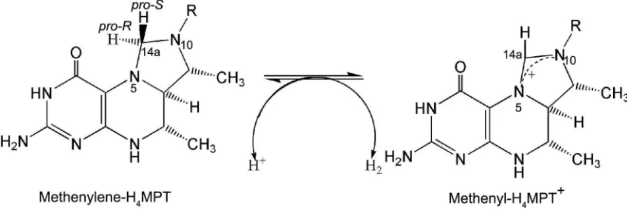
The sequence identity of these various mtd genes is at least 51 %, determined by pairwise sequence alignment, which is high for different genera. The se-quence analysis of the mtd gene of M. thermoautotro-phicum indicates that the coenzyme F420 issituated at the N-terminus. The Mth, a novel type of hydrogenase, lacks iron-sulfur clusters, nickel, and flavins, but does contain an iron–guanylypyridinol cofactor (FeGP) as the prosthetic group, and is composed of one type of subunit with a molecular mass of approximately 43 kDa, and is highly O2-sensitive in dimeric form (Shima and Ermler, 2011). The catalytic mechanism of Mth is that a hydride from H2 is transferred to the pro-R site of the C-14a methenyl group of methenyl-H4MPT, which
leads to changes in hydrogen at the site of the C-14a methylene group of methylene-H4MPT (Figure 3). The gene encoding Mth from M. thermolithotrophicus has been cloned and sequenced. Its genomic sequence is 1059 bp long with ATG and TAA as initiation and ter-mination codons, and includes 39 mol % G+C content (Hartmann et al., 1996).
The methylene-H4MPT reductase catalyzes the re-duction from Methylene-H4MPT to methyl-H4MPT. Its TIM barrel structure contains a nonprolyl cis-peptide bond, but the binding sites of the substrates remain elu-sive, therefore only its crystal structure in a complex with F420 has been reported (Aufhammer et al., 2005). As a tetramer, it is composed of one type of subunit, each with a molecular mass of approximately 37 kDa with the absence of a chromophoric prosthetic group, such as flavins or iron-sulfur complexes. The gene encoding this reductase from M. thermoautotrophicum is 963-966 bp in length with ATG as start codon and TAA as end codon, and encodes a protein consisting of 321 amino acids with a molecular mass of 33.5 kDa and an approxi-mate isoeletric point of 4.5. The transcript of this gene is monocistronic (Nolling et al., 1995).
Reduction of Methyl-H4MPT to Methyl-S-CoM The coenzyme M (HS-CoM) and methyl-H4MPT:HS-CoM methyltransferase involve the reduc-tive process of Methyl-H4MPT to Methyl-S-CoM. The HS-CoM, as a methyl group carrier, takes part in the cru-cial step of CH4 formation through accepting the methyl group from methylcobalamin to form Methyl-S-CoM and is the smallest known organic cofactor. The structure of HS-CoM is 2, 2 - dithiodiethanesulfonic acid (Figure 2), and sulfopyruvate decarboxylase plays an important role in its biosynthesis (Sarmiento et al., 2013). In the rumen, some methanogenic archaea, such as M. ruminantium, cannot synthesize HS-COM, whereas they can satisfy the growth requirement for the coenzyme by the supply of other methanogens having the ability to produce HS-COM (Krishnakumar et al., 2008).
The methyl-H4MPT:HS-CoM methyltransferase consisting of eight different subunits (MtrABCDEFGH) and containing 5-hydroxybenzimidazolyl cobamide as a prosthetic group is an integral membrane-bound enzyme complex, which catalyzes the methyl transfer through the generation of a sodium ion gradient across the membrane (Gottschalk and Thauer, 2001). The meth-yl transfer occurs in two steps; a methmeth-yl group is first transferred from Methyl-H4MPT to an enzyme-bound cobamide prosthetic group to form methylated cobam-ide and subsequently HS-CoM. Its transfer depends on the sodium ion gradient in the second step. Both cloning and sequencing of the corresponding genes reveal that the eight mtr genes form an operon (mtrEDCBAFGH). The operon is located between the methyl-S-CoM re-ductase I operon (mcr) and a downstream open read-ing frame predicted to encode a Na+/Ca+, K+ exchanger
(Harms et al., 1995), while the proposed function of the
latter is consistent with the methyltransferase in the gen-eration of a sodium gradient. The deduced sequences of MtrB, MtrC, MtrD, and MtrE suggest that they are extremely hydrophobic membrane proteins (Stupperich et al., 1993). On the contrary, MtrA containing corrinoid is thought to have a hydrophobic helical conformation and is only partially integrated into the cytoplasmic face of the cell membrane, based on the deduced sequence (Sauer and Thauer, 1998).
Reduction of methyl-S-CoM to CH4
The Methyl-CoM reductase catalyzes the reduction process from methyl-S-CoM to CH4, in which coenzyme B acts as the electron donor and coenzyme F430 as the prosthetic group (Figure 2). Coenzyme B is a colorless cofactor that contains a thiol group and an L-threonine phosphate group which can be specifically recognized by Methyl-CoM reductase. The thiol group displaces CH4 from M of methyl-CoM and L-threonine phosphate group binds to basic amino acids in Methyl-CoM reduc-tase (Mcr). The Methyl-CoM reducreduc-tase is a membrane-associated enzyme that contains two genetically distinct isozymes (Mcr I, Mcr II), both are composed of three dif-ferent subunits (McrABG), whereas the Mcr G subunit from Mcr I is 5 kDa smaller than that from Mcr II (Al-drich et al., 1987). The relative amounts of Mcr I, MCR II present in the cells are changed by concentrations of H2 and CO2 in the culture vessels. In general, Mcr I eas-ily adapts to the environment of low H2 concentration while Mcr II can exist in an environment of high H2 con-centration. In the genomic sequences of M. ruminants and M. AbM4, only Mcr I has been found to date.
Mcr I plays an important role sensitive to supplied H2 in the pathway of CO2 reduction for methanogens (i.e. Ruminococcus albus) (Ntaikou et al., 2008; Leahy et al., 2010; Leahy et al., 2013). At present, the gene encoding McrA has often been used for the assessment of methanogen diversity in the rumen because Mcr is specific to methanogens (St-Pierre and Wright, 2013). The genes encoding the Mcr enzyme form an operon (mcrBDCGA) which contains two additional genes with unknown functions. In the gene sequence, there are five open reading frames, of which the largest is located at the 3' end and the second largest at the 5' end while the others are situated between these two open reading frames. The known genes (mcrA, mcrB, mcrG) have a strong preference for the codon with a C in the wobble position (Bokranz et al., 1988).
Pathway of formate oxidation
Formate utilization starts with oxidation to CO2 and subsequently enters the pathway of CO2 reduction. Formate dehydrogenase (Fdh) catalyzes the process of formate to CO2+H2 and the enzyme contains molybde-num, as a part of the molybdopterin cofactor, flavin ad-enine diuncleotide, zinc, iron, inorganic sulfur and two distinct subunits (FdhA, FdhB) in an ab configuration. The genes encoding the two subunits from M. formicium overlap by 1 bp. In the entire genomic sequence, the content of adenine phosphate + thymine (A+T) is 59 mol %, while in the fdhA gene sequence the A+T con-tent is 71 mol % (Shuber et al., 1986). In the process of gene expression, the fdhA and fdhB genes are co-transcribed and the starting site lies in 4.3 kb upstream of the fdhA gene (Patel and Ferry, 1988). Recently, the genes for the dehydrogenase from M. maripaludis have begun to attract the attention of researchers. The fdh ge-nome contains two important gene clusters (fdh1, fdh2), one of which (fdh1) plays an important role in the trans-fer process of formate to CO2+H2 (Lupa et al., 2008) and can encode two subunits and a putative formate trans-porter while the other (fdh2) encodes only two subunits (Wood et al., 2003).
Pathway of acetate dissimilation
Acetate is not only an important intermediate in the anaerobic fermentation of carbohydrate but also a key precursor of methanogenesis by methanogens, in which only two methanogenic genera are currently known to be capable of utilizing acetate to produce CH4.
One is Methanosarcina and the other is Methanosaeta (Methanothrix). The distinction between them is that Methanosarcina has the ability to utilize acetate, H2 + CO2 and methanol, whereas Methanosaeta is capable of utilizing acetate only. Furthermore, the two genera have different affinities. Methanosaetamainly grows in an en-vironment in which the acetate concentration is below l mM, but Methanosarcina prefer higher acetate concen-trations. Thus, the pathway of acetate dissimilation can be further divided into two branches via Methanosarcina and Methanosaeta. Additionally, the physiological range of acetate concentration is from 15 mM to 55 mM in the rumen, which should favor the growth of Methano-sarcina rather than Methanosaeta. To date, Methanosaeta has not been found in the rumen but might exist in the rumen, which is somewhat surprising due to its wide existence in natural habitats (Smith and Ingram-Smith, 2007) and its optimum growth temperature (35-45 oC),
similar to that in the rumen.
Methanogenesis from acetate in Methanosarcina
The pathway of utilizing acetate to produce CH4 for Methanosarcina is shown in Figure 4. In this path-way, acetate is firstly transformed to acetyl-CoA by ac-etate kinase (Ack) and phosphotransacetylase (Pta) and is then cleaved to enzyme-bound methyl and carbonyl groups by the CO dehydrogenase/acetyl-CoA synthase complex (Codh). In this cleavage process the carbonyl group is oxidized to CO2 and electrons are transferred to ferredoxin. Meanwhile, the Codh transfers the methyl group to H4MPT, yielding methyl-H4MPT. The
H4MPT:HS-CoM methyltransferase catalyzes the reac-tion from methyl-H4MPT to Methyl-S-CoM which is then reductively to CH4 by methyl-CoM reductase. The process from methyl group to CH4 is a reduction reac-tion with electrons originating from the oxidareac-tion of car-boxyl group of the CH3CO-S-CoA (acetyl-CoA) to CO2. The proton gradient produced by a membrance-bound electron transport chain is used to drive ATP synthesis. The methyltransferase and methyl-CoM reductase have been described in detail earlier as part of the pathway of CO2 reduction and, therefore, only Ack, Pta and Codh are described here.
Acetate kinase and phosphotransacetylase
In the pathway, acetate kinase catalyzes the synthesis of acetyl phosphate by transfer of the ATP
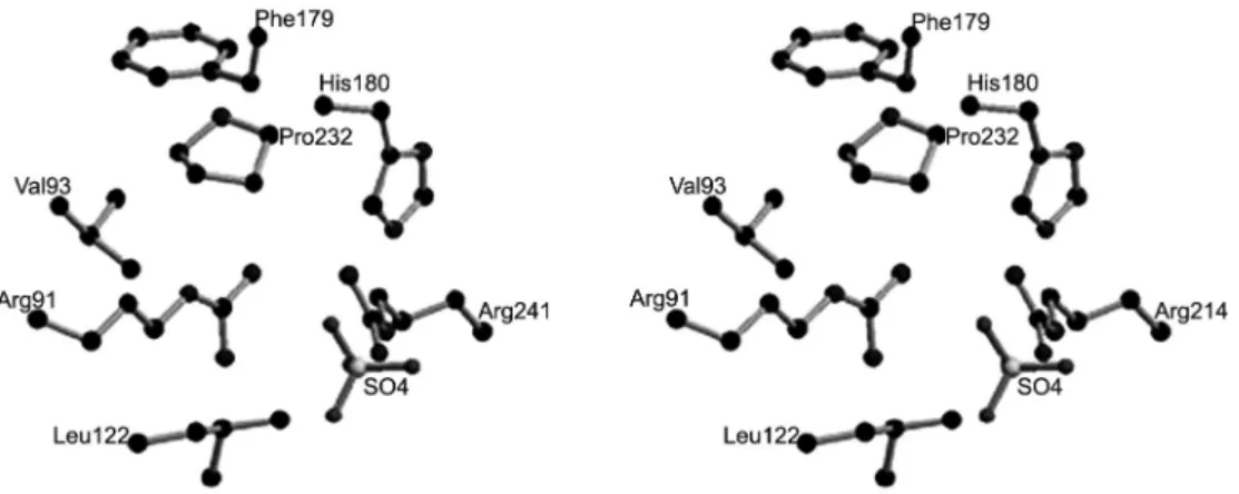
g-phosphoryl group to acetate, phosphotransacetylase catalyzes the transfer of the acetyl moiety to CoA to form CH3CO-S-CoA (acetyl-CoA) and orthophosphate. Acetate kinase contains a hydrophobic pocket for bind-ing the methyl group of acetate and forms the residues Val93,Phe179,Pro232 and Leu122 (Figure 5) (Ingram-Smith et
al., 2005). The catalytic mechanism of acetate kinase is a direct in–line transfer (Figure 6) (Ferry, 2011), and the catalytic process can be completed by nucleophilic
at-Figure 5 − Stereo view of the proposed acetate binding site in the acetate kinase from M. thermophila. Adapted from Ingram-Smith (2005).
tack of the carboxyl group of acetate on the g-phosphate of ATP.
The phosphotransacetylase with a molecular weight of 71 kDa is a dimer, classified as an AB di-mer and a CD didi-mer in the crystalline and soluble state respectively. Each of four monomers is composed of two domains, named I and II. The two domains are approximately the same size and have an ab structure. Domain I is made up of a five-stranded b sheet (b3, b2,
b1, b4 and b11) which is surrounded by four A heli-ces and a small helix on each side. However, domain II includes a six-stranded b sheet (b10, b5, b6, b9, b7 and b8) surrounded on one side by three helices and on the opposite side by two helices (Iyer et al., 2004). The genes (pta and ack) encoding Pta and Ack from M. thermophila are arranged in an operon with pta up-stream of ack, similar to the situation with M. mazei (Tonouchi et al., 2002). The pta and ack genes encode polypeptides with a molecular weight of 35 kDa and 44 kDa respectively and are co-transcribed as a 2.4 kb polycistronic message.The molecular mass of the poly-peptide encoded by the ack gene is identical with that from M. mazei (44.5 kDa) (Tonouchi et al., 2002). The transcriptional starting site of the genes locates 27 bp upstream from the translational start of the pta gene and 24 bp downstream from a consensus archaeal boxA promoter sequence. The regulation of Pta and Ack syn-thesis occurs, at least in part, at the mRNA level based on northern blot analyses. At the same time, southern blot analysis indicates that only one copy of each gene is present per chromosome.
Carbon monoxide dehydrogenase/acetyl-CoA syn-thase complex (Codh)
The Codh is made of five different subunits (Cod-hABCDE) and plays a major role in the pathway. Through detergent treatment, the enzyme complex has been di-vided into two components, one of which is a nickel/ iron-sulfur enzyme including CodhA, CodhB and CodhE subunits (Murakami and Ragsdale, 2000), the other is a corrinoid/iron-sulfur enzyme consisting of CodhC and CodhD. The former mainly catalyzes CO oxidation and decomposes acetyl-CoA into the methyl, carbonyl and CoA moieties. The latter is involved in the transmeth-ylation reaction. Three metal clusters (A, B and C) are identified from the abe component by electron paramag-netic resonance spectroscopy. Cluster A, a novel Ni-X-[Fe4-S4]cluster in which X is an unknown bridging atom, functions as a synthesizer or splitter of the C-C and C-S bonds of acetyl-CoA. Cluster C, possessing CO dehydro-genase activity, is also a bimetallic Ni-X-[Fe4-S4] cluster. Cluster B, a conventional Fe4S4 center thought to shuttle electrons, has the same function as cluster A.
The codh genes encoding the five subunits of the Codh complex from M. thermophila have been cloned, sequenced and expressed. The sequence analysis of the codh genes indicates that the proteins encoding these five subunits have different molecular masses and
iso-electric points, and further suggests they do not belong to a same kind (Maupin-Furlow and Ferry, 1996). The codh genes and an additional open reading frame with unknown function are co-transcribed based on northern blot analysis. The result suggests the sixth subunit may be required for acetyl-CoA cleavage or maturation of the five subunit complex. The regulation of the Codh complex is at the level of transcription via three mecha-nisms, one is differential transcription initiation, another is early elongation termination and the last is transcrip-tion elongatranscrip-tion (Anderson et al., 2009).
Methanogenesis from acetate in Methanosaeta
The core step of the Methanosaeta pathway (Figure 4) is similar to that of Methanosarcina. The only distinc-tion between these two pathways is the use of different enzymes in the step from acetyl moiety of acetate to CoA forming CH3CO-S-CoA(acetyl-CoA). The former is cata-lyzed by acetyl-CoA synthetase (Acs), whereas Ack and Pta are involved in the latter.
Acetyl-CoA synthetase (Acs)
This enzyme comprises ADP-forming Acetyl-CoA synthetase involved in the formation of acetate and AMP-forming Acetyl-CoA synthetase being operative in the activation of acetate to acetyl-CoA. In the rumen, acetate is constantly produced by bacterial decomposi-tion of cellulose and the acetate concentradecomposi-tion is usu-ally high. Hence, activation of acetate to acetyl-CoA by AMP-forming Acetyl-CoA plays an important role in methanogenesis. The AMP-forming Acetyl-CoA from M. sothngenii is composed of three different subunits of 18-75 kDa, and sited in the same direction in the gene sequence (Eggen et al., 1991). However, in the genome of M. tharmophila, four distinct subunits forming the en-zyme are found (Berger et al., 2012). The subunit with a molecular mass of 75 kDa among all the subunits from both methanogens has the potential to synthesize Acetyl-CoA. According to the deduced sequence of Acetyl-CoA synthetase from both methanogens, the sequence simi-larity is high, and can reach 80 % identity for the 1990 amino acids.
Pathway of methanol conversion to methane This pathway is composed of two separate steps (Figure 1). First, the methanol:coenzyme methyltransfer-ase system catalyzes the methyl group of methanol and coenzyme M to form methyl-S-CoM directly. Second, the reduction of methyl-S-CoM to CH4 is catalyzed by meth-yl-CoM reductase. The second step is identical to that which previously describes the reduction of CO2 and, therefore, only the methanol:coenzyme methyltransfer-ase system is detailed here.
methanol:5-hydroxy-benzimidazolycobamide methyltransferase (Mt1) into 2-(methylthio)-ethanesulfonate. In the second reac-tion, 2-(Methylthio)-ethanesulfonate is reductively cleaved into CH4 and 2-mercaptoethanesulfonate by methylcobamide:coenzymeM methyltransferase (Mt2). The Mt1 component consists of two subunits (MtaB and MtaC). The MtaC harbors corrinoids as prosthetic groups and the MtaB transferring the methyl group of methanol to the corrinoid prosthetic group of MtaC con-tains a mole of zinc, and its activity is zinc-dependent (Sauer and Thauer, 1997). The Mt1 component binding the methyl group of methanol, when the cobalt atom of its corrinoid prosthetic groups is in the highly reduced state, can be reactivated under the oxidized state. The Mt2 component is composed simply of a subunit MtaA with a molecular mass of 36 kDa but without any pros-thetic groups.
The mtaA, mtaB and mtaC genes from M. barkeri encoding MtaA, MtaB and MtaC respectively, have been cloned and sequenced (Harms and Thauer, 1996). In the sequence, not only do the genes (mtaB and mtaC ) locate in the same area and form an operon mtaCB, but also the gene mtaB is situated directly downstream of mtaC, suggesting both subunits have a similar function. The mtaCB operon does not contain the gene mtaA, indicat-ing the mtaA has a function that is different from the other subunit. The same results have been demonstrated by northern blot analysis indicating that the gene mtaA is monocistronically transcribed while the other genes (mtaB and mtaC) are co-transcribed.
For three methanogenesis pathways (i.e., formate oxidation, acetate dissimilation and methanol dispor-tionation), information about their mechanisms is rela-tively limited in the rumen. Therefore, identification of some specific methanogens (i.e., Methanosarcina and Methanosaeta) and their genomic sequences, validation of acetate dissimilation and methanol disportionation in the rumen, and the contribution of each pathway to total enteric CH4 production should be emphasized at differ-ent growth and physiological stages and under differdiffer-ent dietary conditions in ruminants.
Final Remarks
The pathways of CO2 reduction and formate oxi-dation assuredly exist in the rumen, and they involve a process of complex reactions due to the many coen-zymes and encoen-zymes involved. The pathways of acetate and methanol metabolism might be present in the rumen dependent on species or genera of methanogens and the presence of precursors which can form CH4. In the fu-ture, the existence of unconfirmed pathways (pathways of acetate dissimilation as well as methanol conversion), proportional contribution to enteric CH4 production from each pathway at different physiological or growth stages, and their regulatory processes in ruminants need to be clarified. Meanwhile, more methanogens from the rumen should be isolated, identified and cultured to
fur-ther understand their specific functions. The genome se-quences or the cloning and sequencing of genes encod-ing the relevant enzymes for more specific methanogens should be studied to provide new insights into enzyme evolution, regulation of gene expression, mechanisms of catalysis, relationships between host species or diet and population structure of symbiotic methanogens, and to explore more effective technologies of mitigating enteric CH4 emissions.
Acknowledgement
The authors wish to thank the "Strategic Priority Research Program - Climate Change: Carbon Budget and Relevant Issues" (Grant No. XDA05020700) and Knowl-edge Innovation Program of CAS (Grant No. KZCX2-YW-455) of the Chinese Academy of Sciences, and Na-tional Natural Science Foundation of China (Grant No. 31320103917 and No.31001023) for their joint financial support.
References
Aldrich, H.C.; Beimborn, D.B.; Bokranz, M.; Schonheit, P. 1987. Immunocytochemical localization of methyl-coenzyme M reductase in Methanobacterium thermoautotrophicum. Archives of Microbiology147: 190-194.
Anderson, K.L.; Apolinario, E.E.; MacAuley, S.R.; Sowers, K.R. 2009. A 5' leader sequence regulates expression of Methanosarcinal CO dehydrogenase/acetyl coenzyme A synthase. Journal of Bacteriology191: 7123-7128.
Attwood, G.T.; Altermann, E.; Kelly, W.J.; Leahy, S.C.; Zhang, L.; Morrison, M. 2011. Exploring rumen methanogen genomes to identify targets for methane mitigation strategies. Animal Feed Science and Technology166-167: 65-75.
Aufhammer, S.W.; Warkentin, E.; Ermler, U.; Hagemeier, C.H.; Thauer, R.K.; Shima, S. 2005. Crystal structure of methylenetetrahydromethanopterin reductase (Mer) in complex with coenzyme F420: Architecture of the F420/FMN binding site of enzymes within the nonprolyl cis-peptide containing bacterial luciferase family. Protein Science14: 1840-1849.
Beauchemin, K.A.; Kreuzer, M.; O'Mara, F.; McAllister, T.A. 2008. Nutritional management for enteric methane abatement: a review. Australian Journal of Experimental Agriculture48: 21-27.
Berger, S.; Welte, C.; Deppenmeier, U. 2012. Acetate activation in Methanosaeta thermophila: characterization of the key enzymes pyrophosphatase and acetyl-CoA synthetase. Archaea 2012: 1-10.
Bokranz, M.; Baumner, G.; Allmansberger, R.; Ankelfuchs, D.; Klein, A. 1988. Cloning and characterization of the methyl-coenzyme M reductase genes from Methanobacterium thermoautotrophicum. Journal of Bacteriology170: 568-577. Denman, S.E.; Tomkins, N.W.; McSweeney, C.S. 2007.
Deppenmeier, U.; Johann, A.; Hartsch, T.; Merkl, R.; Schmitz, R.A.; Martinez-Arias, R.; Henne, A.; Wiezer, A.; Baumer, S.; Jacobi, C.; Brüggemann, H.; Lienard, T.; Christmann, A.; Bömeke, M.; Steckel, S.; Bhattacharyya, A.; Lykidis, A.; Overbeek, R.; Klenk, H.P.; Gunsalus, R.P.; Fritz, H.J.; Gottschalk, G. 2002. The genome of Methanosarcina mazei: evidence for lateral gene transfer between bacteria and archaea. Journal of Molecular Microbiology and Biotechnology4: 453-461.
Eggen, R.I.L.; Geerling, A.C.M.; Boshoven, A.B.P.; Devos, W.M. 1991. Cloning, sequence analysis, and functional expression of the acetyl coenzyme-A synthetase gene from Methanothrix soehngenii in escherichia coli. Journal of Bacteriology173: 6383-6389.
Ferry, J.G. 1999. Enzymology of one-carbon metabolism in methanogenic pathways. FEMS Microbiology Reviews 23: 13-38.
Ferry, J.G. 2011. Acetate kinase and phosphotransacetylase. Methods in Enzymology 494: 219-231.
Franzolin, R.; St-Pierre, B.; Northwood, K.; Wright, A.D.G. 2012. Analysis of rumen methanogen diversity in water buffaloes (bubalus bubalis) under three different diets. Microbial Ecology 64: 131-139.
Gottschalk, G.; Thauer, R.K. 2001. The Na+-translocating
methyltransferase complex from methanogenic archaea. Biochimica Et Biophysica Acta1505: 28-36.
Harms, U.; Thauer, R.K. 1996. Methylcobalamin: coenzyme M methyltransferase isoenzymes MtaA and MtbA from Methanosarcina barkeri: Cloning, sequencing and differential transcription of the encoding genes, and functional overexpression of the mtaA gene in Escherichia coli. European Journal of Biochemistry235: 653-659.
Harms, U.; Weiss, D.S.; Gartner, P.; Linder, D.; Thauer, R.K. 1995. The energy conserving N5-methyltetrahydromethanopterin:
coenzyme-M methyltransferase complex from Methanobacterium thermoautotrophicum is composed of eight different subunits. European Journal of Biochemistry228: 640-648.
Hartmann, G.C.; Klein, A.R.; Linder, M.; Thauer, R.K. 1996. Purification, properties and primary structure of H2-formingN5,
N10-methylenetetrahydromethanopterin dehydrogenase from Methanococcus thermolithotrophicus. Archives of Microbiology 165: 187-193.
Hendrickson, E.L.; Leigh, J.A. 2008. Roles of coenzyme F420 -reducing hydrogenases and hydrogen- and F420-dependent methylenetetrahydromethailopterin dehydrogenases in reduction of F420 and production of hydrogen during methanogenesis. Journal of Bacteriology190: 4818-4821.
Hochheimer, A.; Schmitz, R.A.; Thauer, R.K.; Hedderich, R. 1995. The tungsten formylmethanofuran dehydrogenase from
Methanobacterium thermoautotrophicum contains sequence motifs characteristic for enzymes containing molybdopterin dinucleotide. European Journal of Biochemistry 234: 910-920. Hungate, R.E.; Smith, W.; Bauchop, T.; Yu, I.; Rabinowitz,
J.C. 1970. Formate as an intermediate in the bovine rumen fermentation. Journal of Bacteriology102: 389-397.
Ingram-Smith, C.; Gorrell, A.; Lawrence, S.H.; Iyer, P.; Smith, K.; Ferry, J.G. 2005. Characterization of the acetate binding pocket in the Methanosarcina thermophila acetate kinase. Journal of Bacteriology187: 2386-2394.
Ingram-Smith, C.; Gorrell, A.; Lawrence, S.H.; Iyer, P.; Smith, K.; Ferry, J.G. 2005. Characterization of the acetate binding pocket in the Methanosarcina thermophila acetate kinase. Journal of Bacteriology187: 2386-2394.
Iyer, P.P.; Lawrence, S.H.; Luther, K.B.; Rajashankar, K.R.; Yennawar, H.P.; Ferry, J.G.; Schindelin, H. 2004. Crystal structure of phosphotransacetylase from the methanogenic archaeon Methanosarcina thermophila. Structure12: 559-567. Janssen, P.H.; Kirs, M. 2008. Structure of the archaeal community
of the rumen. Applied and Environmental Microbiology 74: 3619-3625.
Krishnakumar, A.M.; Sliwa, D.; Endrizzi, J.A.; Boyd, E.S.; Ensign, S.A.; Peters, J.W. 2008. Getting a handle on the role of coenzyme M in alkene metabolism. Microbiology and Molecular Biology Reviews72: 445-456.
Kumar, S.; Dagar, S.S.; Puniya, A.K. 2012. Isolation and characterization of methanogens from rumen of Murrah buffalo. Annals of Microbiology62: 345-350.
Kunow, J.; Shima, S.; Vorholt, J.A.; Thauer, R.K. 1996. Primary structure and properties of the formyltransferase from
the mesophilic Methanosarcina barkeri: Comparison with
the enzymes from thermophilic and hyperthermophilic methanogens. Archives of Microbiology165: 97-105.
Leahy, S.C.; Kelly, W.J.; Altermann, E.; Ronimus, R.S.; Yeoman, C.J.; Pacheco, D.M.; Li, D.; Kong, Z.H.; McTavish, S.; Sang, C.; Lambie, S.C.; Janssen, P.H.; Dey, D.; Attwood, G.T. 2010. The genome sequence of the rumen methanogen Methanobrevibacter ruminantium reveals new possibilities for controlling ruminant methane emissions. PLoS One5: 1-17.
Leahy, S.C.; Kelly, W.J.; Li, D.; Li, Y.; Altermann, E.; Lambie, S.C.; Cox, F.; Attwood, G.T. 2013. The complete genome sequence of
Methanobrevibacter sp. AbM4. Standards in Genomic Sciences8: 215-227.
Liu, Y.C.; Whitman, W.B. 2008. Metabolic, phylogenetic, and ecological diversity of the methanogenic archaea. Annals of the New York Academy of Sciences 1125: 171-189.
Lupa, B.; Hendrickson, E.L.; Leigh, J.A.; Whitman, W.B. 2008. Formate-dependent H2 production by the mesophilic methanogen Methanococcus maripaludis. Applied and Environmental Microbiology 74: 6584-6590.
Mamat, B.; Roth, A.; Grimm, C.; Ermler, U.; Tziatzios, C.; Schubert, D.; Thauer, R.K.; Shima, S. 2002. Crystal structures and enzymatic properties of three formyltransferases from archaea: environmental adaptation and evolutionary relationship. Protein Science11: 2168-2178.
Maupin-Furlow, J.A.; Ferry, J.G. 1996. Analysis of the CO dehydrogenase/ acetyl-coenzyme A synthase operon of Methanosarcina thermophila. Journal of Bacteriology178: 6849-6856.
Morgavi, D.P.; Forano, E.; Martin, C.; Newbold, C.J. 2010. Microbial ecosystem and methanogenesis in ruminants. Animal 4: 1024-1036.
Mukhopadhyay, B.; Purwantini, E.; Pihl, T.D.; Reeve, J.N.; Daniels, L. 1995. Cloning, sequencing, and transcriptional
analysis of the coenzyme F420-dependent
methylene-5,6,7,8-tetrahydromethanopterin dehydrogenase gene from
Murakami, E.; Ragsdale, S.W. 2000. Evidence for intersubunit communication during acetyl-CoA cleavage by the multienzyme CO dehydrogenase/acetyl-CoA synthase complex from
Methanosarcina thermophila: Evidence that the beta subunit catalyzes C-C and C-S bond cleavage. Journal of Biological Chemistry275: 4699-4707.
Neumann, L.; Weigand, E.; Most, E. 1999. Effect of methanol on methanogenesis and fermentation in the rumen simulation technique (RUSITEC). Journal of Animal Physiology and Animal Nutrition82: 142-149.
Nolling, J.; Pihl, T.D.; Reeve, J.N. 1995. Cloning, sequencing, and growth phase-dependent transcription of the coenzyme F420-dependent N5, N10-methylenetetrahydromethanopterin
reductase-encoding genes from Methanobacterium
thermoautotrophicumdelta H and methanopyrus kandleri. Journal of Bacteriology177: 7238-7244.
Ntaikou, I.; Gavala, H.N.; Kornaros, M.; Lyberatos, G. 2008. Hydrogen production from sugars and sweet sorghum biomass using Ruminococcus albus. International Journal of Hydrogen Energy33: 1153-1163.
Oppermann, R.; Nelson, W.O.; Brown, R.E. 1961. In vivo studies of methanogenesis in bovine rumen: dissimilation of acetate. Journal of General Microbiology25: 103-111.
Patel, P.S.; Ferry, J.G. 1988. Characterization of the upstream region of the formate dehydrogenase operon of Methanobacterium formicicum. Journal of Bacteriology170: 3390-3395.
Pei, C.X.; Mao, S.Y.; Cheng, Y.F.; Zhu, W.Y. 2010. Diversity, abundance and novel 16S rRNA gene sequences of methanogens in rumen liquid, solid and epithelium fractions of Jinnan cattle. Animal4: 20-29.
Rea, S.; Bowman, J.P.; Popovski, S.; Pimm, C.; Wright, A.D.G. 2007. Methanobrevibacter millerae sp nov and Methanobrevibacter olleyae sp nov., methanogens from the ovine and bovine rumen that can utilize formate for growth. International Journal of Systematic and Evolutionary Microbiology57: 450-456. Samuel, B.S.; Hansen, E.E.; Manchester, J.K.; Coutinho, P.M.;
Henrissat, B.; Fulton, R.; Latreille, P.; Kim, K.; Wilson, R.K.; Gordon, J.I. 2007. Genomic and metabolic adaptations of
Methanobrevibacter smithii to the human gut. Proceedings of the National Academy of Sciences of the United States of America 104: 10643-10648.
Sarmiento, F.; Ellison, C.K.; Whitman, W.B. 2013. Genetic confirmation of the role of sulfopyruvate decarboxylase in
coenzyme M biosynthesis in Methanococcus maripaludis.
Archaea 1-7. DOI: http://dx.doi.org/10.1155/2013/185250 Sauer, K.; Thauer, R.K. 1997. Methanol:coenzyme M
methyltransferase from Methanosarcina barkeri: zinc
dependence and thermodynamics of the methanol:cob(I)alamin methyltransferase reaction. European Journal of Biochemistry 249: 280-285.
Sauer, K.; Thauer, R.K. 1998. His(84) rather than His(35) is the active site histidine in the corrinoid protein MtrA of the energy conserving methyltransferase complex from Methanobacterium thermoautotrophicum. Febs Letters436: 401-402.
Shima, S.; Ermler, U. 2011. Structure and function of [Fe]-hydrogenase and its iron-guanylylpyridinol (FeGP) cofactor. European Journal of Inorganic Chemistry 7: 963-972.
Shuber, A.P.; Orr, E.C.; Recny, M.A.; Schendel, P.F.; May, H.D.; Schauer, N.L.; Ferry, J.G. 1986. cloning, expression, and nucleotide sequence of the formate dehydrogenase genes from
methanobacterium formicicum. Journal of Biological Chemistry 261: 12942-12947.
Sirohi, S.K.; Pandey, N.; Singh, B.; Puniya, A.K. 2010. Rumen methanogens: a review. Indian Journal of Microbiology 50: 253-262.
Smith, K.S.; Ingram-Smith, C. 2007. Methanosaeta, the forgotten methanogen? Trends In Microbiology15: 150-155.
St-Pierre, B.; Wright, A.D.G. 2013. Diversity of gut methanogens in herbivorous animals. Animal7: 49-56.
Stupperich, E.; Juza, A.; Hoppert, M.; Mayer, F. 1993. Cloning, sequencing and immunological characterization of the corrinoid-containing subunit of the N5-methyltetrahydromethanopterin:
coenzyme-M methyltransferase from Methanobacterium
thermoautotrophicum. European Journal of Biochemistry217: 115-121.
Tonouchi, A.; Nishizaki, Y.; Tohyama, H.; Takeda, K. 2002. Cloning of a gene encoding acetate kinase from Methanosarcina mazei 2-P isolated from a Japanese paddy field soil. Current Microbiology45: 390-393.
Tymensen, L.D.; McAllister, T.A. 2012. Community structure analysis of methanogens associated with rumen protozoa reveals bias in universal archaeal primers. Applied and Environmental Microbiology78: 4051-4056.
Upadhyay, V.; Demmer, U.; Warkentin, E.; Moll, J.; Shima, S.; Ermler, U. 2012. Structure and catalytic mechanism of N5, N10-methenyltetrahydromethanopterin cyclohydrolase.
Biochemistry51: 8435-8443.
Vogt, S.; Lyon, E.J.; Shima, S.; Thauer, R.K. 2008. The exchange activities of Fe hydrogenase (iron-sulfur-cluster-free hydrogenase) from methanogenic archaea in comparison with the exchange activities of FeFe and NiFe hydrogenases. Journal of Biological Inorganic Chemistry13: 97-106.
Vorholt, J.A.; Vaupel, M.; Thauer, R.K. 1996. A polyferredoxin with eight 4Fe-4S clusters as a subunit of molybdenum
formylmethanofuran dehydrogenase from Methanosarcina
barkeri. European Journal of Biochemistry236: 309-317. Woese, C.R.; Magrum, L.J.; Fox, G.E. 1978. Archaebacteria.
Journal of Molecular Evolution11: 245-251.
Wood, G.E.; Haydock, A.K.; Leigh, J.A. 2003. Function and regulation of the formate dehydrogenase genes of the methanogenic Archaeon Methanococcus maripaludis. Journal of Bacteriology185: 2548-2554.