Inbred F344 rats as a biologic model of
intra-abdominal sepsis
Ratos isogênicos F344 como modelo biológico de
sepsis intra-abdominal
Sueli Blanes DAMY1; Luci EBISUI1; Marilda Osti SPINELLI1;
Junko Takano OSAKA1; Erasmo Magalhães Castro de TOLOSA1;
Silvia Colleta Barreto da Costa ORTIZ1
CORRESPONDENCE TO: SUELI BLANES DAMY
DTAEP - CENTRO DE BIOTERISMO Faculdade de Medicina da Universidade de São Paulo
Av. Dr. Arnaldo, 455 01246-903 - São Paulo - SP e-mail: damy@biot.fm.usp.br
1- CENTRO DE BIOTERISMO da Faculdade de Medicina da Universidade de São Paulo, São Paulo – SP
SUMMARY
Aiming to design a biologic model of acute intra-abdominal sepsis for experimental studies, conventional, isogenic rats F344 were infected with the bacteria Escherichia coli (E.coli), strain 11775, serotype H7:01:K1. Six hours after inoculation with E. coli, the animals - males and females - showed the following symptoms: piloerection, hyperpnea and decreased motor activity. The lethal dosis (LD50), was 6 x 10⁵ CFU/ml, analyzed in 32 males and 32 females. The highest mortality rate was observed on the first 24 hours. Liver dysfunction, common in intra-abdominal sepsis, was evaluated at 0, 24, 48 and 168 hours after inoculation, by means of serum enzyme activities. Study of the migration of polymorphonuclear-neutrophil cells (PMN) and mononuclear-macrophage ones (MN) showed a significant increase of PMN in E. coli inoculated males (z > 4.7; P<0.003) and females (z > 6.2; P < 0.0003), when compared to control groups. As for MN cells, there were no differences between inoculated and control groups, in males (z = 2.3; P = 0.0107) and in females (z = 1.8; P = 0.0359) as well. In conclusion, these results show that inbred F344 rats are adequate biologic models for studies of acute, intra-abdominal sepsis.
KEY-WORDS: SEPSIS. Rats. Models, Biological.
INTRODUCTION
I
ntra-abdominal sepsis occurs in those patients with altered susceptibility resulting either from the implanting of a microorganism in the peritoneum during a bacteremia, or as consequence of lesions that injure the integrity of a viscus and give entrance to microorganisms which are part of the normal flora in the lesioned areas, or even due to irritants within the peritoneal cavity8.Study of the pathogenesis, prevention and treatment of intra-abdominal sepsis, by making use of biologic models that mimic some features of the disease in human beings, has been reported in several previous work3,5,11,14,15. The necessary
pre-requirements for the use of these work are easy handling, small length and both easy obtention and reproducibility of responses. The most numerous and well-known of the facultative, aerobic microorganisms in the stools of human beings and animals - E. coli - has a relevant role on the maintenance of normal, physiologic status of the host’s intestine, and may appear as an opportunistic pathogen when outside of its natural habitat9,12. There are chances for this microorganism to cause
primary peritonitis, via trans-wall migration, and studies of intra-abdominal sepsis in mankind showed E. coli as more frequently isolated, when compared with other intestinal bacteria8.
Microbial pathogenes within the host’s intra-abdominal cavity encounter three basic defense mechanisms: removal of particles through the lymphatic system; in situ elimination of the bacteria, by opsonization and phagocytosis; kidnapping of the bacteria, thus preventing its export to the bloodstream, through the products of the inflammatory response, whose fibrinogen-rich exsudate retains the bacteria on its meshes6. In early onset of the bacterial infection, the
prevalence of macrophages, in association with translymphatic absorption, represent the host’s first defense line, which works to eliminate the bacteria in their initial stage7.
Liver dysfunction is a frequent finding in bacterial sepsis. Andersson et al.1 and Martínková and Horák10 studied hepatic
dysfunction in experimental peritonitis in the rat by measuring serum levels of alkaline phosphatase (AP), aspartate aminotransferase (AST) and alanine aminotransferase (ALT).
MATERIAL AND METHOD
Animals
Males and females, inbred, F344 rats (FCFUSP, São Paulo, Brazil), weighing 100-150 g and 10-12 weeks old were used in all experiments. The animals, kept on a 12-hour, light-dark cycle, 22o C ± 2o C temperature, were fed standard grain
pellets (Nuvital, Curitiba, Brazil) and given free access to water. They were housed four per cage of polypropylene, on sterile hardwood bedding. All the animals used in this study were treat according to international ethic rules.
Inoculation with E. coli
An initial study of the lethal dosis (LD50) was undertaken by intra-peritoneal inoculation of 1.5 ml of E. coli suspensions, in different dilutions, in 08 rats per dilution. The used E. coli strain was the ATCC 11775, serotype H7:O1:K1. The bacteria was cultivated in heart-brain agar. For preparation of the suspensions, the bacteria were collected during the exponential growth stage and diluted in sterile 0,9% saline. Adjustment of the final concentration was achieved by Mac Farlands Scale. Control of units forming colonies (UFC/ml) was performed by the seeding of 10 µl of the bacteria suspended in Mac Conkey agar, and by reading after 18 - 24 hours of incubation, at 370C.
In parallel, and constituting the controls, groups of rats were inoculated with 1.5 ml of saline, via intra-peritoneal route. The animals were observed daily, for registrations of symptoms and deaths. All survivors were killed in ether chamber, after 7 days.
Quantification of leukocytes in the peritoneal wash
In order to study the migration of PMN and MN into the abdominal cavity, male and female groups of inbred, F344 rats were inoculated with 1 LD 50 of E. coli suspension. For liver function tests, at time intervals of 0, 24, 48 and 168 hours following infection, rats were anaesthetized with an association of Ketamine/Xylazine 25/5mg/kg, intramuscularly, and blood samples collected, by cardiac puncture. The same animals were then killed in carbon dioxide chamber and had 10 ml of saline injected into the peritoneal cavity. This dilution factor was included on the final calculation. After massaging the abdominal wall for one minute, a Pasteur pipette was introduced in the midline for the collecting of peritoneal wash. Total leukocytes count was performed on a CELM CC-510 cells counter. Samples were centrifuged for five minutes at 2000 rpm, surnatant was despised, and the sediment re-suspended in 0.5 ml of saline was homogenized and distributed in two slides. Cells were
RESULTS
Inbred, males and females, F344 rats, six hours after E. coli inoculation, showed the following symptoms: piloerection, hyperpnea and decreased motor activity. The dosis LD50 after 07 days was determined by the Reed and Muench method4, that found 6 x 10⁵ CFU/ml, in 32 males and
32 females analyzed. The greatest mortality rate was observed on the first 24 hours.
The results for the liver function tests are represented in Tab. 1, 2 and 3. The mean values reached for AP, AST and ALT serum levels were submitted to the non-parametric Mann-Whitney test, in order to verify the hypothesis of medians of the tested groups being equal to those of the controls, witha = 0.01.
There were no differences between E. coli infected males and controls, with regard to AP (z= 2.07; P>0.0192), AST (z=2.06; P>0.197) and ALT (z=1.3; P>0.0968) values. E. coli infected females showed significantly higher values for AP (z=2.64; P=0.0041) and AST (z=5.74; P< 0.00003) values. No differences were found between infected animals and controls, in regard to ALT (z=1.3; P>0.0968) values. of them in different periods of time following the
intra-peritoneal infection.
were counted, and neutrophils (PMN) and monocytes (MN) were differentiated. Final calculation of number of cells at each collecting point was achieved by multiplying the number of PMN or MN by the total number of cells registered on the counter, followed by division of the result by 100.
In parallel, control groups of males and females were inoculated with 1.5 ml of saline and submitted to the same procedures described for the infected groups of animals.
Liver function tests
From the described, same groups of animals, serum levels of AP, AST and ALT were determined by analyses of enzyme activities using the Cobas-Mira/Plus* (Roche, Germany) automated system. The collected blood was centrifuged at 2000 rpm for 10 minutes, serum was separated in tubes and stored at -200 C.
Statistical Analysis
Within each group and among groups, the means obtained were compared, by the variance analysis method3.
As some of the parameters were not normally distributed, or displayed unequal variance, the non-parametric, Mann-Whitney test, for comparison among groups, was then used, thereby testing the hypothesis that the values found in E. coli innoculated groups were the same as those in the control lots13. For uniformity in the presentation, the data on the
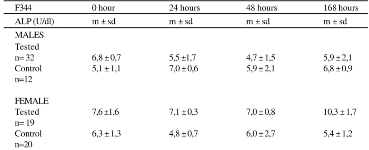
Table 1
Serum levels of AP of F344 rats inoculated with E.coli. São Paulo, 1999
In regard to males, there were no differences between tested animals and controls (z=2.07; P>0.0192). The tested females showed higher values (z=2.64; P=0.0041)
m ± sd = median, standard deviation; U/dl = Unit/deciliter; n=number of rats
F344 0 hour 24 hours 48 hours 168 hours
ALP (U/dl) m ± sd m ± sd m ± sd m ± sd
MALES Tested
n= 32 6,8 ± 0,7 5,5 ±1,7 4,7 ± 1,5 5,9 ± 2,1
Control 5,1 ± 1,1 7,0 ± 0,6 5,9 ± 2,1 6,8 ± 0,9
n=12
FEMALE
Tested 7,6 ±1,6 7,1 ± 0,3 7,0 ± 0,8 10,3 ± 1,7
n= 19
Control 6,3 ± 1,3 4,8 ± 0,7 6,0 ± 2,7 5,4 ± 1,2
n=20
F344 0 hour 24 hours 48 hours 168 hours
AP (U/dl) m ± sd m ± sd m ± sd m ± sd
MALES Tested
n=32 31,9 ± 7,0 32,7 ± 9,5 31,7 ± 9,2 30,3 ± 6,8
Control 35,1 ± 10,6 31,5 ± 10,2 30,7 ± 17,8 35,2 ± 2,9
n=12
FEMALE
Tested 23,0 ± 1,5 31,7 ± 9,3 40,8 ± 6,8 47,9 ± 1,4
n=19
Control 21,7 ± 3,6 24,8 ± 2,1 25,3 ± 2,2 23,2 ± 2,7
n=22
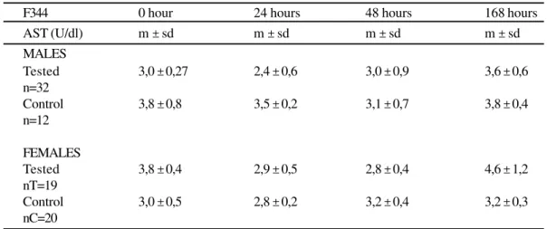
Table 2
Serum levels of AST of F344 rats inoculated with E.coli. São Paulo, 1999
In regard to males, there were no differences between tested animals and controls (z=2.06; P>0.0197). The tested females showed higher values (z=5.74; P=0.00003)
m ± sd = median, standard deviation; U/dl = Unit/deciliter; n=number of rats
peritoneal cavity is represented in Tab. 4 and 5. The Mann-Whitney, non-parametric test was applied in order to verify the hypothesis that medians of PMN and MN cell counts from the peritoneal wash could be equal to the medians found in the control groups, witha = 0.01. In the groups of
E. coli infected males the PMN cells count was higher than the count of PMN cells in the controls (z > 4.7; P < 0.003 ), and likewise for the females (z > 6.2; P< 0.0003). As for the count of MN cells, the same test showed no differences between E. coli tested groups and controls, considering both males ( z=2.3; P=0.0107) and females (z=1.8; P= 0.0359).
DISCUSSION
Oriented to experimental projects, biological models of acute intra-abdominal sepsis have been previously presented, making use of outbred Wistar and Sprague-Dawley rats. The Wistar ones are resistant to E. coli infection, having need of inocula containing bacterial pool associated to fecal contents and/or barium sulfate, or of pre-treatment with corticosteroids in order to become susceptible2,14,15. Sprague-Dawley rats are susceptible and
Table 3
Serum levels of ALT of F344 inoculated with E.coli. São Paulo, 1999
F344 0 hour 24 hours 48 hours 168 hours
AST (U/dl) m ± sd m ± sd m ± sd m ± sd
MALES
Tested 3,0 ± 0,27 2,4 ± 0,6 3,0 ± 0,9 3,6 ± 0,6
n=32
Control 3,8 ± 0,8 3,5 ± 0,2 3,1 ± 0,7 3,8 ± 0,4
n=12
FEMALES
Tested 3,8 ± 0,4 2,9 ± 0,5 2,8 ± 0,4 4,6 ± 1,2
nT=19
Control 3,0 ± 0,5 2,8 ± 0,2 3,2 ± 0,4 3,2 ± 0,3
nC=20
In regard to males and females, there were no differences between tested animals and controls (z=1.3; P>0.0968 for both, males and females)
m ± sd = median, standard deviation; U/dl = Unit/deciliter; n=number of rats
MALES
HOUR TESTED CONTROL
n=32 n=12
M sd m sd
0 hour 1,4 x 10⁵ ±0.7 0,9 x 10⁵ ±0.3
24 hours 3,3 x 10⁵ ±0.9 1,0 x 10⁵ ±0.3
48 hours 4,9 x 10⁵ ±1.1 1,0 x 10⁵ ±0.2
196 hours 2,8 x 10⁵ ±0.6 1,3 x 10⁵ ±0.1
FEMALE
HOUR TESTED CONTROL
n=20 n=20
M sd m sd
0 hour 0.8 x 10⁵ ±0.15 2.0 x 10⁵ ±0.96
24 hours 1.9 x 10⁵ ±0.4 1.8 x 10⁵ ±0.54
48 hours 5.3 x 10⁵ ±0.2 1.3 x 10⁵ ±0.42
168 hours 1.8 x 10⁵ ±0.4 1.0 x 10⁵ ±0.30
Table 4
Results of the differential count of PMN in F344 rats inoculated with E.coli. São Paulo, 1999
·In regard to males and females, there was significant difference between PMN of tested animals and PMN of controls (z³ 4,7; P< 0,0003; z³ 6,2; P < 0,0003)
·m ± sd = median, standard deviation; U/dl = Unit/deciliter; n= number of rats
In this study, the tested, inbred, F344 rats, males and females, showed susceptibility to infection by E. coli, strain ATCC 11775, serotype H7:01K1, therefore representing a new alternative for studies.
Besides the clinical symptoms, sepsis may be tracked through liver function tests as well. In accordance with data
in the studies of other researchers1,10, amongst the E. coli
MALES
HOUR TESTED CONTROL
n=32 n=20
M sd m sd
0 hour 0.6 x 10⁴ ±0.2 0.4 x 10⁴ ±0.3
24 hours 1.3x 10⁴ ±0.4 0.3 x 10⁴ ±0.15
48 hours 1.5 x 10⁴ ±0.6 0.4 x 10⁴ ±0.2
168 hours 0.9x 10⁴ ±0.3 0.2 x 10⁴ ±0.07
FEMALES
HOUR TESTED CONTROL
n=20 n=20
M sd m sd
0 hour 0.3 x 10⁴ ±0.1 0.5 x 10⁴ ±0.2
24 hours 0.6x 10⁴ ±0.2 0.6 x 10⁴ ±0.3
48 hours 2.0x 10⁴ ±1.3 0.5 x 10⁴ ±0.3
168 hours 0.3x 10⁴ ±0.2 0.5 x 10⁴ ±0.2
Table 5
Results of the differential count of MN in F344 rats inoculated with E.coli. São Paulo, 1999
In regard to males and females, there was no significant difference between MN of tested animals and MN of controls (z=2.3; P= 0.0107; z=18;P=0.0359).
m ± sd = median, standard deviation; U/dl = Unit/deciliter; n=number of rats
intra-abdominal sepsis in females of inbred, F344 rats, these two, last, liver function tests may be used to trace hepatic endangerment.
In relation to components of peritoneal defense, the PMN and MN migration into the peritoneal cavity was quantified at 0, 24, 48 and 168 hours post E. coli inoculation. The differences between the mean number of PMN cells in inoculated rats and the number of cells in those animals injected with saline were significant, with a greater increase detected on the 2nd day after inoculation (48 hours). As for
the mean number of MN cells in both inoculated groups and controls, no significant differences were found. The role of polymorphonuclear cells in phagocytosis and consequent cleaning of the intra-abdominal cavity has been shown by other authors2,6,7,14. However, the bacteria, as well as rats
strains used were different from the reported in this study. From Wistar rats14, infected with intra-abdominal,
fibrin clots-implant containing B. fragilis and E. coli, peritoneal cells were quantified by means of monoclonal antibodies, in order to characterize macrophages and neutrophils. Due to the presence of bacteria, PMN number increased rapidly, for when these were not associated to the fibrin clot, PMN influx was smaller. Six hours post-inoculation there was decrease of the PMN absolute number, returning to normality on the 7th day. MN cells were overcome
by PMN during the first hours.
The study done by Dunn et al.6, on self-cleaning of
the peritoneal cavity of Sprague-Dawley, E. coli inoculated
rats -unviable- reported that lymphatic absorption functions as a pump to withdraw suspended particles. Concurrently, bacteria also are phagocytized, which was confirmed by the use of non-viable bacteria, radioactively marked, that caused alterations in the leukocytes population within the peritoneal cavity. MN cells population remained relatively constant, whereas the influx of PMN was found to be larger one hour post-inoculation. Although MN and PMN cells within the peritoneal cavity exhibited similar phagocytosis capacity, the prevalence of macrophages in the beginning of the bacterial infection led to conclude that these cells, in addition to the lymphatic absorption, represent the host’s first defense line, which works to eliminate the bacteria, on their initial stages.
Altogether with the studies quoted in the previous paragraphs, the analysis of these results suggests that there may be a correlation between the time in which the host’s organism performs self-clean of the peritonium and the resistance to the infecting agent, once resistant, Wistar rats showed, after inoculation, an almost immediate augment of polymorphonuclear cells, Sprague-Dawley and F344 rats presented a later increase.
REFERENCES
1- ANDERSSON, A.; POULSEN, H. E.; AHRÉN, B. Effect of bile on liver function tests in experimental E.coli peritonitis in the rat.
Hepato-Gastroenterology, v. 38, p.388-390, 1991.
2- BARTLETT, J. G.; ONDERDONK, A. B.; LOUIE, T.; KASPER, D. L.; GORBACH, S. L. Lessons from an animal model of
intra-abdominal sepsis. Archives of Surgery, v. 113, p. 853-857, 1978.
3- BEIGUELMAN, B. Curso prático de estatística. Ribeirão Preto:
Revista Brasileira de Genética, 1991. p. 153-183.
4- BIER, O. Bacteriologia e imunologia. São Paulo, Editora
Melhoramentos - EDUSP, 1975. p. 171-172.
5- DAMY, S. B.; EBISUI, L.; SPINELLI, M. O.; SORBELLO, A.; OSAKA, J. T.; TOLOSA, E. M. C. Inbred mice A/SNELL, BALB/c
and C57BL/6 as animal models of intra-abdominal sepsis. Revista
de Medicina do Hospital Universitário, v.7, n. 2, p.36-42, 1997. 6- DUNN, D. L.; BARKE, R. A.; KNIGHT, N. B.; HUMPHREY, E. W.; SIMMONS, R. L. Role of resident macrophages, peripheral neutrophils, and translymphatic absorption in bacterial clearance
from peritoneal cavity. Infection and Immunity, v. 49, n. 3,
p.257-264, 1985.
7- DUNN, D. L.; BARKE, R. A.; EWALD, D. C.; SIMMONS, R. L. Macrophages and translymphatic absorption represent the first line of host defense of the peritoneal cavity. Archives of Surgery, v. 122, p. 105-110, 1987.
8- KASPER, D. L. Peritonitis. In: BRAUDE, A. I.; DAVIS, C. E.;
FIERER, J. Infectious diseases and medical microbiology.
Philadelphia: W.B.Saunders, 1987. p. 1004-1009.
RESUMO
Com o objetivo de estudar um modelo biológico de sepsis intra-abdominal aguda para estudos experimentais, foram infectados ratos isogênicos F344, convencionais, com a bactéria Escherichia coli (E.coli), cepa ATCC 11775, sorotipo H7:O1:K1. Os animais inoculados, machos e fêmeas, apresentaram 6 horas após a inoculação por E.coli os seguintes sintomas: arqueamento do dorso, piloereção, hiperpnéia e diminuição das atividades motoras. A dose que produziu 50% de mortalidade (DL50) após 7 dias, determinada pelo método Reed & Muench, foi de 6 x 10⁵ CFU/ml (analisado em 32 machos e 32 fêmeas). A maior concentração de mortalidade foi observada nas primeiras 24 horas. A disfunção hepática, comum em sepsis intra-abdominal, foi avaliada por provas enzimáticas, em 0, 24, 48 e 168 horas após a inoculação. O estudo da migração de células polimorfonucleares-neutrófilos (PMN) e mononucleares-macrófagos (MN) apontou um aumento significante de PMN entre o grupos de machos (z ³ 4,7; p < 0,003) e de fêmeas (z ³ 6,2; p < 0,0003) inoculados E.coli, quando comparados ao grupos controles. Quanto às células MN, não houve diferença entre os grupos inoculados e os controles, tanto para os machos (z=2,3; p = 0,0107), como para as fêmeas (z=1,8; p =0,0359). Em conclusão, estes resultados demonstram que os ratos isogênicos F344 são modelos biológicos adequados para estudos de sepsis intra-abdominal aguda.
PALAVRAS-CHAVE: SEPSE. Ratos. Modelos biológicos.
Received: 11/01/2001
ACKNOWLEDGEMENTS
We want to thank Dr. N. M. Sumita, from the Service of Clinical Biochemistry of Hospital das Clínicas; V.L.D.Urbano, I.Nishitokukado, A. V. Silva, R. J. Da Cruz for laboratorial support, and A. H. Griman for support in Informatics.
9- LEVINE, M. M. Escherichia coli that cause diarrhea: enterotoxigenic, enteropathogenic, enteroinvasive,
enterohemorrhagic and enteroadherent. The Journal of Infectious
Diseases, v. 155, n. 3, p. 377-389, 1987.
10-MARTÍNKOVÁ, I. R.; HÓRAK, V. A contribution to the model
of biliary infection in rats. Experiment and Toxicologic
Pathology, v.44, p. 102-104, 1992.
11-MATLOW, A. G.; BOHNEN, J. M. A.; NOHR, C.; CHRISTOU, N.; MEAKINS, J. Pathogenicity of enterococci in a rat model of
fecal peritonitis. The Journal of Infectious Diseases, v.160, n.
1, p.142-145, 1989.
12-SACK, B. Enterotoxigenic Escherichia coli: Identification and
characterization. The Journal of Infectious Diseases, v.142, n.
2, p. 279-286, 1980.
13-SIEGEL, S. Estatística não-paramétrica. São Paulo:
McGraw-Hill, 1975. p. 131-144.
14-VERWEIJ, W. R.; NAMAVAR, F.; SCHOUTEN, W. F.; MACLAREN, D. M. Early events after intra-abdominal infection with Bacteroides fragilis and Escherichia coli. Journal of Medical Microbiology, v. 35, p. 18-22, 1991.
15-WEINSTEIN, W. M.; ONDERDONK, A. B.; BARTLETT, J. G.; LOUIE, T. J.; GORBACH, S. L. Antimicrobial therapy of
experimental intra-abdominal sepsis. The Journal of Infectious
Diseases, v. 132, p. 282-286, 1975.