Effects of bromopride on expression of
metalloproteinases and interleukins
in left colonic anastomoses:
an experimental study
S.M. Silva
1, M.S. Jeroˆnimo
2, I. Silva-Pereira
3, A.L. Bocca
3and J.B. Sousa
4 1Programa de Po´s-Graduac¸a˜o em Cieˆncias Me´dicas, Faculdade de Medicina, Universidade de Brası´lia, Brası´lia, DF, Brasil 2Programa de Po´s-Graduac¸a˜o em Patologia Molecular, Faculdade de Medicina, Universidade de Brası´lia, Brası´lia, DF, Brasil 3Departamento de Biologia Celular, Instituto de Biologia, Universidade de Brası´lia, Brası´lia, DF, Brasil 4Departamento de Clı´nica Ciru´rgica, Faculdade de Medicina, Universidade de Brası´lia, Brası´lia, DF, BrasilAbstract
Anastomotic dehiscence is the most severe complication of colorectal surgery. Metalloproteinases (MMPs) and interleukins (ILs) can be used to analyze the healing process of anastomosis. To evaluate the effects of bromopride on MMP and cytokine gene expression in left colonic anastomoses in rats with or without induced abdominal sepsis, 80 rats were divided into two groups for euthanasia on the third or seventh postoperative day (POD). They were then divided into subgroups of 20 rats for sepsis induction or not, and then into subgroups of 10 rats for administration of bromopride or saline. Left colonic anastomosis was performed and abdominal sepsis was induced by cecal ligation and puncture. A colonic segment containing the anastomosis was removed for analysis of gene expression of MMP-1a, MMP-8, MMP-13, IL-b, IL-6, IL-10, tumor necrosis factor-a(TNF-a), and interferon-c(IFN-c). On the third POD, bromopride was associated with increased MMP-1a, MMP-13, IL-6, IFN-c, and IL-10 gene expression. On the seventh POD, all MMP transcripts became negatively modulated and all IL transcripts became positively modulated. In the presence of sepsis, bromopride administration increased MMP-8 and IFN-c
gene expression and decreased MMP-1, TNF-a, IL-6, and IL-10 gene expression on the third POD. On the seventh POD, we observed increased expression of MMP-13 and all cytokines, except for TNF-a. In conclusion, bromopride interferes with MMP and IL gene expression during anastomotic healing. Further studies are needed to correlate these changes with the healing process.
Key words: Bromopride; Matrix metalloproteinases; Interleukins; Surgical anastomosis; Sepsis; Rats
Introduction
Anastomotic dehiscence is the most feared complica-tion of colorectal surgery and may affect 2.4% to 3.8% of patients undergoing this procedure (1,2). Several experi-mental studies have been conducted to assess colonic anastomotic healing as well as factors contributing to anastomotic failure, such as fecal contamination and drug administration (3,4).
Prokinetic agents are often used in the early post-operative period to assist in gastric emptying and to ac-celerate the resolution of paralytic ileus, especially after intestinal surgery (5,6). The prokinetic drug class includes substituted benzamides, such as metoclopramide (meth-oxy-2-chloro-5-procainamide) and bromopride (4-amino-5-bromo-N-[2-(diethylamino)ethyl]-2-methoxybenzamide),
which act as dopamine antagonists. These drugs have both antiemetic and motility-stimulating effects (7). Bromopride is considered a good alternative to metoclopramide and other antidopaminergic drugs, with fewer reports of extrapyrami-dal side effects.
There is no consensus in the literature about the mechanism of action of prokinetic agents on anastomotic healing. Garcı´a-Olmo et al. (8) investigated the effects of pharmacological manipulation of postoperative intestinal motility on the resistance of colonic anastomoses and concluded that the use of metoclopramide was associated with fewer adhesions to the anastomosis and, conse-quently, with a significant decrease in anastomotic resis-tance 4 days after surgery.
Correspondence: S.M. Silva, SQS 405, Bloco B, Apto 205, 70239-020 Brası´lia, DF, Brasil. Fax: ++55-61-3244-4271. E-mail: [email protected]
Conversely, in previous studies we found that admin-istration of bromopride in the presence or absence of sepsis was associated with decreased tensile strength of left colonic anastomoses in rats 3 days after surgery, with no changes in adhesion formation (9,10), suggesting that other mechanisms may be involved. Therefore, the study of other healing parameters to elucidate the real action of bromopride on the healing process is needed.
Wound repair activates multiple biological pathways that can restore tissue integrity in a sequence of events and consists of three overlapping phases: inflammation, formation of new tissues, and remodeling (11).
In the inflammatory phase that dominates the healing process within the first days after wounding, leukocytes produce large amounts of cytokines that interact to deter-mine the vigor of the inflammatory response (12). These interactions are complex and produce both pro- and anti-inflammatory cytokines. The former include tumor necrosis factor-a(TNF-a), interleukin (IL)-1b, IL-6, and IL-8, while the latter include IL-4 and IL-10. The imbalance of cytokine action during healing could explain anastomotic failure.
Collagen deposition is a further key mechanism of wound healing (13). It is the most essential basal, skeletal protein involved in the healing process (11). Metallo-proteinases (MMPs) are a family of endopeptidases involved in the degradation of extracellular matrix compo-nents (14), coagulation, immunity, inflammation, angio-genesis, tissue removal, connective tissue remodeling, and epithelial cell and fibroblast migration (15,16). MMP-1, MMP-8, and MMP-13 are collagenases that cleave types I and III collagen fibers (14). An increased presence of active forms of MMPs may lead to localized matrix
degradation and a transient loss of anastomotic strength, thus leading to anastomotic dehiscence (17).
This study aimed to investigate whether the effect of bromopride on anastomotic healing could be explained by changes in MMP or cytokine gene expression caused by the drug.
Material and Methods
The study was approved by the Animal Ethics Committee of the Instituto de Cieˆncias Biologicas, Universidade de Brası´lia, (UnBDOC protocol No. 67336/ 2009), Brazil. All procedures were conducted in accordance with the Brazilian Guidelines for Use of Animals in Research.
Animals
Eighty healthy male Wistar rats (Rattus norvegicus) were used in the study. The animals were 90 to 120 days of age at the beginning of the experiment. For 2 weeks preoperatively, the animals were housed in cages with 5 animals each, kept under a standard 12:12-h artificial light-dark cycle, and had free access to water and standard chow. There was no preoperative fasting period.
Experimental protocol
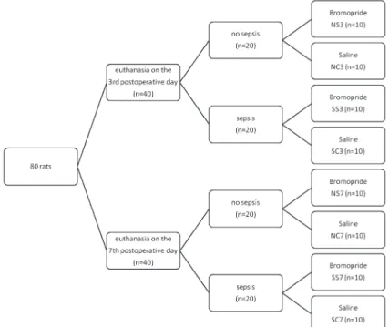
The rats were randomly divided into two groups of 40 animals each for euthanasia on the third or seventh postoperative day (POD). Further randomization was performed, and the animals were divided into subgroups of 20 rats each for sepsis induction or not, and then into subgroups of 10 rats each for administration of bromo-pride or saline (Figure 1).
General anesthesia was performed with 10 mg/kg xylazine hydrochloride and 75 mg/kg ketamine hydro-chloride administered intramuscularly. All surgical proce-dures were performed by the same surgeon.
The surgical site was shaved and disinfected with povidone-iodine. Clean, nonsterile surgical instruments were used. A 4.0-cm midline laparotomy incision was made, starting 1.0 cm above the external genitalia. The distal colon was exposed, and a 0.5-cm segment of the left colon (2.5 to 3.0 cm above the peritoneal reflection) was resected, followed by end-to-end anastomosis of the resulting segment, in a single transmural plane, using a continuous 6-0 polypropylene suture and a cylindrical needle.
After anastomosis, abdominal sepsis was experimen-tally induced in all rats in the sepsis group using the cecal ligation and puncture (CLP) method, as described by Rittirsch et al. (18), where one-half of the cecum was ligated using 3-0 silk suture (Shalon, Brazil). The end of the cecum, located distal to the ligation, was punctured at 10 random sites using an 18G61.50(40612 mm)
venipunc-ture needle, and some fecal content was extruded. The abdominal wall was closed in two planes with a simple running suture (3-0 silk suture).
Animals that died within the first 24 postoperative hours were excluded from the study. Metamizole sodium monohydrate (20 mg/kg) was added to the animals’ water for analgesia until the day of euthanasia. No antibiotics were used.
After the operations, animals in the experimental subgroups received bromopride at a dose of 1 mg/100 g subcutaneously every 12 h until the day of euthanasia. Control animals received identical amounts of saline subcutaneously every 12 h.
Reoperation was performed on the scheduled day for each subgroup. After exposure of the abdominal cavity, signs of anastomotic dehiscence were evaluated. A 4.0-cm colonic segment, with the anastomosis at the center, was removed as a whole together with any adherent structures. For euthanasia, the rats received an overdose of 150 mg/kg sodium thiopental injected directly into the inferior vena cava.
Specimen preparation
After resection of the adherent structures, specimens were opened at the antimesenteric border and divided into longitudinal sections for later analysis of gene expression of MMP-1a, MMP-8, MMP-13, IL-1b, IL-6, IL-10, TNF-a, and interferon-c(IFN-c).
The anastomotic segment was macerated in TRI Reagent solution (Applied Biosystems, USA), and RNA was isolated by a single extraction with a phenol-chloroform mixture and treated with Promega DNAse in the presence of an RNAse inhibitor (Promega, catalog#M6101; Lot#
262753; USA) in order to remove all traces of residual genomic DNA contamination, according to the protocol. Equal amounts of 0.4mg RNA were subjected to reverse
transcription with High Capacity cDNA Reverse Transcri-ption kits (Applied Biosystems).
Real-time quantitative reverse transcription polymer-ase chain reaction (PCR) was performed using a 7500 Fast Real-Time PCR System (Applied Biosystems). Amplification of cDNA sequences was performed using the PCR Master Mix kit (Applied Biosystems) containing the SYBR Green intercalating agent, a fluorescent mole-cule that intercalates into double-stranded DNA after initial denaturation at 956C for 15 s, followed by 606C for 1 min. To confirm amplification specificity, PCR products were analyzed by denaturation curves or melting curves. Comparisons were made using the threshold cycle or crossing point (19) method with cyclophilin to normalize values in order to assess the variation in fold-change expression of each gene. The fold-change expression represents the ratio of the measured value for the treated animals to the value for the nontreated cohorts. Values greater than 2.0 were considered positively modulated, whereas values less than 0.5 were negatively modulated. We considered that there was no change in gene expression in the range between these values. Reactions were performed in triplicate for all genes analyzed. All oligonucleotide pairs were designed using the Primer Express software (Applied Biosystems; listed in Table 1).
Results
Effects of bromopride on nonseptic (NS) rats
There were no deaths in either group on the third or seventh POD. One animal in the NS3 group and one in the NS7 group had anastomotic leakage blocked by adjacent organs, which was noticed only after opening the surgical specimen. No animals in the control group had anastomotic leakage.
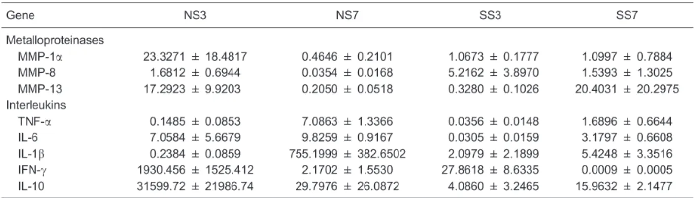
Administration of bromopride was associated with increased expression of MMP-1aand MMP-13 on the third POD. On the seventh POD, transcripts of all MMPs of all treated animals became negatively modulated (Table 2).
Administration of bromopride positively modulated IL-6, IFN-c, and IL-10 gene expression on the third POD and gene expression of all ILs on the seventh POD, especially IL-1b(Table 2).
Effects of bromopride on septic rats
There were no deaths among animals euthanized on the third POD. There was one death in each subgroup of animals scheduled to be euthanized on the seventh POD, none of them because of complications related to anastomosis.
On the third POD, administration of bromopride was associated with increased MMP-8 gene expression and decreased MMP-13 gene expression. On the seventh POD, only MMP-13 gene expression was increased (Table 2).
On the third POD, administration of bromopride was associated with increased IFN-c gene expression and decreased TNF-a, IL-6, and IL-10 gene expression. On the seventh POD, gene expression of all cytokines was positively modulated, except for TNF-a(Table 2).
Discussion
Bromopride is a prokinetic agent that is often used in the postoperative period, but its effect on the healing process has not been completely elucidated.
In the present study, administration of bromopride increased expression of MMP-1a and MMP-13 in the absence of abdominal sepsis and of MMP-8 in the presence of abdominal sepsis on the third POD. During the same period, MMP-13 gene expression decreased under septic conditions. On the seventh POD, transcripts of all MMPs of bromopride-treated animals became negatively modulated in the absence of abdominal sepsis, while MMP-13 gene expression increased in the presence of sepsis.
Increased rates of anastomotic dehiscence have been reported in patients with increased MMP-1, MMP-2, and MMP-9 levels in perioperative colonic biopsies (20). Stumpf et al. (21) demonstrated increased MMP-13 levels in intestinal samples of patients with anastomotic dehiscence compared to patients who did not have this complication, with no differences in MMP-1 levels between groups. Pas-ternak et al. (22) measured 1, 2, 3, MMP-7, MMP-8, MMP-9, and MMP-13 gene expression in the postoperative intraperitoneal fluid of patients after anterior resection of the rectum for cancer and found increased MMP-8 and MMP-9 levels in patients who developed
Table 2. Fold-change expression of metalloproteinases and interleukins assessed in septic and non-septic rats.
Gene NS3 NS7 SS3 SS7
Metalloproteinases
MMP-1a 23.3271 ± 18.4817 0.4646 ± 0.2101 1.0673 ± 0.1777 1.0997 ± 0.7884
MMP-8 1.6812 ± 0.6944 0.0354 ± 0.0168 5.2162 ± 3.8970 1.5393 ± 1.3025
MMP-13 17.2923 ± 9.9203 0.2050 ± 0.0518 0.3280 ± 0.1026 20.4031 ± 20.2975
Interleukins
TNF-a 0.1485 ± 0.0853 7.0863 ± 1.3366 0.0356 ± 0.0148 1.6896 ± 0.6644
IL-6 7.0584 ± 5.6679 9.8259 ± 0.9167 0.0305 ± 0.0159 3.1797 ± 0.6608
IL-1b 0.2384 ± 0.0859 755.1999 ± 382.6502 2.0979 ± 2.1899 5.4248 ± 3.3516
IFN-c 1930.456 ± 1525.412 2.1702 ± 1.5530 27.8618 ± 8.6335 0.0009 ± 0.0005 IL-10 31599.72 ± 21986.74 29.7976 ± 26.0872 4.0860 ± 3.2465 15.9632 ± 2.1477
The fold-change expression represents the ratio of the measured value for the treated animals to the value for the nontreated cohorts. Values greater than 2.0 were considered positively modulated, whereas values less than 0.5 were negatively modulated. MMP: matrix metalloproteinase; TNF-a: tumor necrosis factor-a; IFN-c: interferon-c; IL: interleukin; NS3, NS7: non-septic rats receiving bromopride euthanized on the 3rd and 7th postoperative day, respectively; SS3, SS7: septic rats receiving bromopride euthanized on the 3rd and 7th postoperative day, respectively.
Table 1. Genes and oligonucleotides used in real-time quantita-tive reverse transcription polymerase chain reaction (qRT-PCR).
Genes Oligonucleotides
Cyclophilin
Forward 59-TAT CTG CAC TGC CAA GAC TGA GTG-39 Reverse 59-CTT CTT GCT GGT CTT GCC ATT CC-39 TNF-a
Forward 59-AAA TGG GCT CCC TCT CAT CAG TCC-39 Reverse 59-TCT GCT TGG TGG TTT GCT ACG AC-39 IL-6
Forward 59-TCC TAC CCC AAC TCC CAA TGC TC-39 Reverse 59-TTG GAT GGT CTT GGT CCY YAG CC-39 IL-1b
Forward 59-CAC CTC TCA AGC AGA GCA CAG-39 Reverse 59-GGG TTG CAT GGT GAA GTC AAC-39 IL-10
Forward 59-CAG ACC CAC ATG CTC CGA GA-39 Reverse 59-CAA GGC TTG GCA ACC CAA GTA-39 IFN-c
Forward 59-GAT CCA GCA CAA AGC TGT CA-39 Reverse 59-GAC TCC TTT TCC GCT TCC TT-39 MMP-1a
Forward 59-AGG AGC ACT AAT GTT CCC CAG CT-39 Reverse 59-TGG GAT TTG GGG AAG GCC CAT A-39 MMP-8
Forward 59-GGA GTG TGC CAT CAA CCC TGA CC-39 Reverse 59-TGT CAC CAT GGT CTC TTG AGA CGA-39 MMP-13
Forward 59-ACC CAG CCC TAT CCC TTG ATG CC-39 Reverse 59-GGT GCA GAC GCC AGA AGA ATC TGT-39
dehiscence. Evaluations based on immunohistochemistry and zymography demonstrated increased levels of MMP-2, MMP-3, MMP-8, and MMP-9 in colonic anastomoses of rats (23), whereas MMP-10 and MMP-13 were not associated with the healing of intestinal anastomoses (24).
In a previous study, we found that positive modulation of MMP-1aand negative modulation of MMP-8 gene expres-sion were associated with loss of anastomotic strength of left colonic anastomoses in rats after CLP-induced abdom-inal sepsis on the third POD. On the seventh POD, anastomotic breaking strength in the same group was higher than that in saline-treated animals, but MMP-1aand MMP-8 gene expression remained unaltered (25). We can assume that the MMP effect on anastomotic healing is not dependent on the action of an isolated MMP, but is the result of the interaction of different classes of endopepti-dases. Moreover, the study of MMP activity by immunohis-tochemical staining or mRNA analysis does not necessarily mean that matrix degradation will occur, since strict control of MMP activity is preserved when MMPs are released as inactive proenzymes or by the presence of inhibitors (26).
Several authors have analyzed the variation in cytokine levels postoperatively and the occurrence of complications (27,28). Herwig et al. (28) reported a marked increase in IL-6 and TNF-alevels, which may be regarded as a precursor event that occurs days before the secondary clinical signs of anastomotic leakage. The authors also reported that the increase in IL-1b cytokine levels became statistically significant on the third POD in patients with anastomotic
dehiscence (28). In addition, a positive correlation has been described between IL-1b, IL-6, and TNF-alevels and prolonged operative time, blood loss, and bacterial contamination of peritoneal fluid (29).
In the present study, in the absence of sepsis, administration of bromopride positively modulated IL-6, IFN-c, and IL-10 gene expression on the third POD and gene expression of all ILs on the seventh POD. In the group of septic animals, administration of bromopride was associated with increased IFN-c gene expression and decreased TNF-a, IL-6, and IL-10 gene expression on the third POD. On the seventh POD, gene expression of all cytokines was positively modulated, except for TNF-a.
Therefore, we were not able to define a cytokine profile (pro- or anti-inflammatory) that could be respon-sible for the loss of strength of colonic anastomoses in the rat on the third POD during bromopride administration, since there is no consensus on which ILs are the most important cytokines in each phase of the healing process. It is worth mentioning, however, that detection of cytokine gene expression or even cytokine levels in tissues or secretions does not always correlate with its biological activity. Instead of being produced, cytokines may have been released as inactive proenzymes or may have already been degraded (30).
In conclusion, bromopride interferes with MMP and IL gene expression during anastomotic healing. Further studies are needed to correlate these changes with the healing process.
References
1. Platell C, Barwood N, Dorfmann G, Makin G. The incidence of anastomotic leaks in patients undergoing colorectal surgery.Colorectal Dis2007; 9: 71-79, doi: 10.1111/j.1463-1318.2006.01002.x.
2. Buchs NC, Gervaz P, Secic M, Bucher P, Mugnier-Konrad B, Morel P. Incidence, consequences, and risk factors for anastomotic dehiscence after colorectal surgery: a pro-spective monocentric study.Int J Colorectal Dis2008; 23: 265-270, doi: 10.1007/s00384-007-0399-3.
3. Thornton FJ, Barbul A. Healing in the gastrointestinal tract. Surg Clin North Am1997; 77: 549-573, doi: 10.1016/S0039-6109(05)70568-5.
4. de Sousa JB, Soares EG, Aprilli F. Effects of diclofenac sodium on intestinal anastomotic healing. Experimental study on the small intestine of rabbits.Dis Colon Rectum 1991; 34: 613-617, doi: 10.1007/BF02049903.
5. Verlinden M, Michiels G, Boghaert A, de Coster M, Dehertog P. Treatment of postoperative gastrointestinal atony. Br J Surg1987; 74: 614-617, doi: 10.1002/bjs.1800740727. 6. Jian R, Ducrot F, Piedeloup C, Mary JY, Najean Y, Bernier
JJ. Measurement of gastric emptying in dyspeptic patients: effect of a new gastrokinetic agent (cisapride).Gut1985; 26: 352-358, doi: 10.1136/gut.26.4.352.
7. Longo WE, Vernava AM III. Prokinetic agents for lower gastrointestinal motility disorders.Dis Colon Rectum1993; 36: 696-708, doi: 10.1007/BF02238599.
8. Garcı´a-Olmo D, Paya J, Lucas FJ, Garcı´a-Olmo DC. The effects of the pharmacological manipulation of postoperative intestinal motility on colonic anastomoses. An experimental study in a rat model.Int J Colorectal Dis1997; 12: 73-77, doi: 10.1007/s003840050084.
9. Marques e Silva S, Ferreira VM, Carneiro FP, Feres O, de Oliveira PG, de Sousa JB. Effects of bromopride on the healing of left colon anastomoses of rats.Rev Col Bras Cir 2011; 38: 429-434.
10. Silva SM, Carneiro FP, Oliveira PG, Morais PH, Silva NG, Sousa JB. Effects of bromopride on the healing of left colonic anastomoses in rats with induced abdominal sepsis.Acta Cir Bras 2012; 27: 370-375, doi: 10.1590/S0102-865020120 00600003.
11. Krzesniak N, Paziewska A, Rubel T, Skrzypczak M, Mikula M, Dzwonek A, et al. Gene expression alterations induced by low molecular weight heparin during bowel anastomosis healing in rats.Acta Biochim Pol2011; 58: 79-87. 12. Bertram P, Junge K, Schachtrupp A, Gotze C, Kunz D,
Schumpelick V. Peritoneal release of TNFalpha and IL-6 after elective colorectal surgery and anastomotic leakage.J Invest Surg2003; 16: 65-69, doi: 10.1080/08941930390194398. 13. Kuper MA, Scholzl N, Traub F, Mayer P, Weinreich J, Coerper
14. Stamenkovic I. Extracellular matrix remodelling: the role of matrix metalloproteinases. J Pathol 2003; 200: 448-464, doi: 10.1002/path.1400.
15. Agren MS, Jorgensen LN, Delaisse JM. Matrix metallopro-teinases and colon anastomosis repair: a new indication for pharmacological inhibition?Mini Rev Med Chem 2004; 4: 769-778.
16. Parks WC. Matrix metalloproteinases in repair.Wound Repair Regen 1999; 7: 423-432, doi: 10.1046/j.1524-475X.1999. 00423.x.
17. de Hingh I, Siemonsma MA, de Man BM, Lomme RM, Hendriks T. The matrix metalloproteinase inhibitor BB-94 improves the strength of intestinal anastomoses in the rat. Int J Colorectal Dis2002; 17: 348-354, doi: 10.1007/s00384-002-0418-3.
18. Rittirsch D, Huber-Lang MS, Flierl MA, Ward PA. Immunodesign of experimental sepsis by cecal ligation and puncture. Nat Protoc 2009; 4: 31-36, doi: 10.1038/ nprot.2008.214.
19. Livak KJ, Schmittgen TD. Analysis of relative gene expression data using real-time quantitative PCR and the 2(-Delta Delta C(T)) Method.Methods2001; 25: 402-408, doi: 10.1006/meth.2001.1262.
20. Stumpf M, Klinge U, Wilms A, Zabrocki R, Rosch R, Junge K, et al. Changes of the extracellular matrix as a risk factor for anastomotic leakage after large bowel surgery.Surgery 2005; 137: 229-234, doi: 10.1016/j.surg.2004.07.011. 21. Stumpf M, Cao W, Klinge U, Klosterhalfen B, Kasperk R,
Schumpelick V. Collagen distribution and expression of matrix metalloproteinases 1 and 13 in patients with anasto-motic leakage after large-bowel surgery.Langenbecks Arch Surg2002; 386: 502-506, doi: 10.1007/s00423-001-0255-9. 22. Pasternak B, Matthiessen P, Jansson K, Andersson M, Aspenberg P. Elevated intraperitoneal matrix metalloprotei-nases-8 and -9 in patients who develop anastomotic
leakage after rectal cancer surgery: a pilot study. Colorectal Dis2010; 12: e93-e98.
23. Shaper KR, Savage FJ, Hembry RM, Boulos PB. Regulation of matrix metalloproteinases in a model of colonic wound healing in a rabbit.Dis Colon Rectum2001; 44: 1857-1866, doi: 10.1007/BF02234468.
24. Vaalamo M, Karjalainen-Lindsberg ML, Puolakkainen P, Kere J, Saarialho-Kere U. Distinct expression profiles of stromelysin-2 (MMP-10), collagenase-3 (MMP-13), macro-phage metalloelastase (MMP-12), and tissue inhibitor of metalloproteinases-3 (TIMP-3) in intestinal ulcerations.Am J Pathol1998; 152: 1005-1014.
25. Silva SM, Jeronimo MS, Silva-Pereira I, Bocca AL, Sousa JB. Expression of metalloproteinases and interleukins on anastomoses in septic rats.J Surg Res2013; 183: 777-782, doi: 10.1016/j.jss.2013.02.012.
26. Brew K, Dinakarpandian D, Nagase H. Tissue inhibitors of metalloproteinases: evolution, structure and function. Biochim Biophys Acta2000; 1477: 267-283, doi: 10.1016/ S0167-4838(99)00279-4.
27. Baker EA, El-Gaddal S, Williams L, Leaper DJ. Profiles of inflammatory cytokines following colorectal surgery: rela-tionship with wound healing and outcome.Wound Repair Regen2006; 14: 566-572, doi: 10.1111/j.1743-6109.2006. 00163.x.
28. Herwig R, Glodny B, Kuhle C, Schluter B, Brinkmann OA, Strasser H, et al. Early identification of peritonitis by peritoneal cytokine measurement. Dis Colon Rectum 2002; 45: 514-521, doi: 10.1007/s10350-004-6231-z. 29. Tsukada K, Katoh H, Shiojima M, Suzuki T, Takenoshita S,
Nagamachi Y. Concentrations of cytokines in peritoneal fluid after abdominal surgery.Eur J Surg1993; 159: 475-479. 30. Witte MB, Barbul A. Repair of full-thickness bowel injury.