Adre no ce pto rs o f the me dial se ptal
are a mo dulate wate r intake and re nal
e xcre to ry functio n induce d by ce ntral
administratio n o f angio te nsin II
1Departamento de Fisiologia e Patologia, Faculdade de O dontologia,
Universidade Estadual Paulista, Araraquara, SP, Brasil
2Departamento de O dontologia, Universidade de Taubaté, Taubaté, SP, Brasil 3Departamento de Anestesiologia, Hospital Médico 9 de Julho, São Paulo, SP, Brasil 4Departamento de Cirurgia, Faculdade de Medicina, Universidade de São Paulo,
São Paulo, SP, Brasil W.A. Saad1,2,
I.F.M.S. Guarda3,
L.A.A. Camargo1,
T.A.F.B. Santos3,
S. Simões2 and
Willian A. Saad4
Abstract
We investigated the role of α-adrenergic antagonists and clonidine injected into the medial septal area (MSA) on water intake and the decrease in Na+, K+ and urine elicited by ANGII injection into the third
ventricle (3rdV). Male Holtzman rats with stainless steel cannulas implanted into the 3rdV and MSA were used. ANGII (12 nmol/µl) increased water intake (12.5 ± 1.7 ml/120 min). Clonidine (20 nmol/ µl) injected into the MSA reduced the ANGII-induced water intake (2.9 ± 0.5 ml/120 min). Pretreatment with 80 nmol/µl yohimbine or prazosin into the MSA also reduced the ANGII-induced water intake (3.0 ± 0.4 and 3.1 ± 0.2 ml/120 min, respectively). Yohimbine + prazosin + clonidine injected into the MSA abolished the ANGII-induced water intake (0.2 ± 0.1 and 0.2 ± 0.1 ml/120 min, respec-tively). ANGII reduced Na+ (23 ± 7 µEq/120 min), K+ (27 ± 3 µEq/120
min) and urine volume (4.3 ± 0.9 ml/120 min). Clonidine increased the parameters above. Clonidine injected into the MSA abolished the inhibitory effect of ANGII on urinary sodium. Yohimbine injected into the MSA also abolished the inhibitory effects of ANGII. Yohim-bine + clonidine attenuated the inhibitory effects of ANGII. Prazosin injected into the MSA did not cause changes in ANGII responses. Prazosin + clonidine attenuated the inhibitory effects of ANGII. The results showed that MSA injections of α1- and α2-antagonists
de-creased ANGII-induced water intake, and abolished the Na+, K+ and
urine decrease induced by ANGII into the 3rdV. These findings suggest the involvement of septal α1- and α2-adrenergic receptors in
water intake and electrolyte and urine excretion induced by central ANGII.
Co rre spo nde nce
W.A. Saad
Departamento de Fisiologia e Patologia
Faculdade de O dontologia, UNESP Rua Humaitá, 1680
14801-903 Araraquara, SP Brasil
Fax: + 55-16-201-6488 E-mail: silvana@ foar.unesp.br
Research supported by FAPESP, CNPq and FUNDUNESP.
Received February 8, 2001 Accepted June 11, 2002
Ke y words
•α-Adrenoceptors
•Angiotensin II
•Water intake
•Sodium
•Potassium
•Water excretion
Intro ductio n
Central administration of angiotensin II (ANGII) induces thirst in satiated animals by interacting with neurotransmitters, especially catecholamines (1,2). Adrenergic neurotrans-mitters from several hypothalamic areas may participate in the effect of ANGII regulating hydromineral fluid intake and renal
electro-lyte excretion in a process that involves α1
-and α2-adrenoceptors (3-5). Central
injec-tion of an α-adrenergic antagonist
suppres-ses water intake induced by intracerebroven-tricular (icv) ANGII (6,7). Several areas of the limbic system participate in the regula-tion of sodium, potassium and water excre-tion (8). Previous studies have demonstrated
the effects of α-antagonists and agonists
in-jected into the lateral hypothalamus on the water and sodium intake induced by ANGII injection into the subfornical organ (3). The adrenergic pathways of the septal area play an important role in the regulation of
electro-lyte and water excretion. α-Adrenoceptors
present an excitatory effect, whereas ß-adre-noceptors present an inhibitory effect (9,10). Rats with electrolytic lesions of the septal area drink more water than normal ones in response to thirst stimuli mediated by ANGII (11). Extensive neural pathways from cir-cumventricular structures to the septal area are involved in the regulation of fluid intake and cardiovascular regulation (12,13).
Clo-nidine, an α2-adrenergic receptor agonist,
has a potent and well-known antidipsogenic action (14-17). Peripheral or central injec-tion of clonidine reduces water intake in-duced by peripheral or central administra-tion of ANGII (18,19). Central treatment
with yohimbine (an α2-adrenergic receptor
antagonist) reduces the antidipsogenic ef-fect of clonidine, suggesting the
participa-tion of α2-adrenergic receptors in this effect
(18). Idazoxan (an α2-adrenergic and
imid-azoline receptor antagonist) reduced the ef-fect of clonidine on hypertonic NaCl and water intake (20). The natriuretic-kaliuretic
response elicited by cholinergic stimulation of the lateral hypothalamic area depends in part on synapses located in the medial septal area (MSA), the response elicited by cholin-ergic stimulation of the MSA also utilizes synapses located in the lateral hypothalamic area (21). Increased renal sodium and potas-sium excretion has been obtained with nor-adrenaline and other adrenergic drugs in-jected into the septal area (9). There is some evidence that fibers from the subfornical organ converge to the nucleus medianus and also project to the supraoptic nucleus, para-ventricular nucleus and throughout the lat-eral preoptic area-latlat-eral hypothalamic area (22). The central part of the subfornical or-gan, which is linked to circulating ANGII (23), also contains ANGII-immunoreactive terminal fields which seem to come from cells in the MSA (24). In view of the impor-tance of the circumventricular structures and MSA for the hydromineral balance in rats (9,25), as well as the important interac-tions between areas of the circumventricular structures and the MSA, we determined the effect of the injection of clonidine, yohim-bine and prazosin into the MSA on water intake and on the antinatriuretic, antikaliuretic and antidiuretic effects induced by the ad-ministration of ANGII into the third ven-tricle (3rdV).
Mate rial and Me thods
Anim als
Male Holtzman rats weighing 240-280 g at the beginning of the experiments were housed in individual metabolic cages.
Stan-dard Purina pellets (Na+ content 5 nmol/100
in four experiments at most.
Brain surge ry
After an acclimatization period of 7 days, the animals were restrained in a stereotaxic apparatus (Kopf model) and maintained un-der intraperitoneal tribromoethanol (20 mg/ 100 g body weight; Aldrich Chemical Com-pany Inc., Milwaukee, WI, USA) anesthesia throughout surgery. A longitudinal incision was made in the skin of the head of each animal, the subcutaneous tissue was pulled back and the skull was drilled with a spheri-cal drill. A stainless steel cannula (14 x 0.7 mm OD) was introduced into the MSA and another cannula (10 x 0.7 mm OD) was introduced into the 3rdV. The skull was positioned by having the bregma and lambda at the same level. The coordinates for ap-proaching the MSA and 3rdV were obtained from the Paxinos and Watson atlas (26). For the MSA the following coordinates were used: AP, 0.9 mm caudal to the bregma; L, 0.0 mm in the midline, and V, 4.6 mm below the dura mater. For the 3rdV, the following coordinates were used: AP, 0.2 mm caudal to bregma; L, 0.0 mm on the sagittal line, and V, 7.8 mm below the dura mater. The can-nula was fixed to the skull with screws and acrylic resin. A prophylactic dose of penicil-lin (30,000 IU) (Pentabiótico, Fontoura Wyeth, São Paulo, SP, Brazil) was given intramuscularly and presurgically. The in-sertion of a close-fitting stylet kept the lu-men free of debris and clots.
Intrace re bral inje ction te chnique s
Bolus intracranial injections were made after gently removing the animal from its cage, replacing the stylet with an injector that protruded 1.0 mm beyond the tip of the guide cannula in order to reach the MSA and 3rdV. This injector was connected by a PE-10 tubing to a PE-10-µl microsyringe, and a total volume of 1.0 µl was injected over a period
of 30-60 s. The stylet and injector were constantly wiped with cotton soaked in 70% alcohol. After the injection, the injector was removed, the stylet was replaced and the animals were returned to their cages so that we could observe the water intake, as well as renal sodium, potassium and water excre-tion.
D rugs
The drugs were dissolved in sterile0.15
M NaCl and injected into the MSA or 3rdV with a Hamilton microsyringe (10 µl) con-nected by PE-10 polyethylene tubing (25 cm) to a needle (0.3 mm OD), and intro-duced into the brain through the cannula previously fixed to the animals’ heads. The volume of injection was always 1 µl injected over a 30-60-s period.
The drugs used were clonidine
hydro-chloride (Boehringer-Ingelheim Ltd., Lon-don, UK), prazosin hydrochloride (Pfizer, Guarulhos, SP, Brazil), yohimbine hydro-chloride (Sigma, St. Louis, MO, USA), and
Ile5-angiotensin II (Sigma). Saline (0.15 M
NaCl) was used as control.
Histo lo gy
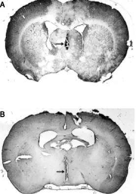
After the experiments, the animals were anesthetized with ether and perfused through the heart with saline and 10% formalin. The brain was removed and stored in 10% forma-lin for at least 1 week. It was then frozen and coronal sections (20-30 µm) were cut and stained with hematoxylin-eosin for exami-nation by light microscopy. Only the results of rats whose MSA and 3rdV were reached by the injection were used. Figure 1A and B shows the site of injection into the MSA and 3rdV as indicated by the arrows.
Statistical analysis
vari-ance. Values were considered to be statisti-cally significant when P<0.05. The New-man-Keuls post hoc test was used to assess the difference between individual means.
Expe rime ntal protocol
The study of water ingestion, as well as renal sodium, potassium and water excretion produced by injecting ANGII into the 3rdV was started 5 days after brain surgery. Water ingestion and the renal parameters were de-termined during different experimental ses-sions and in several groups of animals after the injection of the following drugs into the MSA and 3rdV of satiated rats: saline into the MSA and 3rdV (control); saline into the MSA and ANGII (12 nmol) into the 3rdV; clonidine (20 nmol) or yohimbine or prazo-sin (80 nmol) into the MSA and saline into the 3rdV; clonidine (20 nmol) or yohimbine or prazosin (80 nmol) into the MSA and ANGII into the 3rdV; yohimbine or prazosin (80 nmol) + clonidine (20 nmol) into the MSA and ANGII (12 nmol) into the 3rdV.
Water intake was recorded for 2 h after the injection of ANGII into the 3rdV. Prazo-sin and yohimbine were also injected 20 min before ANGII. When associated with cloni-dine, prazosin and yohimbine were injected 20 min before clonidine. The volume of water was measured using graduated (0.1 ml markings) tubes fitted with metal drinking spouts. No solid food was made available to the animals during the experiments.
Urine was collected after a period of solid food deprivation, and the animals were weighed and received a 5% water overload by gavage, consisting of a volume of water at 37ºC equal to their body weight. The animals were returned to their cages, with no water or solid food available. Excreted urine was collected into graduated tubes through a funnel located at the bottom of the cage. After 60 min the rats were submit-ted to a second water overload (5% of body weight) by gavage. Twenty minutes later, a control urine sample was collected and the drug was diluted in 1 µl 0.15 M NaCl solution and injected into the MSA/3rdV. Urine samples were collected 2 h after this injection. Sodium and potassium concentra-tions in the urine samples were measured with an IL-143 flame spectrophotometer (In-strumental Laboratories, Lexington, MA, USA).
Re sults
Effe ct of pre tre atme nt with intrase ptal
yohimbine and prazosin on the wate r intake
induce d by ANGII inje cte d into the 3rdV
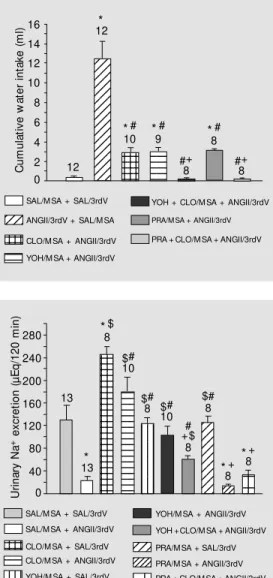
The injection of ANGII (12 nmol) into the 3rdV produced a progressive increase in water intake starting 30 min after injection. After injection of 0.15 M NaCl (control) into the MSA/3rdV, water intake was 0.4 ± 0.1 ml/120 min. The injection of ANGII into the 3rdV produced an increase in water intake to 12.5 ± 1.7 ml/120 min. Clonidine (20 nmol) injection into the MSA before ANGII
injec-Figure 1. Photomicrographs of a hematoxylin-eosin-stained trans-verse section of the rat brain show ing the sites (arrow s) of in-jection into the medial septal area (A) and into the third ven-tricle (B).
A A A A A
tion into the 3rdV decreased water intake to 2.9 ± 0.5 ml/120 min. Yohimbine (80 nmol) and prazosin (80 nmol) injected into the MSA before injection of ANGII into the 3rdV decreased the dipsogenic effect of ANGII, with water intake of 3.0 ± 0.4 and 3.1 ± 0.2 ml/120 min, respectively. Yohim-bine and prazosin injected before clonidine into the MSA, and before injection of ANGII into the 3rdV abolished the effect of ANGII,
reducing water intake to controlvalues, i.e.,
0.2 ± 0.1 and 0.2 ± 0.1 ml/120 min, respec-tively (Figure 2).
Effe ct of the inje ction of yohimbine or
prazosin be fore clonidine into the MSA
on re nal sodium e xcre tion afte r the
administration of ANGII into the 3rdV
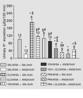
Injection of 0.15 M NaCl into the MSA/ 3rdV induced a urinary sodium excretion of 129 ± 27 µEq/120 min. Intracerebroven-tricular ANGII reduced sodium excretion to 23 ± 7.0 µEq/120 min. Clonidine injected into the MSA increased sodium excretion to 246 ± 14 µEq/120 min. Treatment with clo-nidine into the MSA reduced the inhibitory effect of ANGII on sodium excretion (179 ± 27 µEq/120 min). Sodium excretion after yohimbine administration into the MSA was 123 ± 11 µEq/120 min. Treatment with yo-himbine attenuated the inhibitory effect of ANGII on renal sodium excretion (103 ± 16 µEq/120 min). Sodium excretion after pra-zosin injection into the MSA was 125 ± 12 µEq/120 min. Prazosin produced no signifi-cant change in the inhibitory effect of ANGII on sodium excretion (14 ± 2 µEq/120 min). Treatment with yohimbine before clonidine and ANGII attenuated the decrease in so-dium excretion induced by ANGII, but with less intensity compared to clonidine injected before ANGII, with values of 59 ± 8 µEq/ 120 min. Treatment with prazosin before clonidine and ANGII produced no signifi-cant change in sodium excretion (33 ± 8 µEq/120 min) (Figure 3).
123 123 123 123 123 123 123 123 123 123 123 123 123 123 123 123 123 123 123 123 123 123 123 123 123 123 1234 1234 1234 1234 1234 1234 1234 1234 1234 1234 1234 1234 1234 1234 1234 1234 1234 1234 1234 123 123 123 123 123 123 123 123 123 123 123 123 123 123 12 12 12 12 12 12 12 12 12 12 123 123 123 123 123 U ri n a ry N a
+ e
x c re ti o n ( µ E q /1 2 0 m in ) 0 40 80 120 160 200 240 280 13 * 13 *$ 8 $# 10 $# 8 $# 10 *+ 8 *+ 8 # +$ 8 $# 8
SAL/M SA + SAL/3rdV
1234 1234 1234 1234 1234
CLO/M SA + SAL/3rdV
1234 1234
12345 12345
12345PRA/M SA + ANGII/3rdV 123
123
PRA + CLO/M SA + ANGII/3rdV YOH/M SA + SAL/3rdV
SAL/M SA + ANGII/3rdV
YOH/M SA + ANGII/3rdV
123 123
123PRA/M SA + SAL/3rdV
Figure 3. Urinary sodium excre-tion induced by intracerebroven-tricular injection of angiotensin II (ANGII, 12 nmol/µl) into the third ventricle (3rdV) of rats treated w ith intraseptal clonidine (CLO, 20 nmol/µl), yohimbine (YOH, 80 nmol/µl) or prazosin (PRA, 80 nmol/µl). The results are reported as means ± SEM . The number of animals is indicated at the top of each column. * P<0.05 com-pared to SAL/M SA + SAL/3rdV.
#P<0.05 compared to CLO/M SA
+ SAL/3rdV. $P<0.05 compared
t o SAL/M SA + ANGII/3rdV.
+P<0.05 compared to CLO/M SA
+ ANGII/3rdV (New man-Keuls
post hoc test). SAL, saline; M SA, medial septal area.
YOH + CLO/M SA + ANGII/3rdV
CLO/M SA + ANGII/3rdV
Figure 2. Cumulative w ater in-take induced by intracerebroven-tricular injection of angiotensin II (ANGII, 12 nm ol/µl) in rat s treated w ith intraseptal injection of clonidine (CLO, 20 nmol/µl), yohimbine (YOH, 80 nmol/µl) or prazosin (PRA, 80 nmol/µl). The number of animals is indicated at the top of each column. The results are reported as means ± SEM . * P< 0.05 com pared t o SAL/M SA + SAL/3rdV. #P<0.05
compared to SAL/M SA + ANGII/ 3rdV. +P< 0.05 com pared t o
CLO/M SA + ANGII/3rdV (New -man-Keuls post hoc test). SAL, saline; M SA, medial septal area; 3rdV, third ventricle.
Effe ct of the inje ction of yohimbine or
prazosin be fore clonidine into the MSA
on re nal potassium e xcre tion afte r the
application of ANGII into the 3rdV
Potassium excretion after injection of 0.15 M NaCl into the MSA/3rdV was 101 ± 12 µEq/120 min. ANGII injected into the 3rdV caused a reduction in renal potassium excretion to 27 ± 3 µEq/120 min. Treatment with clonidine into the MSA increased po-tassium excretion (191 ± 15 µEq/120 min). Intraseptal clonidine reduced icv ANGII-inhibited potassium excretion (117 ± 5 µEq/ 120 min). Potassium excretion after
yohim-C u m u la ti v e w a te r in ta k e ( m l) 16 123 123 123 123 123 123 123 123 123 123 123 123 123 123 123 123 123 123 123 123 123 123 123 1234 1234 1234 1234 1234 1234 14 12 10 8 6 4 2 0
SAL/M SA + SAL/3rdV
12 * 12 *# 10 * # 9 * # 8 #+ 8 #+ 8 123 123 1234 1234 1234 1234
ANGII/3rdV + SAL/M SA
CLO/M SA + ANGII/3rdV YOH/M SA + ANGII/3rdV
PRA/M SA + ANGII/3rdV
bine injection into the MSA was 106 ± 16 µEq/120 min. The decrease in renal potas-sium excretion after ANGII injection into the 3rdV was attenuated by yohimbine, with values of 91 ± 11 µEq/120 min. Yohimbine injected before clonidine and ANGII attenu-ated potassium excretion (52 ± 11 µEq/120 min). Potassium excretion after treatment with prazosin was 32 ± 7 µEq/120 min. Prazosin produced no change in the inhibi-tory effect of ANGII on renal potassium excretion (24 ± 6 µEq/120 min). Potassium excretion after treatment with prazosin in-jected before clonidine and ANGII was
45 ± 6 µEq/120 min (Figure 4).
Effe ct of the inje ction of yohimbine or
prazosin be fore clonidine into the MSA
on urine volume afte r inje ction of ANGII
into the 3rdV
Urine volume after injection of 0.15 M NaCl into the MSA/3rdV was 9.0 ± 2.0 ml/ 120 min. ANGII injected into the 3rdV caused a reduction of urine volume to 4.3 ± 0.9 ml/ 120 min. Treatment with clonidine into the MSA increased urine volume to 17.1 ± 0.9 ml/120 min. Clonidine abolished the inhibi-tory effect of ANGII on urine volume (11.9 ± 1.3 ml/120 min). Urine volume after intraseptal treatment with yohimbine was 9.7 ± 1.2 ml/120 min. Yohimbine attenuated the reduction of urine volume caused by ANGII (10.3 ± 0.9 ml/120 min). Yohimbine injected before clonidine and ANGII abol-ished the effect of clonidine on urine volume (5.9 ± 1.0 ml/120 min). The urine volume after intraseptal prazosin was 3.1 ± 0.8 ml/ 120 min. Prazosin produced no change in the urine volume caused by ANGII (2.8 ± 0.3 ml/120 min). Prazosin injected before cloni-dine and ANGII abolished the effect of clo-nidine on urine volume (5.8 ± 1.1 ml/120 min) (Figure 5).
D iscussio n
The present results show that the α2
-adrenoceptor agonist clonidine injected into the MSA attenuated the dipsogenic response produced by ANGII injection into the 3rdV.
Prior injection of the α1- and α2-adrenergic
receptor antagonists, prazosin and yohim-bine, into the MSA reduced the icv ANGII-induced water intake. However, it has been shown that central treatment with an adre-nergic agent also increases the water intake induced by ANGII in rats (5). Prazosin and phentolamine injected into the rostral hypo-thalamus attenuate the drinking response in-duced by icv ANGII injection (6). Thus, the
SAL/M SA + SAL/3rdV
1234 1234 1234 1234 1234 1234
CLO/M SA + SAL/3rdV CLO/M SA + ANGII/3rdV
1234 1234
1234YOH/M SA + SAL/3rdV
SAL/M SA + ANGII/3rdV 1234 1234 1234 1234 1234 1234 1234 1234 1234 1234 1234 1234 1234 1234 1234 1234 1234 1234 1234 1234 1234 1234 1234 1234 1234 1234 1234 123 123 123 123 123 123 123 123 123 123 123 123 123 123 123 123 123 123 123 123 123 123 123 123 123 123 123 123 123 123 123 123 123 123 123 12 12 12 12 12 123 123 123 123 123 123 12 12 12 12 12 12 U ri n a ry v o lu m e ( m l/ 1 2 0 m in ) 0.0 3.0 6.0 9.0 12.0 15.0 18.0 13 * 13 *$ 8 $# 10 $# 8 $# 10 +# 8 +# 8 +
*8#
1234 1234
1234PRA/M SA + ANGII/3rdV 123
YOH/M SA + ANGII/3rdV
123
123PRA/M SA + SAL/3rdV YOH + CLO/M SA + ANGII/3rdV
PRA + CLO/M SA + ANGII/3rdV
+
*8#
Figure 5. Urinary volume excre-tion induced by intracerebroven-tricular injection of angiotensin II (ANGII, 12 nmol/µl) into the third ventricle (3rdV) of rats treated w ith intraseptal clonidine (CLO, 20 nmol/µl), yohimbine (YOH, 80 nmol/µl) or prazosin (PRA, 80 nmol/µl). The results are reported as means ± SEM . The number of animals is indicated at the top of each column. * P<0.05 compared t o SAL/M SA + SAL/3rdV.
#P<0.05 compared to CLO/M SA
+ SAL/3rdV. $P<0.05 compared
t o SAL/M SA + ANGII/3rdV.
+P<0.05 compared to CLO/M SA
+ ANGII/3rdV (New man-Keuls
post hoc test). SAL, saline; M SA, medial septal area.
Figure 4. Urinary potassium ex-cretion induced by intracerebro-ventricular injection of angio-tensin II (ANGII, 12 nmol/µl) into the third ventricle (3rdV) of rats treated w ith intraseptal clonidine (CLO, 20 nmol/µl), yohimbine (YOH, 80 nmol/µl) or prazosin (PRA, 80 nmol/µl). The number of animals is indicated at the top of each column. The results are report ed as m eans ± SEM . * P<0.05 compared to SAL/M SA + SAL/3rdV. #P<0.05 compared
t o CLO/M SA + SAL/3rdV.
$P<0.05 compared to SAL/M SA
+ ANGII/3rdV. +P< 0.05
com-pared to CLO/M SA + ANGII/ 3rdV (New man-Keuls post hoc
test). SAL, saline; M SA, medial septal area.
SAL/M SA + SAL/3rdV
1234 1234 1234 1234 1234
CLO/M SA + SAL/3rdV CLO/M SA + ANGII/3rdV
1234 1234
YOH/M SA + SAL/3rdV SAL/M SA + ANGII/3rdV
123 123 123 123 123 123 123 123 123 123 123 123 123 123 123 123 123 123 123 123 123 123 123 123 123 123 123 123 123 123 123 123 123 123 123 123 123 123 123 123 123 123 123 123 123 123 12 12 12 12 1234 1234 1234 1234 12 12 12 12 U ri n a ry K
+ e
x c re ti o n ( µ E q /1 2 0 m in ) 0 40 80 120 160 200 240 280 13 * 13 *$ 8 $# 10 $#8 $#
10
+$ 8 +#
*$
8 $#+ 8
1234 1234
1234PRA/M SA + ANGII/3rdV 123
123
YOH/M SA + ANGII/3rdV
123
123PRA/M SA + SAL/3rdV YOH + CLO/M SA + ANGII/3rdV
PRA + CLO/M SA + ANGII/3rdV
*
inhibitory and facilitatory effects of nor-adrenaline injected into the central nervous system on ingestive behavior suggest a dual role for noradrenaline in the central control of water and salt intake induced by ANGII (27). The present results also show that ANGII injection into the 3rdV decreased sodium, potassium and water excretion in water-loaded rats. Injection of clonidine into the MSA abolished the antinatriuresis, anti-kaliuresis and antidiuresis induced by ANGII injected into the 3rdV. The decrease in so-dium, potassium and water excretion induced by ANGII injection into the 3rdV in water-loaded rats (with volume expansion) was abolished by pretreatment with yohimbine injected into the MSA. Although several studies have shown the natriuretic effect of ANGII, the present results showed the anti-natriuretic effect of ANGII injected into the 3rdV. Intracerebroventricular injection of ANGII produced a decrease in sodium ex-cretion. The different effects of ANGII on natriuresis suggest that central control of natriuresis may involve more than one mech-anism depending on the excitatory stimuli that activate different areas of the central nervous system. Urine volume was also re-duced by icv administration of ANGII (28). Other studies have demonstrated several in-teractions between adrenergic, cholinergic and angiotensinergic pathways in the central nervous system (3). Adrenergic stimulation of the MSA influences sodium and potas-sium excretion (29). Natriuresis resulting
from α2-adrenoceptor activation has been
demonstrated by the application of nor-adrenaline, clonidine, but not phenylephrine (30). The same occurs with injection of adre-nergic agonists or antagonists into the MSA on the renal excretory function response to icv ANGII. Taken together, these results show
that the central injection of an α-adrenergic
agonist or antagonist can disrupt the renal effect of ANGII in rats and suggest that the adrenergic pathways of the MSA can pro-duce a dual effect on electrolyte excretion.
Clonidine induced an increase in sodium, potassium and water excretion which was reduced by treatment with yohimbine and
prazosin by blocking the α2-adrenoceptor
subtype. The release of norepinephrine may be an excitatory step along the ANGII-induced sodium, potassium and water excre-tion pathway and when an adrenergic an-tagonist is used it can block these responses. In spite of this excitatory effect, the adrener-gic pathways could also be involved in an inhibitory system for electrolyte and water excretion. The participation of the lateral hypothalamic area in these mechanisms of water intake was also demonstrated (21). The autoradiographic localization of [3]-clonidine binding to non-adrenergic sites was
similar to, but distinct from, α2-adrenergic
receptors in human brain (31). Prazosin is
effective in blocking the natriuretic and
kali-uretic response to the α1-adrenoceptor
ago-nist phenylephrine (32). The present study shows that injection of prazosin into the MSA did not modify the inhibitory responses in-duced by ANGII injected into the 3rdV on sodium, potassium and water excretion. Clo-nidine induced natriuresis, kaliuresis and diuresis when injected into the MSA. Evi-dence has shown that clonidine, when cen-trally injected alone, produces diuresis, kali-uresis and natrikali-uresis (33-35). The dikali-uresis produced by the central administration of clonidine has been attributed to the inhibi-tion of vasopressin release (35) and natriure-sis has been attributed to renal sympatho-inhibition (34,36). The present results sug-gest that the facilitatory effect of clonidine on natriuresis and kaliuresis is mediated by
the activation of α1-adrenoceptors and the
inhibitory effects are mediated by α2A
di-uresis, kalidi-uresis, and pressor responses in a
process that involves α1- and α2
-adrenocep-tors (38). Thus, the α2-receptors of the MSA
play an inhibitory role and the α1-receptors
play an excitatory role in sodium, potassium and water excretion. Another important fact
is that the effect of α1- and α2-adrenoceptors
on ANGII, which affects water and electro-lyte regulation, is due to the release or inhibi-tion of vasopressin or atrial natriuretic fac-tor. The administration of clonidine has been reported to increase the circulating levels of atrial natriuretic factor (29). Vasopressin or atrial natriuretic factor has some influence on sodium, potassium and water.
The participation of imidazoline
recep-tors in the effect of α2-adrenoceptors has
been postulated. It has been demonstrated that rilmenidine, an imidazoline agonist, when injected into the paraventricular nu-cleus of the hypothalamus, decreases the hypertensive effect of ANGII injected into the 3rdV (39). It has also been observed that rilmenidine injection into the paraventricu-lar nucleus of the hypothalamus, prior to ANGII injection into the subfornical organ blocks the dipsogenic effect of ANGII (33).
These observations support the notion that yohimbine reversed the effect of ANGII when injected alone by blocking the imidazoline receptors. ANGII may produce antinatriure-sis, antikaliuresis and antidiuresis by acting on imidazoline receptors. This response was confirmed by yohimbine blocking the dine effect. Yohimbine injected with cloni-dine failed to fully reverse the effect of ANGII, probably due to the action of cloni-dine on the imidazoline receptors. These findings agree with the results of other stud-ies reporting that imidazoline drugs such as clonidine, rilmenidine and the catecholamine
α2-adrenoceptor agonist α
-methylnorepi-nephrine have distinct mechanisms of action (40).
Ackno wle dgm e nts
The authors thank José Vanderlei Menani and Laurival Antonio de Luca Jr. for helping to improve the manuscript. They also appre-ciate the technical assistance of Reginaldo C. Queiróz, Silas P. Barbosa, Silvana A.D. Malavolta and Silvia Foglia. We would also like to thank Ana V. Oliveira for animal care.
Re fe re nce s
1. Epstein AN, Fitzsimons JY & Rolls BJ (1970). Drinking induced by injection of angiotensin into the brain of the rat. Jour-nal of Physiology, 210: 457-474. 2. Summers C (1992). Norepinephrine
in-creases angiotensin II binding in the rat synaptosomes. Brain Research Bulletin, 28: 411-415.
3. Almeida NAA, Antunes VR, Saad WA & Camargo LAA (1999). Effects of alpha an-tagonists and agonists injected into lat-eral hypothalamus on the w ater and so-dium intake induced by angiotensin II in-jection into the subfornical organ. Brain Research Bulletin, 48: 521-525. 4. Bastos R, Saad WA, M enani JV, Renzi A,
Silveira JEN & Camargo LAA (1994). Role of adrenergic pathw ay of the medial pre-optic area in ANG II-induced w ater and renal excretion in rats. Brain Research, 636: 81-86.
5. Chan JYH, Pan S & Chan SHH (1991). Participation of adrenergic neurotransmis-sion in angiotensin II-induced dipsogenic behavior in the rat. Life Sciences, 48: 1293-1301.
6. Jones DL (1988). Hypothalamic alpha-adrenergic blockade modifies drinking and blood pressure responses to central an-giotensin II in conscious rats. Canadian Journalof Physiology and Pharmacology, 66: 1270-1277.
7. Severs W B, Sum m y-Long J, Danils-Severs A & Connor JD (1971). Influence of adrenergic blocking drugs on central angiotensin effects. Pharmacology, 5: 205-214.
8. Pillar AX, Silva-Netto CR, Camargo LAA, Saad WA, Antunes Rodrigues J & Covian M R (1977). Adrenergic stimulation of the lateral hypothalamic area on sodium and potassium excretion. Pharmacology,
Bio-chemistry and Behavior, 6: 147-149. 9. Camargo LAA, Saad WA, Silva-Netto CR,
Gentil CG, Antunes-Rodriques J & Covian M R (1976). Effects of catecholamines in-jected into the septal area of the rat brain on natriuresis, kaliuresis and diuresis. Ca-nadian Journal of Pharmacology, 54: 219-228.
10. Rocha M JA, Franci CR & Ant unes-Rodrigues J (1985). Participation of cho-linergic and adrenergic synapses of the medial septal area (M AS) in the natriuret-ic, kaliuretic and diuretic response to in-traventricular hypertonic saline (NaCl).
Physiology and Behavior, 34: 23-28. 11. Blass EM , Nussbaum IA & Hanson DG
A, De Luca Jr LA & M enani JV (1991). Pressor, natriuretic and kaliuretic re-sponse to central carbachol in rats w ith lesion of the medial septal area. Neurosci-ence Letters, 132: 195-198.
13. Saad WA, Antunes-Rodrigues J, Gentil CG & Covian M R (1972). Interaction betw een hypothalamus, amygdala and septal area in the control of sodium chloride intake.
Physiology and Behavior, 9: 629-636. 14. Callera JC, Camargo LAA, De Luca Jr LA,
M enani JV, Renzi A & Saad WA (1993). Clonidine and phenylephrine injected into the lateral preoptic area reduce w ater in-take in dehydrated rats. Journal of Phar-macology and Experimental Therapeutics, 46: 39-43.
15. Callera JC, Saad WA, Camargo LAA, Renzi A, De Luca Jr LA & M enani JV (1994). Role of the adrenergic pathw ays of the lateral hypothalamus on w ater intake and pressor response induced by the cholin-ergic activation of the medial septal area in rats. Neuroscience Letters, 167: 153-155.
16. Colombari E, Saad WA, Camargo LAA, Renzi A, De Luca Jr LA & M enani JV (1990). Role of the central α1 and α2
-adrenoceptors on the dipsogenic and car-diovascular effect of angiotensin II. Phar-macology, Biochemistry, and Behavior, 36: 893-896.
17. Ferrari AC, Camargo LAA, Saad WA, Renzi A, De Luca Jr LA & M enani JV (1991). Role of the alpha1- and alpha2-adreno-ceptors of the lateral hypothalamus in the dipsogenic response to central angio-tensin II in rat. Brain Research, 560: 291-296.
18. Fregly M J, Row land N & Greenleaf JE (1984). A role for presynaptic α2
-adreno-ceptors in angiotensin II-induced drinking in rats. Brain Research Bulletin,12: 393
-398.
19. Fregly M J, Row land N & Greenleaf JE (1984). Clonidine antagonist of angio-tensin-related drinking: A central site of action. Brain Research, 298: 321-327. 20. Yada M M , De Paula PM , M enani JV, Renzi
A, Camargo LAA, Saad WA & De Luca Jr LA (1997). Receptor-mediated effects of clonidine on need-induced 3% NaCl and w ater intake. Brain Research Bulletin, 42: 205-209.
21. Franci CR, Silva-Netto CR, Saad WA, Ca-margo LAA & Antunes-Rodrigues J (1980). Interaction betw een the lateral
hypotha-lamic area (LHA) and the medial septal area (M AS) in the control of sodium and potassium excretion in rats. Physiology and Behavior, 25: 801-806.
22. Thrasher TN (1989). Role of the forebrain circumventricular organs in body fluid bal-ance. Acta Physiologica Scandinavica, 583 (Suppl): 141-150.
23. Van Houtten M , Schiffrin EL, M ann JFE, Posner BI & Boucher R (1980). Radioauto-graphic localization of specific binding sites for blood born angiotensin II in rat brain. Brain Research, 186: 480-485. 24. Saad WA, Camargo LAA & Simões S
(1998). Effect of electrolytic and chemical lesion by ibotenic acid of the septal area on w ater and salt intake. Brain Research Bulletin, 47: 163-169.
25. Camargo LAA, M enani JV, Saad WA & Saad WA (1984). Interaction betw een ar-eas of the central nervous system in the control of w ater and arterial pressure in rats. Journal ofPhysiology, 350: 1-8. 26. Paxinos G & Watson C (1986). The Rat
Brain in Stereotaxic Coordinates Atlas. Academic Press, San Diego, CA, USA.
27. De Luca Jr LA, Camargo LAA, M enani JV, Renzi A & Saad WA (1994). On a possible dual role for central noradrenaline in the control of hydromineral fluid intake. Bra-zilian Journal of M edical and Biological Research, 27: 905-914.
28. Thunhorst RL & Johnson AK (1993). Ef-fects of arterial pressure on drinking and urinary responses to intracerebroventricu-lar angiotensin II. American Journal of Physiology, 264: R211-R217.
29. Saad WA, Guarda IFM S, Ferreira AC, Ca-margo LAA, Neto AFS, Santos TAFB & Saad WA (2000). Participation of alpha-1 and alpha-2 adrenoceptors of the hypo-thalamic area in w ater intake, and renal sodium, potassium and urinary volume ex-cretion induced by central administration of angiotensin II. Brain Research Bulletin, 52: 491-497.
30. Leite DF, Camargo LAA, Saad WA, Renzi A, Foglia S, De Luca Jr LA & M enani JV (1992). Role of cholinergic and adrenergic pathw ays of the medial septal area in the control of w ater intake and renal excre-tion in rats. Pharmacology, Biochemistry and Behavior, 42: 1-8.
31. Piletz JE, Ordw ay GA, Zhu H, Duncan BJ & Halaris A (2000). Autoradiographic com-parison of [3]-clonidine binding to non-adrenergic sites and alpha (2)-non-adrenergic
receptors in human brain. Neuropsycho-pharmacology,23: 697-708.
32. Saad WA, Camargo LAA & Saad WA (1984). Effect of application of alpha-1 and alpha-2 adrenoceptor agonists and an-tagonists into the ventromedial hypothala-mus on the sodium and potassium renal excretion. Pharmacology, 28: 228-234. 33. Arrais CAG, Saad WA, Camargo LAA,
Renzi A, Silveira JEN & Saad R (1997). Effect of rilmenidine injection into the paraventricular nucleus of the hypothala-mus on the w ater intake induced by appli-cation of angiotensin II to the subfornical organ. Journal of Physiology, 91: 97-98. 34. Patel KP & Zeigler DW (1993). Renal
nerves are involved in the natriuresis and diuresis produced by central administra-tion of clonidine in the rat. Proceedings of the Society for Experimental Biology and M edicine, 202: 81-87.
35. Roman R, Cow ley AW & Lechene C (1979). Water diuretic and antidiuretic ef-fect of clonidine in the rat. Journal of Phar-macology and Experimental Therapeutics, 211: 385-393.
36. Koepke JP & Dibona GF (1986). Central adrenergic control of renal function in con-scious hypertensive rats. Hypertension, 8: 133-141.
37. Saad WA, Camargo PAC & Camargo LA (1996). On a dual role for clonidine stimu-lation of the ventromedial nucleus of the hypothalamus in sodium and potassium renal excretion of rats. Pharmacology, 53: 281-288.
38. Silva RKP, Saad WA, Renzi A, M enani JV & Camargo LAA (1995). Effect of lateral hypothalamus lesion on the w ater and salt intake and sodium and urine excre-tion induced by activaexcre-tion of the medium preoptic nucleus in conscious rats. Jour-nal of the Autonomic Nervous System, 53: 195-204.
39. Saad WA, Camargo LAA, Silveira JEN, Saad WA & Camargo GM PA (1998). Imid-azoline receptors of the paraventricular nucleus on the pressor response induced by stimulation of the subfornical organ.
Journal of Physiology, 92: 25-30. 40. Sy GY, Bruban V, Bousquet P & Feldman